Budras Klaus-Dieter, Habel Robert E. Bovine Аnatomy
Подождите немного. Документ загружается.

h'
29 Cavernous venous plexuses
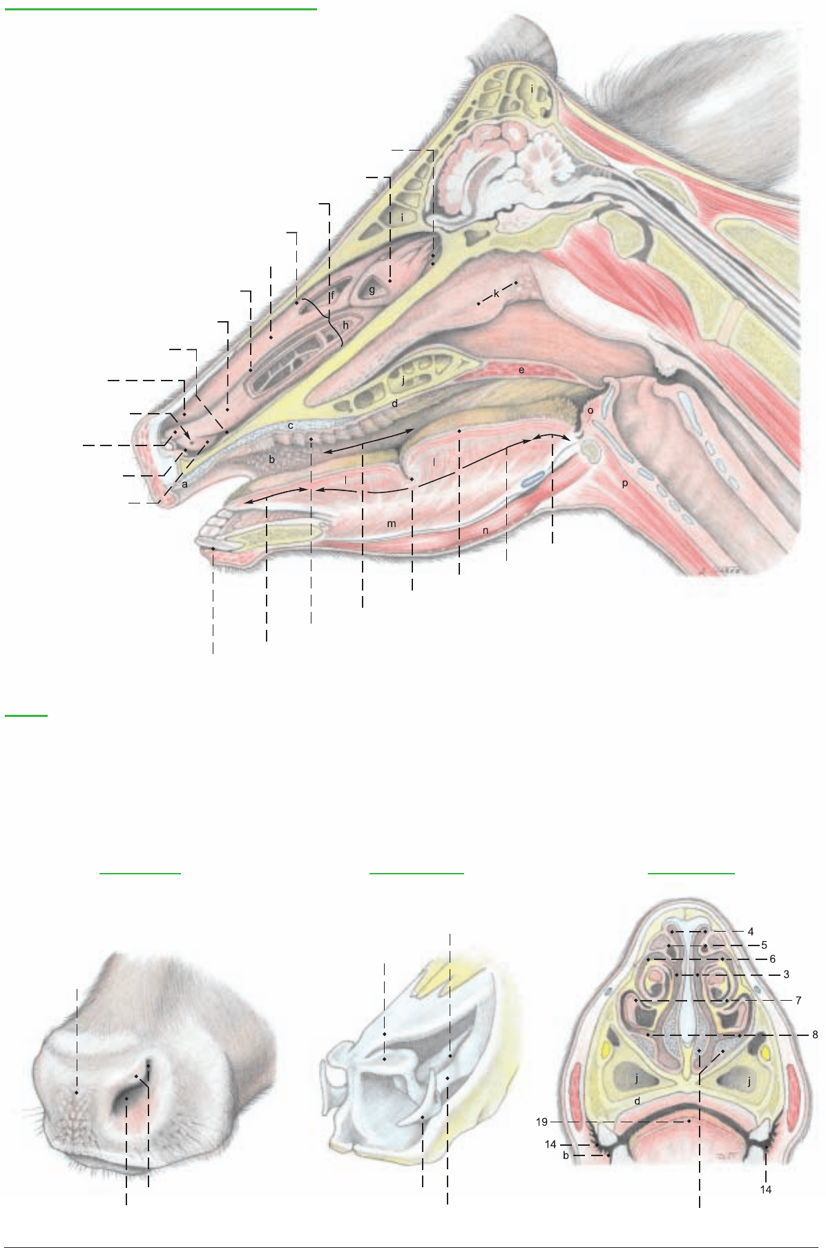
Nasal cavity, Oral cavity, and External nose
(Paramedian section)
1 Ethmoid conchae
2 Middle concha
3 Common meatus
4 Dorsal meatus
5 Dorsal concha
6 Middle meatus
7 Ventral concha
8 Ventral meatus
9 Straight fold
10 Nasolacrimal orifice
11 Alar fold
12 Nasal vestibule
13 Basal fold
14 Oral vestibule
15 Apex of tongue
16 Palatine ridges
17 Oral cavity proper
18 Lingual fossa
19 Torus of tongue
20 Body of tongue
21 Root of tongue
(See pp. 47, 49)
Legend:
a Dental pad
b Labial and buccal papillae
(See also text figure)
c Palatine venous plexus
d Hard palate
e Soft palate
f Dorsal conchal sinus
g Middle conchal sinus
h Ventral conchal sinus
h' Bulla and cells of
ventral concha
i Frontal sinus
j Palatine sinus
k Pharyngeal septum and
pharyngeal tonsil
l Proper lingual muscle
m Genioglossis
n Geniohyoideus
o Hyoepiglotticus
p Sternohyoideus
External nose
22 Planum
nasolabiale
23 Nostril
24 Alar groove and
ala nasi
Nasal cartilages
25 Med. accessory nasal cartilage
26 Dorsal lateral
nasal cartilage
27 Lat. accessory nasal cartilage
28 Ventral lat. nasal cartilage
Nasal conchae
45
Anatomie des Rindes englisch 09.09.2003 13:30 Uhr Seite 45
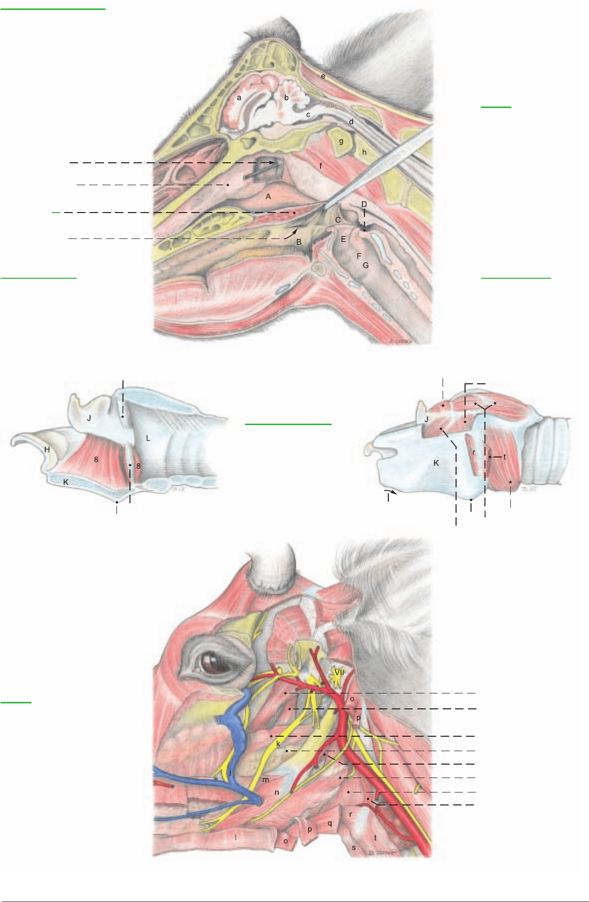
a) The cavity of the PHARYNX consists of three parts: the
oropharynx, laryngopharynx, and nasopharynx. The oropharynx
(pars oralis, B) communicates with the oral cavity through the isth-
mus of the fauces, which is bounded dorsally by the soft palate
(velum palatinum), ventrally by the tongue, and laterally by the
palatoglossal arches (p. 44, text fig.). The oropharynx extends to
the base of the epiglottis, and its lateral wall contains the palatine
tonsil (4, 14). The laryngopharynx (pars laryngea, D) lies below the
intrapharyngeal ostium, which is surrounded by the free border of
the soft palate (3) (raised by forceps) and the right and left
palatopharyngeal arches. The arches meet on the caudal wall over
the arytenoid cartilages. When the animal is breathing, the larynx
projects through the ostium into the nasopharynx, and the cavity of
the laryngopharynx is obliterated, except for the lateral piriform
recesses, which conduct saliva around the larynx to the esophagus
without the necessity of swallowing. In swallowing, the intrapha-
ryngeal ostium and the larynx are closed, and the function of the
laryngopharynx changes from respiratory to digestive. The caudal
part of the laryngopharynx (D) joins the esophagus over the cricoid
lamina without visible demarcation. The nasopharynx (pars
nasalis, A) extends from the choanae (p. 31, F) to the intrapharyn-
geal ostium, and is separated from the oropharynx by the soft
palate (3). The choanae are divided dorsally by the crest of the
vomer, covered by mucosa with a thick submucosal cavernous
venous plexus. Caudal to the vomer in ruminants, the membranous
pharyngeal septum (2) divides the dorsal part of the nasopharynx
lengthwise, and extends to the caudodorsal wall, where it contains
the pharyngeal tonsil (p. 45, k). On the wall of the nasopharynx lat-
eral to the tonsil, is a slit—the pharyngeal orifice of the auditory
tube (1), leading to the middle ear.
I. The pharyngeal muscles are identified from the lateral surface,
sparing the arteries and the pharyngeal branches of cranial nerves
IX and X, which innervate the muscles and the mucosa. (See p. 49.)
Muscles of the soft palate: The tensor veli palatini (11), has a super-
ficial part originating from the muscular process of the temporal
bone and terminating in a tendon that passes around the hamulus
of the pterygoid bone. The deep part originates on the pterygoid
bone and works in the opposite direction to open the auditory tube
by pulling on its cartilage.* The levator veli palatini (12) also orig-
inates from the muscular process. With the contralateral muscle it
forms a sling in the soft palate. The palatinus (not illustrated)
comes from the choanal border of the palatine bones and runs
through the median line of the soft palate.* The palatopharyngeus
(p. 49, e) forms a thin band in the palatopharyngeal arch and acts
as a constrictor of the intrapharyngeal ostium. It may also be
classed with the:
Rostral pharyngeal constrictors: The pterygopharyngeus (13),
comes from the hamulus of the pterygoid bone and passes caudal-
ly lateral to the levator. The rostral stylopharyngeus (not illustrat-
ed) lies on the lateral wall of the pharynx rostral to the stylohyoid
bone. It is inconstant in most species, but constant in ruminants. It
arises from the medial surface of the distal half of the bone and ter-
minates with the pterygopharyngeus.
Middle pharyngeal constrictor: The hyopharyngeus (16) originates
mainly from the thyrohyoid, but also from the keratohyoid and the
ventral end of the stylohyoid.
Caudal pharyngeal constrictors: The thyropharyngeus (17) comes
from the oblique line on the thyroid cartilage. The cricopharyngeus
(18) comes from the lateral surface of the cricoid. All pharyngeal
constrictors terminate on the pharyngeal raphe.
The only dilator of the pharynx is the caudal stylopharyngeus (15),
originating from the proximal half of the stylohyoid, it passes
between the rostral and middle constrictors, and in the ox, termi-
nates mainly on the dorsal border of the thyroid cartilage, so that
it draws the larynx upward and forward. Another part turns
around the rostral border of the hyopharyngeus to terminate on the
lateral pharyngeal wall and act as a dilator of the pharynx.
II. The pharyngeal lymphatic ring consists of the palatine, pharyn-
geal, lingual, and tubal tonsils, and the tonsil of the soft palate.
The palatine tonsil (14) is concealed outside the mucosa of the lat-
eral wall of the oropharynx. Only the orifice of the central tonsil-
lar sinus (4), into which the crypts of the follicles open, is visible.
The sides of the pharyngeal tonsil (see p. 45) are marked by long
ridges and grooves, in which the openings of mucous glands can be
seen. The lingual tonsil has been described (p. 44). The tubal ton-
sil, in the lateral wall of the pharyngeal orifice of the auditory tube,
is flat and nonfollicular. The tonsil of the soft palate, on the oral
side, consists of some lymphatic tissue and a few follicles. On the
medial surface, the paired medial retropharyngeal lnn. (p. 49, a),
important clinically and in meat inspection, lie in the fat between
the caudal wall of the pharynx and the longus capitis (f).
III. The auditory tube connects the middle ear with the nasophar-
ynx. The tubal cartilage, unlike that of the horse, does not extend
into the mucosal flap that closes the pharyngeal orifice. The latter
is in a transverse plane just rostral to the temporomandibular joint,
and at the level of the base of the ear. The tube is medial to the ten-
sor veli palatini. Of the domestic mammals, only the Equidae have
a diverticulum of the tube (guttural pouch).
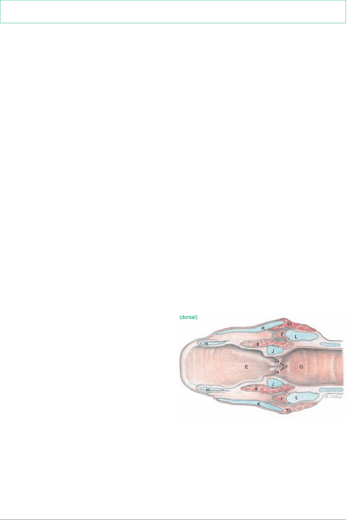
b) The LARYNX (see also text fig.) Because there are no laryngeal
ventricles or vestibular folds, the wall of the the laryngeal vestibule
(E) is smooth. The vestibular lig. of the horse is represented by a
flat, fan-shaped sheet of fibers.
The vocal fold (F) is only a low ridge containing the vocal ligament
(5). The glottis (F) is composed of the vocal folds, arytenoid carti-
lages, and the glottic cleft (rima glottidis). Behind the glottis is the
infraglottic cavity (G).
I. The cartilages of the larynx show the following species differ-
ences in the ox: The epiglottic cartilage (H) is broad and rounded.
The corniculate, vocal, and muscular processes of the arytenoid
cartilages (J) resemble those of the dog and horse, but there is no
cuneiform process. The thyroid cartilage (K) has a rostral notch
(K'), absent in other species, and the caudal notch is not palpable
in the live animal. The laryngeal prominence (K") a landmark, is
not at the rostral end of the cartilage, as is the human “Adam’s
apple”, but two-thirds of the way toward the caudal end. The lam-
ina of the cricoid cartilage (L) is short.
II. The LARYNGEAL MUSCLES act like those of the dog and
horse. The cricoarytenoideus dorsalis (9) is the primary dilator of
the glottis. Because there is no lateral ventricle, the ventricularis
and vocalis are combined in the thyroarytenoideus (8). Other con-
strictors of the glottis are the cricoarytenoideus lateralis (7),
cricothyroideus (10), and arytenoideus transversus (6).
The innervation of the larynx by the cranial and recurrent laryngeal
nn. from the vagus n. corresponds to that of the horse and dog.
46
9. PHARYNX AND LARYNX
* Himmelreich, 1964
Dissection and study are carried out from the medial cut surface as well as the lateral side. Laterally, the pterygoids, digastricus, sty-
lohyoideus, and occipitohyoideus are removed, as well as the remnants of the mandibular and parotid glands.
Anatomie des Rindes englisch 09.09.2003 13:30 Uhr Seite 46
K"
K'
K"
j Deep facial v.
k Lingual n. (V2)
l Mylohyoideus
m Hyoglossus
n Styloglossus
o Digastricus
p Stylohyoideus
q Omohyoideus
r Thyrohyoideus
s Sternohyoideus
t Sternothyroideus
Pharynx and Larynx
(Paramedian section)
1 Pharyngeal orifice
of auditory tube
2 Pharyngeal septum
3 Soft palate
4 Sinus
of palatine tonsil
Legend:
(Brain, see p. 51)
a Cerebrum
b Cerebellum
c Medulla oblongata
d Medulla spinalis (Spinal cord)
e Lig. nuchae
f Longus capitis
g Atlas
h Axis
Pharyngeal cavity
A Nasopharynx
B Oropharynx
C Palatopharyngeal arch
D Laryngopharynx
Laryngeal cavity
E Laryngeal vestibule
F Glottis and vocal fold
G Infraglottic cavity
Cricoarytenoid lig.
(medial)
(lateral)
5 Vocal lig.
6 Arytenoideus transversus
7 Cricoarytenoideus lat.
8 Thyroarytenoideus
9 Cricoarytenoideus dors.
10 Cricothyroideus
11 Tensor veli palatini
12 Levator veli palatini
13 Pterygopharyngeus
14 Palatine tonsil
15 Stylopharyngeus caudalis
16 Hyopharyngeus
17 Thyropharyngeus
18 Cricopharyngeus
(See pp. 36, 37, 39, 45, 49)
(lateral)
Laryngeal cartilages
H Epiglottic
J Arytenoid
K Thyroid
K' Rostral notch
K" Laryngeal prominence
L Cricoid
Legend:
47
Anatomie des Rindes englisch 09.09.2003 13:30 Uhr Seite 47

a) The ARTERIES OF THE HEAD show species-specific charac-
teristics different from the dog and horse (for veins and arteries of
the head, see text fig. p. 36).
The common carotid a. (16, see also p. 61) reaches the head-neck
junction accompanied dorsally by the vagosympathetic trunk, and
ventrally by the recurrent laryngeal n. Here it gives off the ster-
nomastoid brr. (15). At the thyroid gl. it gives off, as in the horse,
the inconstant caud. thyroid a. and the cran. thyroid a. (17). The
latter gives rise to the caud. laryngeal br. which accompanies the
caud. laryngeal n. The cran. laryngeal a. with its laryngeal and pha-
ryngeal brr. comes either directly from the common carotid a. or, as
in the horse, from the cran. thyroid a. Shortly before its termination
the common carotid a. gives off the ascending pharyngeal a. for the
soft palate, tonsils, and pharynx.
The common carotid a. is continued by its largest terminal br., the
external carotid a., whose origin is marked by the origin of the
occipital a. (9) because the smaller terminal br., the internal carotid
a., undergoes atrophy of its extracranial part in the ox. By three
months after birth it is completely closed.
The external carotid a. (11), as it turns dorsally, gives off the lin-
guofacial trunk (4) rostroventrally. This divides into the facial and
lingual aa. The lingual a. (5) runs medial to the mandible along the
stylohyoid bone, gives off the sublingual a., and passes medial to
the hyoglossus into the tongue. The facial a. (6) also runs first
medial to the mandible, and then turns at the vascular groove, cov-
ered by the sternomandibularis, onto the lateral surface at the ros-
tral border of the masseter. After giving off the caudal auricular a.
(8) caudodorsally, the masseteric br. (2) rostroventrally, and the
supf. temporal a. (7) dorsally, the external carotid is continued by
the maxillary a. (1) directed rostrodorsally toward the base of the
skull.
b) The THYROID GL. (18) consists of two flat lobulated irregu-
larly triangular lobes connected by a parenchymatous isthmus. The
lobes are lateral to the trachea, esophagus, and cricoid cartilage,
and the isthmus passes ventral to the trachea at the first or second
cartilage. In old cattle the isthmus may be reduced to a fibrous
band.
c) The PARATHYROID GLL. The external parathyroid gl. is
6–10 mm long and reddish-brown. It is always cranial to the thy-
roid gl., usually dorsomedial to the common carotid a., about 3 cm
caudal to the origin of the occipital a. It may be on the caudal bor-
der of the mandibular gl. The internal parathyroid gl. is 1–4 mm
long, and brown. It is on the tracheal surface of the lobe of the thy-
roid gl., near the craniodorsal border, embedded in the parenchy-
ma.
d) The ESOPHAGUS (23, see also p. 60) in the cranial third of the
neck, is dorsal to the trachea; between the third and sixth vertebrae
it lies on the left side of the trachea; and at the thoracic inlet it is in
a left dorsolateral position.
e) The TRACHEA (24, see also p. 60) of the ox changes the shape
of its cross section in life and after death mainly by the state of con-
traction of the trachealis muscle attached to the inside of the tra-
cheal cartilages. It is relatively small (4 x 5 cm).
f) CRANIAL NERVES OF THE VAGUS GROUP (IX–XI) emerge
through the jugular foramen, as in the horse and dog.
I. The glossopharyngeal n. (IX, 3) innervates mainly the tongue
with its large lingual br. Before it divides into dorsal and ventral
brr., the lingual br. in the ox bears a lateropharyngeal ganglion
medial and rostroventral to the stylohyoid. The pharyngeal br. sup-
plies several branches to the pharynx.
II. The vagus n. (X, 20) has the widest distribution of all the cra-
nial nn. Its nuclei of origin are in the nucleus ambiguus of the
medulla oblongata for the motor fibers and in the parasympathet-
ic nucleus of the vagus for the parasympathetic fibers. The sensory
nuclei are in the nucleus of the solitary tract and in the nucleus of
the spinal tract of C. N. V (see pp. 54, 55). The pseudounipolar
nerve cells of the afferent fibers are in the proximal ganglion and in
the distal ganglion of the vagus, which is very small in the ox, and
lies near the jugular foramen. The vagus, after leaving the skull,
first gives off the pharyngeal brr. (21), whose cranial brr. join those
of C. N. IX in the pharyngeal plexus, supplying pharyngeal muscles
and mucosa. The caudal continuation innervates the thyropharyn-
geus and cricopharyngeus and becomes the esophageal br. This is
motor to the cran. part of the cervical esophagus, and joins the
caud. laryngeal n. The cranial laryngeal n. (13) originates from the
vagus caudal to the pharyngeal brr., and runs cranioventrally, cross-
ing lateral to the pharyngeal brr. Its external br. usually joins the
pharyngeal br., then separates again to innervate the cricothy-
roideus. The internal br. of the cran. laryngeal n. enters the larynx
through the thyroid fissure and innervates the mucosa. It then
courses caudally inside the thyroid lamina and emerges caudal to
the larynx to join the esophageal br. or the caudal laryngeal n. (19)
which comes from the recurrent laryngeal n. In the thorax the vagus
gives off the recurrent laryngeal n., which, on the right side, turns
dorsally around the caudal surface of the subclavian a. and runs
cranially between the common carotid a. and the trachea. On the
left side, the recurrent n. turns medially around the aorta and the
lig. arteriosum, passes medial to the great arteries, and runs cra-
nially between the esophagus and trachea. Both nerves terminate as
the caudal laryngeal nerves which pass deep to the cricopharyngeus
to innervate all of the laryngeal muscles except the cricothyroideus.
After giving off the recurrent laryngeal n., the vagus still carries
parasympathetic and visceral afferent fibers serving the heart,
lungs, and abdominal organs as far as the descending colon. The
visceral afferents greatly predominate (see pp. 65, 73).
III. The accessory n. (XI, 10) divides at the level of the atlas into a
dorsal br. to the cleidooccipitalis and trapezius, and a ventral br. to
the cleidomastoideus and sternocephalicus (see p. 60).
g) The HYPOGLOSSAL N. (XII, 12) emerges through the
hypoglossal canals. It innervates the proper (intrinsic) muscle of the
tongue (f) and the following extrinsic muscles: styloglossus, hyo-
glossus, and genioglossus. The geniohyoideus (h) and thyrohy-
oideus (see p. 47) are also supplied by the hypoglossal n. with a
variable contribution from the first cervical n. via the ansa cervi-
calis.
h) From the SYMPATHETIC TRUNK of the autonomic system,
fibers pass in the region of the thoracic inlet through the cervi-
cothoracic ganglion (p. 65) and middle cervical ganglion and then
in the vagosympathetic trunk (14) to the head. Here in the cran.
cervical ganglion (22), large in the ox, the fibers synapse with gan-
glion cells whose postganglionic sympathetic fibers run in perivas-
cular (mainly periarterial) plexuses in the adventitia of the large
vessels of the head to their distribution in glands and internal eye
muscles.
48
10. ARTERIES OF THE HEAD AND HEAD-NECK JUNCTION, THE CRANIAL NN. OF THE VAGUS
GROUP (IX–XI), AND THE HYPOGLOSSAL N. (XII)
For the demonstration of these aa. and nn.: laterally the dorsocaudal third of the stylohyoid bone, and medially the rectus capitis ven-
tralis and longus capitis are removed.
Anatomie des Rindes englisch 09.09.2003 13:30 Uhr Seite 48
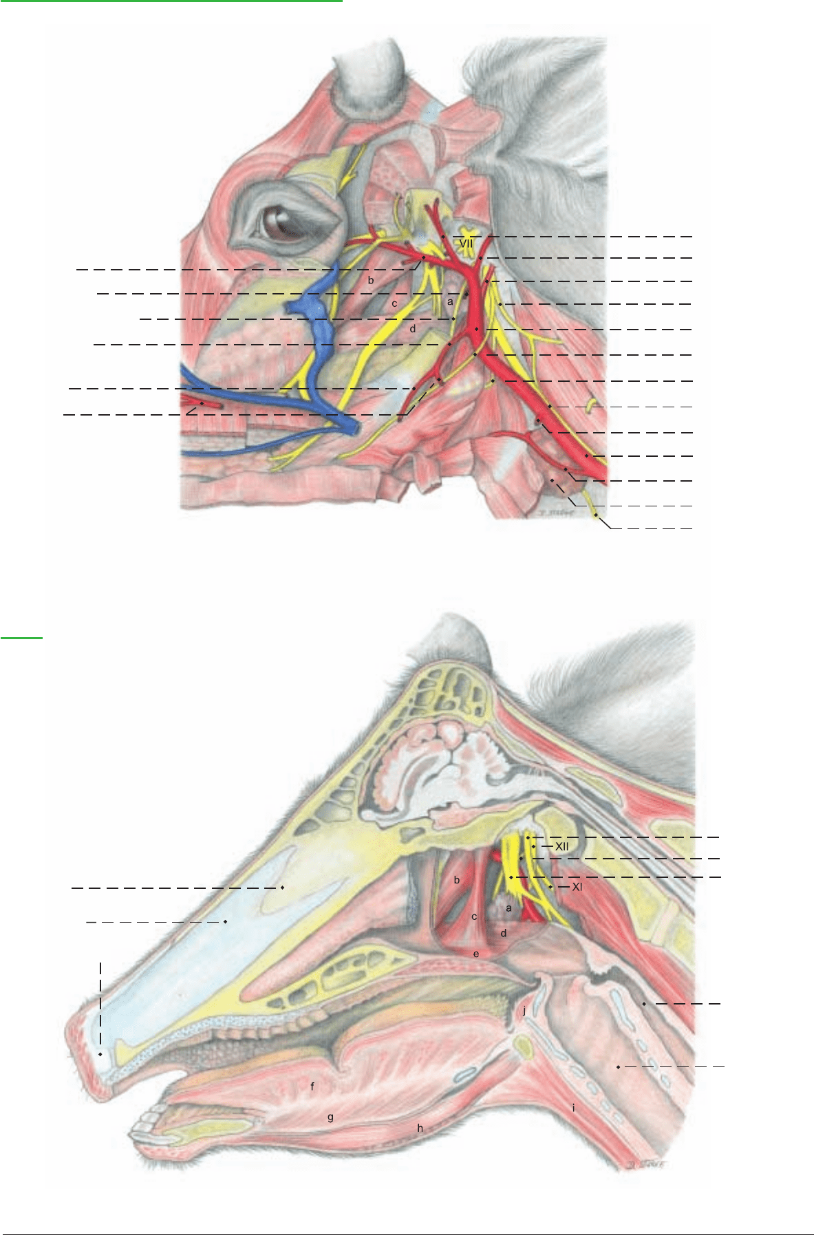
(See pp. 39, 45, 47, 51)
Arteries of the head and Cranial nn. IX, X, XI, XII
(lateral)
1 Maxillary a.
2 Masseteric br.
3 Glossopharyngeal n. (IX)
4
Linguofacial trunk
5 Lingual a.
6 Facial a.
7 Supf. temporal a.
8 Caud. auricular a.
9 Occipital a.
10 Accessory n. (XI)
11 External carotid a.
12 Hypoglossal n. (XII)
13 Cran. laryngeal n.
14 Vagosympathetic trunk
15 Sternomastoid brr.
16 Common carotid a.
17 Cran. thyroid a.
18 Thyroid gl.
19 Caud. laryngeal n.
Legend:
a Med. retropharyngeal ln.
b Tensor veli palatini
c Levator veli palatini
d Pterygopharyngeus
e Palatopharyngeus
f Proper lingual m.
g Genioglossus
h Geniohyoideus
i Sternohyoideus
j Hyoepiglotticus
(medial)
Nasal septum:
Bony part
Cartilaginous
part
Membranous part
20 Vagus n. (X)
21 Pharyngeal br.
22 Cranial cervical
ganglion
23 Esophagus
24 Trachea
49
Anatomie des Rindes englisch 09.09.2003 13:30 Uhr Seite 49
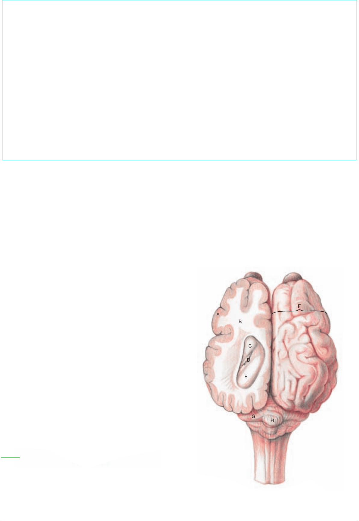
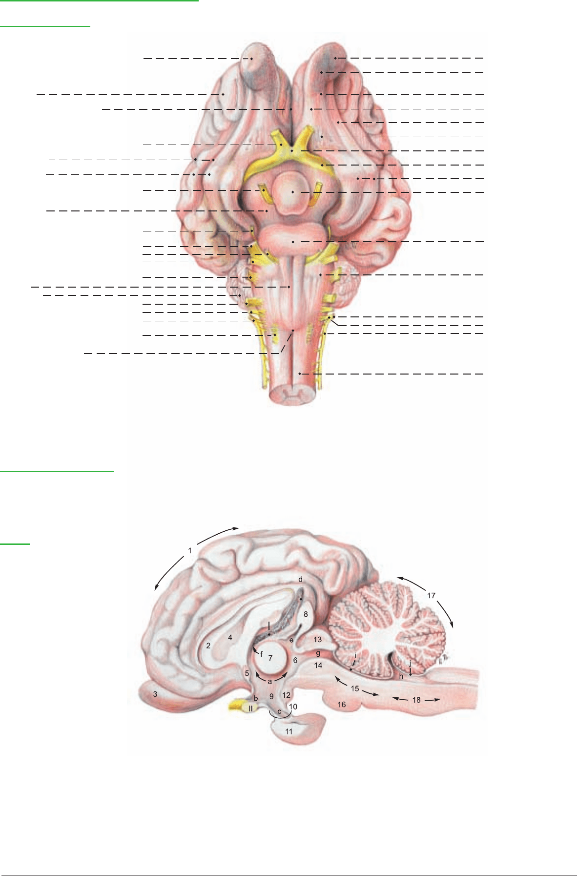
a) The BRAIN is relatively small. Because species-specific differ-
ences are of minor significance among domestic mammals, refer-
ence to a general textbook description is advised. Only a few fea-
tures of the bovine brain will be mentioned here; greater impor-
tance will be given to the illustrations.
I. The dorsal part of the rhombencephalon, the cerebellum (17), is
much more complex and irregular than in man, and the vermis (H)
is not very prominent.
II. The midbrain (mesencephalon) exhibits four dorsal eminences,
the rostral and caudal colliculi. The caudal pair is smaller. On the
ventral surface is the cerebral crus.
III. The diencephalon is connected through its hypothalamus (9)
with the infundibulum (10) of the hypophysis (11). Caudal to the
infundibulum is the mammillary body (12). The pineal gl. (8) proj-
ects dorsocaudally from the diencephalon.
IV. The greatest part of the telencephalon (cerebrum) is the hemi-
sphere (F). It consists of the cortex (A) and the white matter (B). It
is markedly convoluted on the surface, bearing gyri (folds) and sul-
ci (grooves). The herbivora have additional variable and inconstant
sulci which make the brain more complex than the brains of carni-
vores. On the rhinencephalon (3) the olfactory bulb is smaller than
in the dog and horse. It is continuous caudally with the olfactory
peduncle, which branches into lateral and medial olfactory tracts.
b) The VENTRICULAR SYSTEM. In the roof of the fourth ven-
tricle (h) the caudal medullary velum (j) is invaginated by a choroid
plexus. The third ventricle (a) is in the median plane; it encircles the
interthalamic adhesion (7), and with its choroid plexus (a'), extends
over the pineal gl. as the suprapineal recess (d). The third ventricle
also extends into the pineal gl. The cerebral aqueduct (g) connects
the third and fourth ventricles. Rostrally, the third ventricle com-
municates on each side through an interventricular foramen (f)
with a lateral ventricle, which contains a choroid plexus continu-
ous with that of the third ventricle. A long process of the lateral
ventricle extends into the olfactory bulb.
50
CHAPTER 4: CENTRAL NERVOUS SYSTEM AND CRANIAL NERVES
1. THE BRAIN
To remove the half-brain from the bisected head, the cut end of the spinal cord is first lifted from the dura mater, cutting the attach-
ments of the denticulate lig. and the cervical nn. Then the brain is detached by identifying and cutting the cranial nn. in caudorostral
order, midway between the brain and the dura. The roots of the hypoglossal n. (XII) emerge from the ventrolateral groove, lateral to
the decussation of the pyramids, and exit through the dura, and to the hypoglossal canals. The nerves of the vagus group (IX, X, XI)
emerge from the lateral funiculus of the medulla oblongata. The accessory n. (XI) has a long spinal root, which begins at the fifth cer-
vical segment and runs up to unite with the small cranial root. The glossopharyngeal (IX) and vagus (X) nerves originate by a continu-
ous series of rootlets and pass out through the jugular foramen with the accessory n. The vestibulocochlear (VIII) and facial (VII) nerves
also arise close together from the medulla, between the cerebellum and the trapezoid body, with VIII dorsolateral to VII, and run dor-
solaterally to the internal acoustic meatus. The small abducent n. (VI) passes out through the trapezoid body at the lateral edge of the
pyramid, and enters a hole in the dura on the floor of the cranium in the transverse plane of the internal acoustic meatus. The large
trigeminal n. (V) comes from the end of the pons just rostral to the facial n. and runs rostroventrally to the largest aperture in the dura.
Nn. IV and III come from the midbrain (13, 14). The trochlear n. (IV), the only one to emerge from the dorsal surface of the brain
stem, arises behind the caudal colliculus, decussates with the contralateral nerve, and passes around the lateral surface of the midbrain,
on or in the free border of the tentorium cerebelli, to the floor of the cranium. The larger oculomotor n. (III) arises from the crus cere-
bri, caudolateral to the hypophysis, which should be carefully dissected out of the Sella turcica (p. 31, 42) while maintaining its con-
nection with the brain. The internal carotid a. will be cut between the rete mirabile and the arterial circle of the cerebrum. Nerves III,
IV, VI, and the ophthalmic and maxillary nerves join outside the dura and leave the cranium through the orbitoround for. in ruminants
and swine. The optic n. (II) is cut distal to the optic chiasm. The optic tract connects the chiasm to the diencephalon. To free the cere-
bral hemisphere, the median dorsal fold of the dura (falx cerebri) is removed and preserved for study of the enclosed sagittal sinus,
and the membranous tentorium cerebelli is cut at its dorsal attachment. (There is no osseous tentorium in ruminants.) The half-brain
is lifted out of the dura by inserting scalpel handles between the cerebrum and the dura dorsally and between the olfactory bulb and
the ethmoidal fossa, severing the olfactory nn. (I).
E Hippocampus
F Cerebral hemisphere
G Cerebellar hemisphere
H Vermis
Section of cerebrum (dorsal)
Legend:
A Cerebral cortex [Gray matter]
B White matter
C Head of caudate nucleus
D Choroid plexus of lateral ventricle
E Hippocampus
F Cerebral hemisphere
G Cerebellar hemisphere
H Vermis
Legend:
A Cerebral cortex [Gray matter]
B White matter
C Head of caudate nucleus
D Choroid plexus of lateral ventricle
Anatomie des Rindes englisch 09.09.2003 13:30 Uhr Seite 50
a'
Rhombencephalon:
15 Metencephalon
16 Pons
17 Cerebellum
18 Myelencephalon
[Medulla oblongata]
Brain [Encephalon] and Cranial Nerves
Base of brain (ventral)
I
Cerebrum
Longitudinal cerebral fissure
II
Cerebral sulci
Cerebral gyri
III
Cerebral crus
IV
V
VI
VII
VIII
Pyramid of medulla ob-
longata
Cerebellum
IX
X
XI
XII
Decussation of the
pyramids
Olfactory nn.
Olfactory bulb
Olfactory peduncle
Medial olfactory tract
Lateral olfactory tract
Olfactory trigone
Optic chiasm
Optic tract
Piriform lobe
Hypophysis [Pituitary gland]
Pons
Trapezoid body
Accessory nerve (XI)
Cranial roots
Spinal roots
Spinal cord
[Medulla spinalis]
Median section of the brain
Legend:
a Third ventricle
a' Choroid plexus of third ventricle
b Optic recess
c Infundibular recess
d Suprapineal recess
e Pineal recess
f Interventricular foramen
g Cerebral aqueduct
h Fourth ventricle
i Rostral medullary velum
j Caudal medullary velum
Cerebrum:
1 Hemisphere
2 Corpus callosum
3 Rhinencephalon
4 Septum pellucidum
5 Rostral commissure
Diencephalon:
6 Thalamus
7 Interthalamic adhesion
[Intermediate mass]
8 Epiphysis [Pineal gland]
9 Hypothalamus
10 Infundibulum
11 Hypophysis [Pituitary gl.]
12 Mamillary body
Mesencephalon [Midbrain]:
Tectum
13 Lamina tecti
[Rostral and caudal colliculi]
14 Tegmentum
51
Anatomie des Rindes englisch 09.09.2003 13:49 Uhr Seite 51

NERVE PAGE NAME/FIBER FUNCTION DISTRIBUTION REMARKS
I
50 Olfactory nn. (special sensory) Olfactory region in caud. nasal cavity 1st neuron in olfactory mucosa, synapse in
olfactory bulb
II 42, 50 Optic n. (special sensory) Optical part of retina Evagination of diencephalon
III 40, 50 Oculomotor n. (m., psy.)* Orig. mesencephalon, exits by for. orbitoro-
tundum
(1) • Dorsal br. (m.) Dors. rectus, levator palpebrae
superioris, retractor bulbi
(2) • Ventr. br. (m., psy.) Med. and ventr. recti, ventral oblique Psy. neurons synapse in ciliary gangl. and
pass in ciliary nn. to eyeball
IV 40, 50 Trochlear n. (m.) Dorsal oblique Orig. mesencephalon, exits skull by for.
orbitorotundum
V 38, 50 Trigeminal n. Orig. rhombencephalon and mesencephalon.
Nerve of 1st pharyngeal arch
V1 40 • Ophtalmic n. (s.) Dorsum nasi, ethmoid bone, Exits skull by foramen orbitorotundum
lacrimal gl., upper eyelid
(3) 40 •• Nasociliary n. (s.)
(4) 40 ••• Ethmoid n. (s.) Dorsal nasal mucosa Enters nasal cavity through ethmoid for. and
cribriform plate
(5) 40 ••• Infratrochlear n. (s.) Conjunctiva, 3
rd
lid, lacrimal caruncle, Crosses dors. margin of orbit below trochlea;
skin of med. angle of eye may reach cornual process
(6) 40 ••• Long ciliary nn. (s., psy.) Iris and cornea, ciliary muscle Psy. fibers from ciliary ganglion
(7) 40 •• Lacrimal n. (s., psy., sy.) Lacrimal gl., skin and conjunctiva of Thin lat. and med. brr., which, after junction
lat. angle of eye with r. communicans from zygomatic n., join
to form zygomaticotemporal br.
(8) 40 ••• Zygomaticotemporal br. Skin of temporal region
(9) 40 •••• Cornual br. Cornual dermis Dehorning anesthesia!
(10) 40 •• Frontal n. (s.) Skin of frontal region and upper eyelid Ends as supraorbital n. in skin of frontal
region
V2 38 • Maxillary n. (s.) Exits skull from for. orbitorotundum
(11) •• Zygomatic n. (s., psy.) Communicating br. with lacrimal n. (V1)
(12) ••• Zygomaticofacial br. (s.) Lower eyelid Exits orbit at lat. angle of eye
(13) •• Pterygopalatine n. (s., psy.) Psy. fibers from pterygopalatine ganglion
(14) ••• Major palatine n. (s., psy.) Mucosa and gll. of the hard palate Goes through caudal palatine for., palatine
canal, and major palatine for.
(15) ••• Minor palatine nn. (s., psy.) Soft palate with its glands Exit palatine canal through minor palatine
foramina
(16) ••• Caud. nasal n. (s.) Ventr. parts of nasal cavity, palate Enters nasal cavity through sphenopalatine
for.
(17) 38 •• Infraorbital n. (s.) Skin of dorsum nasi, nares, and Traverses maxillary for. and infraorbital canal
upper lip and for.
V3 38 • Mandibular n. (s., m.) Exits skull by oval foramen
(18) 38 •• Masticatory n. (m.)
(19) 38 ••• Deep temporal nn. (m.) Temporalis
(20) 38 ••• Masseteric n. (m.) Masseter Goes through mandibular notch
(21) •• Med. and lat. pterygoid nn. (m.) Med. and lat. pterygoid mm. The otic gangl. (s., psy.) at root of buccal n.,
is large in the ox
(22) •• Tensor tympani n. (m.) Tensor tympani Enters tympanic cavity
(23) •• Tensor veli palatini n. Tensor veli palatini
(24) 38 •• Auriculotemporal n. Skin of auricle and temporal region, Turns around the neck of the mandible, psy.
(s., psy., sy.) Parotid gl. fibers from otic ganglion
(25) 38 ••• Communicating brr. with Connection with dors. buccal br. (VII)
facial n. (s.)
(26) 38 •• Buccal n. (s. psy.) Mucosa of cheek and buccal gll. Psy. fibers from otic gangl.
(27) 38 ••• Parotid br. (psy.) Parotid gl. Follows parotid duct backward through vas-
cular groove
(28) 38 •• Lingual n. (taste, s., psy.) Sensory to floor of mouth and Receives taste, s., and psy. fibers from chorda
tongue, taste from rostral 2/3 tympani (VII). (Psy. fibers synapse in
of tongue mandibular ganglion.)
(29) 38 ••• Sublingual n. (s., psy.) Mucosa of rostral floor of mouth Carries psy. fibers to mandibular and sublin-
gual gll.
(30) 38 •• lnferior alveolar n. (s.) Inferior teeth and gingiva Traverses mandibular foramen and canal
(31) 38 ••• Mylohyoid n. (m.) Mylohyoid, rostral belly of digastricus
(32) 38 ••• Mental n. (s.) Skin and mucosa of chin and lower lip Leaves the mandib. canal at the mental
foramen
52
2. CRANIAL NERVES I–V
* Fiber function: s. = sensory, m. = motor and proprioceptive, sy. = sympathetic, psy. = parasympathetic
Anatomie des Rindes englisch 09.09.2003 13:49 Uhr Seite 52
53
d'
e'
e"
e"
h'
h'
q'
q"
m'
P'
N'
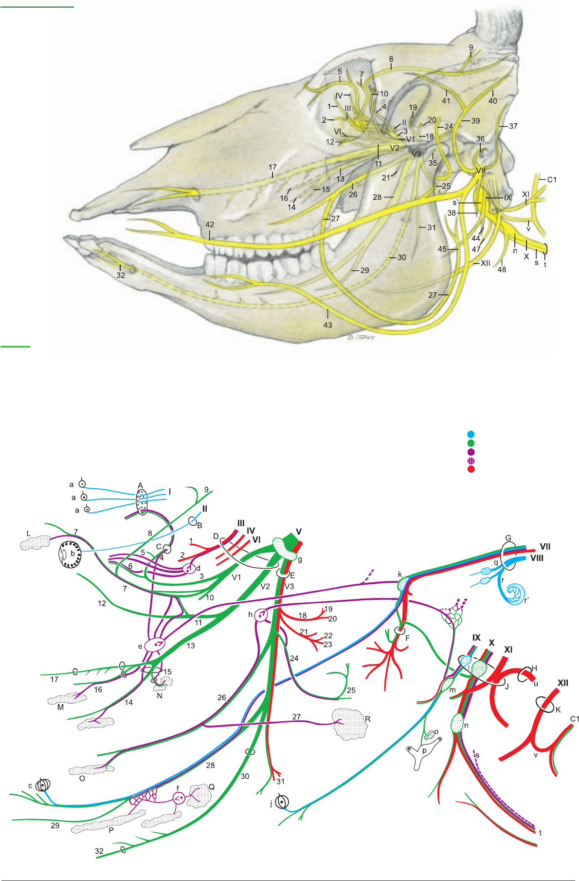
Special sensory neuron
Sensory neuron
Parasympathetic neuron
Sympathetic neuron
Motor neuron
Cranial nerves
Legend:
A Cribriform plate
B Optic canal
C Ethmoid foramen
D For. orbitorotundum
E Oval foramen
F Stylomastoid for.
G Int. acoustic meatus
H Foramen magnum
J Jugular foramen
K Hypoglossal canal
L Lacrimal gl.
M Nasal gll.
N Palatine gll. (soft palate)
N' Palatine gll. (hard palate)
O Buccal gll.
P Monostomatic sublingual gl.
P' Polystomatic sublingual gl.
Q Mandibular gl.
R Parotid gl.
a Olfactory region
b Retina
c Fungiform papillae
d Ciliary ganglion
d' Short ciliary nn.
e Pterygopalatine gangl.
e' Orbital brr.
e" N. of pterygoid canal
(major and deep petrosal nn.)
f Mandibular ganglion
g Trigeminal ganglion
h Otic ganglion
h' Minor petrosal n.
j Vallate papillae
k Geniculate ganglion
l Proximal ganglia
m Distal gangl. (petrosal)
m' Tympanic n.
n Distal gangl. (nodose)
o Carotid glomus
p Carotid sinus
q Vestibular n.
q' Sup. vestibular gangl.
q" Inf. vestibular gangl.
r Cochlear n.
r' Spiral gangl. of cochlea
s Sympathetic trunk
s' Cranial cervical gangl.
t Vagosympathetic trunk
u Spinal root of accessory n.
v Ansa cervicalis
Anatomie des Rindes englisch 09.09.2003 13:49 Uhr Seite 53
UQL|qNyNzKjvWvGT8zRmLENm5w==|1287791586

NERVE PAGE NAME/FIBER FUNCTION DISTRIBUTION REMARKS
VI
40, 50 Abducent n. (m.) Lat. rectus, lat. part of retractor bulbi Orig.: Rhombencephalon; exits skull at for.
orbitorotundum
VII 36, 50 Facial n. (intermediofacial n.) Mm. of face and ear, lacrimal and Goes through int. acoustic meatus into facial
(taste, m., psy.)* salivary gll. canal and leaves through stylomastoid for.;
nerve of 2nd pharyngeal arch
(33) • Major petrosal n. (psy.) Gll. of nose and palate, and lacrimal Joins the deep petrosal n. (sy.) to form the
gll. n. of the pterygoid canal, which goes to
pterygopalatine ganglion
(34) • N. to stapedius (m.) Stapedius
(35) 38 • Chorda tympani (taste, psy.) Mandibular and sublingual gll., Leaves petrous temporal bone through
rostral 2/3 of tongue, taste petrotympanic fissure and joins lingual n.
(V3)
(36) 36 • Int. auricular br. (s.) Int. surface of auricle Passes through auricular cartilage
(37) 36 • Caud. auricular n. (m.) Auricular mm. Communicates with the dors. brr. of first
2 cervical nn.
(38) • Digastric br. (m.) Caud. belly of digastricus
• Parotid plexus (psy.) Parotid gl. Impulses from auriculopalpebral n. (V3)
(39) 36 • Auriculopalpebral n. (m.) Communicates with auriculotemporal n. (V3)
(40) •• Rostral auricular brr. (m.) Rostral auricular mm.
(41) •• Zygomatic br. (m.) Orbicularis oculi, levator anguli Ends with palpebral brr.
oculi med., frontalis
(42) 36 • Dorsal buccal br. (m.) Mm. of upper lip, planum nasale, Communicating br. (s.) with auriculotempo-
and nostril ral n. (V3)
(43) 36 • Ventral buccal br. (m.) Buccinator, depressor labii inferioris Passes through vascular groove with facial a.
and v.
VIII 50 Vestibulocochlear n. Orig.: Medulla oblongata; enters int. acoustic
(special sensory) pore
• Cochlear n. (hearing) Spiral organ of the cochlea 1st neuron: in spiral gangl. of cochlea;
2
nd
neuron: in rhombencephalon
• Vestibular n. (equilibrium) Ampullae of semicircular ducts, 1st neuron: in vestibular gangl.; 2nd neuron
maculae of utriculus and sacculus in rhombencephalon
IX 48, 50 Glossopharyngeal n. Mucosa of tongue and pharynx, Orig.: medulla oblongata; exits skull through
(taste, s., m. psy.) tonsils, tympanic cavity jugular for. Nerve of 3rd pharyngeal arch;
1st n. of vagus group
(44) 48 • Pharyngeal br. (s., m.) Pharyngeal mucosa, caud. Forms pharyngeal plexus with pharyngeal
stylopharyngeus brr. of vagus (X)
(45) • Lingual br. (taste, s., psy.) Mucosa of soft palate and root of Before it divides into dorsal and ventr. brr.
tongue with its taste buds this n. bears the lateropharyngeal ganglion
X 48, 50 Vagus n. (s., m., psy.) Viscera of the head, neck, thorax, Orig.: medulla oblongata; exits skull from
and abdomen jugular foramen; n. of 4th pharyngeal arch;
2nd n. of vagus group
(46) • Auricular br. (s.) Skin of ext. acoustic meatus Enters the facial canal and joins the facial n.
(VII)
(47) 48 • Pharyngeal brr. (s., m.) Pharyngeal mm. and mucosa Caud. contribution to pharyngeal plexus,
ends as esophageal br.
(48) 48 • Cran. laryngeal n. (s., m.) Branches off from distal ganglion
and crosses lat. to pharyngeal br.
(49) 48 •• External br. (m.) Cricothyroideus Joins pharyngeal brr.
(50) 48 •• Internal br. (s.) Laryngeal mucosa rostral to the Passes through the thyroid fissure
rima glottidis
48, 60 • Recurrent laryngeal n. Branches to cardiac plexus, trachea, Separates from the vagus in the thorax and
(s., m., psy.) and esophagus turns cranially
48 •• Caud. laryngeal n. (s., m.) All laryngeal mm. except cricothyroid,
laryngeal mucosa caud. to rima glottidis
XI 48, 50 Accessory n. (m.) Exits skull through jugular for.; 3rd n. of
vagus group
(51) • Cran. root: int. br. (m.) Orig.: medulla oblongata, joins vagus n. and
gives it motor fibers
(52) • Spinal root: ext. br. (m.) Orig. cervical spinal cord
48 •• Dorsal br. (m.) Trapezius and cleidooccipitalis
48 •• Ventral br. (m.) Cleidomastoideus and
sternocephalicus
XII 48, 50 Hypoglossal n. (m.) Proper lingual m., genio-, stylo-, Orig.: Medulla oblongata, leaves the skull via
and hyoglossus; together with ventr. hypoglossal canal, forms the ansa cervicalis
br. of 1st cervical n.: genio- and with 1st cervical n.
thyrohyoideus
54
3. CRANIAL NERVES VI–XII
* Fiber function: s. = sensory, m. = motor and proprioceptive, sy. = sympathetic, psy. = parasympathetic
Anatomie des Rindes englisch 09.09.2003 13:49 Uhr Seite 54
