Budras Klaus-Dieter, Habel Robert E. Bovine Аnatomy
Подождите немного. Документ загружается.

16 Bulbar epidermal tubules
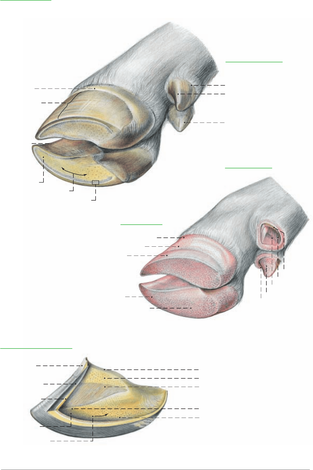
Hoof and Dewclaw
Epidermis of the dewclaw
Perioplic epidermis
Coronary epidermis
Bulbar epidermis
1 Perioplic
epidermis
2 Coronary epidermis
3 Sole epidermis
4 Bulbar epidermis
5 White zone
Dermis of the hoof
Dermis of the declaw
6 Perioplic dermis
7 Coronary dermis
8 Wall (Parietal) dermis
9 Sole dermis
10 Bulbar dermis
Coronary dermis
Wall (Parietal) dermis
Sole dermis
Bulbar dermis
Perioplic dermis
Epidermis (Capsule) of the hoof
Perioplic epidermis
Coronary epidermis
11 Wall (Parietal)
epidermis
Epidermis of the sole
Bulbar epidermis
12 Perioplic epidermal tubules
13 Coronary epidermal tubules
14 Epidermal lamellae
15 Epidermal tubules of the sole
2' Dorsal border
25
Anatomie des Rindes englisch 09.09.2003 12:36 Uhr Seite 25

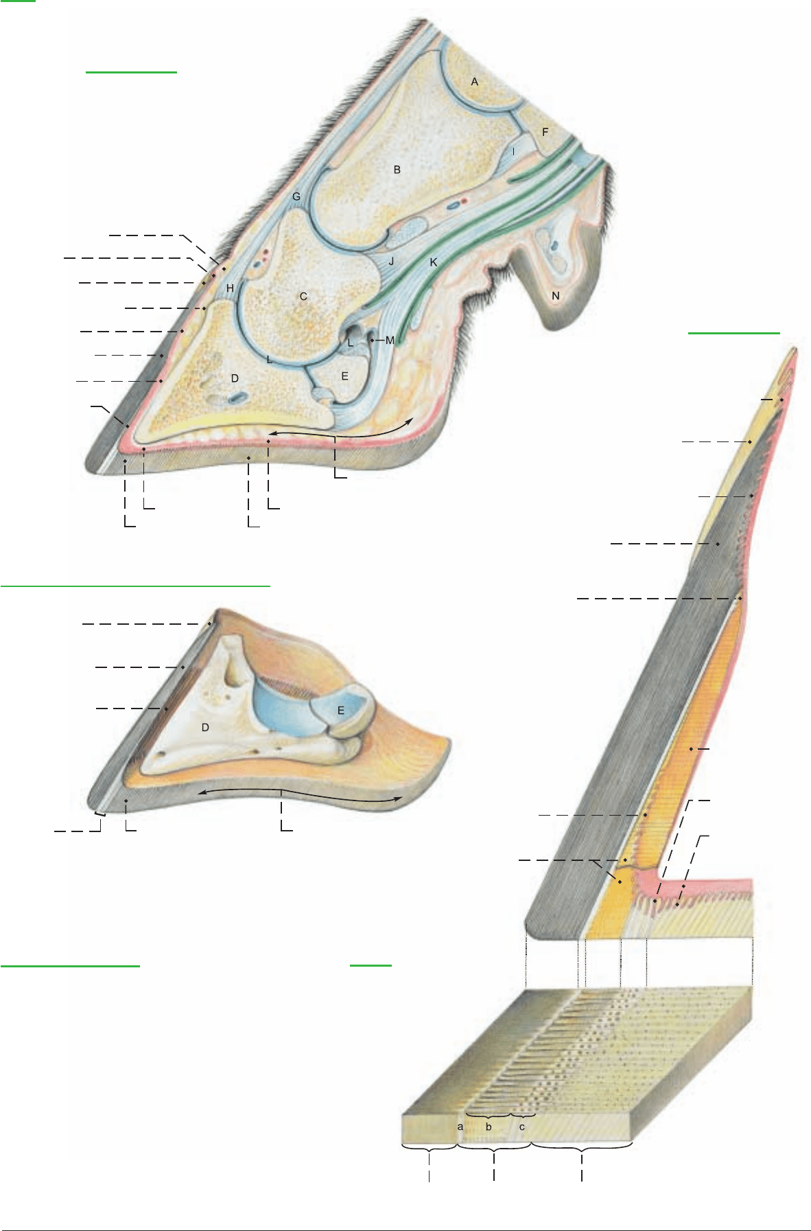
a) The HOOF CAPSULE surrounds: the distal end of the middle
phalanx (C), the distal interphalangeal joint (L), and the distal pha-
lanx (coffin bone, D) with the terminations of the common dig.
extensor tendon (H) on the extensor process and the deep dig. flex-
or tendon (K) on the flexor tubercle. Also enclosed is the distal
sesamoid (navicular) bone (E), which serves as a trochlea for the
deep dig. flexor tendon. The navicular bursa (M) reduces friction
between them.
The cornified hoof capsule consists of the lamina (horny wall) with
an abaxial part, a dorsal border, and an axial part facing the inter-
digital space, as well as the horny sole and horny bulb. The capsule
has a thickness of about 10 mm in the dorsal part and about 5 mm
in the axial part. The growth of the epidermis pushes the cornified
masses distally at a rate of about 5 mm per month. After an exun-
gulation the renewal of the entire hoof capsule would require up to
20 months. Horn formation is more intensive in calves than in
adults and more active on the pelvic than on the thoracic limb. In
the last third of pregnancy and in very high milk production, horn
formation is reduced. That is shown on the superficial surface of
the hoof by the formation of semicircular grooves.
When cattle are kept on soft footing with little or no possibility of
exercise the horn grows faster than it is worn off and therefore the
hoofs must be trimmed regularly.
I. The lamina (Paries corneus) consists of external, middle, and
internal layers, which are bonded together and formed by the peri-
oplic, coronary, and wall segments respectively. The external layer
is very thin; the middle layer constitutes the bulk of the lamina; and
the internal layer bears the horny lamellae that make up the junc-
tional horn.
II. The junctional horn is part of the suspensory apparatus of the
coffin bone. This term includes all of the tissues that attach the cof-
fin bone (distal phalanx) to the inside of the lamina. The suspenso-
ry apparatus of the coffin bone consists of a connective tissue (der-
mal) part and an epidermal part. Collagenous fiber bundles
anchored in the outer zone of the coffin bone run obliquely proxi-
modorsally in the reticular layer and then in the lamellae of the der-
mis. The collagen fibers are attached to the basement membrane.
The tension is then transmitted through the living epidermal cell
layers by desmosomes and bundles of keratin filaments to the junc-
tional (lamellar) horn, which is attached to the lamina. The pres-
sure exerted on the coffin bone by the body weight is transformed
by the shock absorbing suspensory apparatus of the coffin bone
into tension; the tension is transformed in the lamina to pressure;
this pressure weighs upon the ground at the solear border of the
lamina. One part of the body weight is not transformed, but falls
directly on a support of solear and apical bulbar horn. In the basal
bulbar segment the elastic horn and the thick subcutaneous cush-
ion act as a shock absorbing mechanism of the hoof. The cham-
bered cushions work in a manner comparable to the gel cushion
system of modern running shoes. With the exception of a non-
weightbearing concavity at the axial end of the white zone, the sole
and bulb horn form a flat ground surface.
The suspensory apparatus of the coffin bone actuates the hoof
mechanism by traction on the internal surface of the lamina and by
pressure on the sole and bulb. This can be measured with strain
gauges. It concerns the elastic changes in form of the hoof capsule
that occur during loading and unloading. In weight bearing, the
space inside the lamina is reduced, while the palmar/plantar part of
the capsule expands and the interdigital space is widened. During
unloading, the horny parts return to their initial form and position.
III. The rate of horn formation differs greatly among the individual
hoof segments. In the coronary segment horn formation is very
intensive. In the proximal half of the wall segment the rate of horn
formation is low. In the distal half, on the other hand, horn is
formed in measurable amounts and at an increasing rate toward the
apex of the hoof. (The term sterile bed, used in older textbooks for
the wall segment is therefore incorrect.) Proximally in the wall seg-
ment the beginnings of the dermal lamellae bear proximal cap
papillae. From the epidermis on these papillae, nontubular proxi-
mal cap horn is produced. This is applied to the sides of the proxi-
mal parts of the horny lamellae. Distal to the cap horn, as far as the
middle of the wall, not much lamellar horn is added. In the distal
half of the wall segment the horny lamellae become markedly high-
er, up to 5 mm, and, beginning with their middle portion, become
flanked by amorphous distal cap horn that is applied cap-like over
the edges of the dermal lamellae. It is formed on the distal cap
papillae by the living epidermis there (see p. 27, right figure).
Distally on the wall-sole border the almost vertically directed der-
mal lamellae bend into horizontally directed dermal ridges of the
sole segment. At the bend the lamellae are split into terminal der-
mal papillae which have a remarkable diameter of 0.2–0.5 mm.
They are covered by living epidermis from which terminal tubular
horn is formed. As a part of the white zone the terminal horn fills
the spaces between the horny lamellae (see p. 27, right figure).
IV. The white zone (white line) consists only of horn produced by
the wall segment, and presents external, middle, and internal parts.
The external part (a) appears to the naked eye as a shining white
millimeter-wide stripe. It consists of the basal sections of the horny
lamellae and the flanking proximal cap horn, and borders the most-
ly nonpigmented inner coronary horn, which does not belong to the
zona alba. The middle part (b) of the white zone is formed by the
intermediate sections of the horny lamellae with the distal cap horn
that lies between them. The internal part (c) of the white zone con-
sists of the crests of the horny lamellae and, between them, the ter-
minal tubular horn. They cornify in the distal half of the wall or at
the wall-sole border.
The white zone has abaxial and axial crura (b", b'), which lie
between the mostly unpigmented coronary horn and the sole horn.
The axial crus ends halfway between the apex of the hoof and the
palmar/plantar surface of the bulb. The abaxial crus extends far-
ther, to the basal part of the bulb, where the end of the white zone
becomes distinctly wider and turns inward. (See p. 25 above and
text illustration.) The whole white zone and especially the wider
abaxial end are predisposed to “white line disease,” which by
ascending infection can lead to “purulent hollow wall.” The way
for ascending microorganisms is opened by crumbling cap and ter-
minal tubular horn, which technical material testing proves to be
masses of soft horn.
V. Horn quality is the sum of the characteristics of the biomaterial
horn, including hardness or elasticity, resistance to breakage, water
absorption, and resistance to chemical and microbial influences.
Horn quality is adapted to the biomechanical requirements of the
different parts of the hoof. Accordingly, hard horn is found in the
lamina; soft elastic horn in the proximal part of the bulb. Horn
quality can be determined by morphological criteria in combina-
tion with data from physicotechnical material testing.
26
7. THE HOOF (UNGULA)
a White zone
Sole:
b Body of the sole
b' Axial crus
b" Abaxial crus
Bulb of hoof:
c Basal part
c' Apical part
c'
b'
b"
Anatomie des Rindes englisch 09.09.2003 12:36 Uhr Seite 26
Sole epidermis
Hoof
Sagittal section
Subcutaneous
perioplic cushion
Perioplic
dermis
Perioplic
epidermis
Subcutaneous coronary
cushion
Coronary dermis
Coronary epidermis
Wall (Parietal)
dermis
Wall (Parietal)
epidermis
Subcutaneous digital cushion
Sole dermis
Sole epidermis
Bulbar dermis
Bulbar epidermis
Sagittal section
Perioplic dermis
(Perioplic dermal papillae)
Perioplic epidermis
Coronary dermis
(Coronary dermal papillae)
Coronary epidermis
Prox. dermal cap papillae
Hoof capsule and Distal phalanx (Coffin bone)
Perioplic
epidermis
Coronary epidermis
Wall (Parietal)
epidermis
Wall dermis
(Dermal
lamellae)
Terminal dermal
wall papillae
Sole dermis
(Dermal papillae
of sole)
Dist. dermal cap
papillae
Wall epidermis
White zone
Sole epidermis Bulbar epidermis
Legend: (See figure above.)
A Metacarpal bone IV
B Proximal phalanx
C Middle phalanx
D Distal phalanx (coffin bone)
E Distal sesamoid (navicular) bone
F Axial prox. sesamoid bone
G Tendon of lat. dig. extensor
H Tendon of com. dig. extensor
I Cruciate sesamoid lig.
J Tendon of supf. dig. flexor
K Tendon of deep dig. flexor
L Distal interphalangeal (coffin) joint
M Navicular bursa
N Dewclaw
Legend:
White zone:
a External part
b Middle part
c Internal part
Coronary epidermis
White zone
27
Anatomie des Rindes englisch 09.09.2003 12:36 Uhr Seite 27

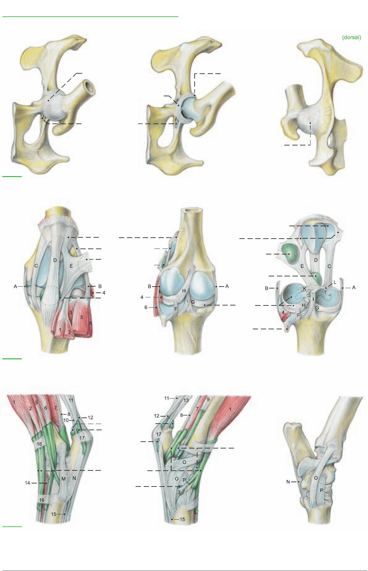
a) JOINTS OF THE PELVIC LIMB
NAME BONES involved TYPE OF JOINT FUNCTION REMARKS
I. Hip joint Ilium, ischium, pubis Composite Restricted to Ligaments: transverse acetabular,
(Art. coxae) in acetabulum, and head spheroidal flexion and labrum acetabulare, lig. of head of
of femur extension femur. Accessory lig. absent.
II. Stifle Composite joint
(Art. genus)
a) Femorotibial joint Tibial condyles and Simple condylar Mainly flexion Ligg.: collateral, cruciate,
femoral condyles and extension transverse, meniscotibial, menisco-
restricted by femoral. Injection: Med. sac,
ligaments same as II b. Lat. sac in extensor
groove of tibia on border of tendon
of peroneus tertius; does not com-
municate with any other sac.*
b) Femoropatellar joint Femoral trochlea and Simple sesamoid Tendon guide Ligg.: med., middle, and lat.
patella patellar, and med. and lat. fem.-
patel. Injection: 4 cm. prox. to tibial
tuberosity, between med. and mid-
dle patellar ligg. Communicates
with med. fem.-tibial sac.
III. Prox. tibiofibular joint. Present in exceptional cases only. Usually the rudimentary fibula is fused with the lateral tibial condyle.
IV. Distal tibiofibular joint is a tight joint. Its cavity communicates with the tarsocrural joint.
V. Tarsal joint (hock) Composite joint
a) Tarsocrural joint Tibial cochlea, prox. Composite Flexion and The collateral ligg. each have long
trochlea of talus, cochlear joint extension, and short parts. Long plantar lig. is
calcaneus, and lat. snap joint divided into medial and lat.
malleolus branches. Many other ligg. are
blended with the fibrous joint
b) Prox. intertarsal joint Distal trochlea of Composite Flexion and capsule.
talus, calcaneus, and trochlear joint extension Injection: Into dorsomed. pouch
T IV + T C. between med. collat. lig. and med.
branch of tendon of cran. tibial
c) Dist. intertarsal joint T C and T I–T III Composite Slight movement muscle
plane joint
d) Tarsometatarsal joint T I–T IV and metatarsal Composite Slight movement
III and IV plane joint
e) Intertarsal joints. Vertical, slightly moveable joints between tarsal bones in the same row.
VI. Digital joints. See thoracic limb.
b) SYNOVIAL BURSAE
Of the inconstant bursae, the iliac (coxal) subcutaneous bursa, uni-
lateral or bilateral over the tuber coxae, and the ischial subcuta-
neous bursa lateral on the tuber ischiadicum, are clinically impor-
tant. Of the important bursae related to the major trochanter, the
inconstant trochanteric bursa of the gluteus medius is on the sum-
mit and mediodistal surface of the trochanter. The constant
trochanteric bursa of the gluteus accessorius is on the lateral sur-
face of the femur just distal to the major trochanter. The clinically
important, but inconstant trochanteric bursa of the biceps femoris
is between the vertebral head of the biceps and the terminal part of
the gluteus medius on the major trochanter. This bursa may be the
cause of a dislocation of the vertebral head of the biceps behind the
major trochanter.
The large, up to 10 cm long, constant distal subtendinous bursa of
the biceps femoris lies between the lat. femoral condyle and the
thick terminal tendon of the biceps attached to the patella and the
lat. patellar lig. Occasionally it communicates with the lat.
femorotibial joint. When inflamed it produces a decubital swelling
on the stifle.
The inconstant subcutaneous bursa of the lat. malleolus, when
inflamed, produces a decubital swelling on the tarsus.
The multilocular subcutaneous calcanean bursa on the calcanean
expansion of the supf. digital flexor tendon is also inconstant and
occurs only in older animals.
The constant, extensive subtendinous calcanean bursa of the supf.
digital flexor lies between that tendon and the termination of the gas-
trocnemius on the tuber calcanei. The navicular (podotrochlear) bur-
sae (p. 27, M) between the terminal branches of the deep digital flex-
or tendon and the navicular bones are like those of the thoracic limb.
c) SYNOVIAL SHEATHS
Dorsally on the hock the tendons of the peroneus longus and the
digital extensors are surrounded by synovial sheaths. The sheaths
of the digital extensors communicate partially with the sheath of
the cranial tibial and the sheath-like bursa of the peroneus tertius.
On the plantar aspect of the hock the lat. digital flexor and the cau-
dal tibial m. have a common sheath, and the med. digital flexor has
a separate sheath. The tendon sheaths in the digits are like those of
the thoracic limb.
28
8. SYNOVIAL STRUCTURES OF THE PELVIC LIMB
* Desrochers et al. 1996
Anatomie des Rindes englisch 09.09.2003 12:36 Uhr Seite 28
M'
O'
O'
Joints, Bursae, and Synovial Sheaths of the Pelvic Limb
15 Interossei III and IV
16 Extensor retinacula
17 Flexor retinaculum
(ventral)
Joint capsule
Acetabular labrum
Transverse acetabular lig.
Lig. of head of femur
Joint capsule
Hip joint Hip joint
Legend:
A Med. collateral lig.
B Lat. collateral lig.
C Med. patellar lig.
D Middle patellar lig.
E Lat. patellar lig.
F Cd. tibial lig. of lat. meniscus
G Cd. tibial lig. of med. meniscus
H Meniscofemoral lig.
I Cd. cruciate lig.
J Cr. cruciate lig.
K Cr. tib. lig. of lat. meniscus
L Cr. tib. lig. of med. meniscus
(cranial)
(caudal)
(caudoproximal)
Med. parapatellar fibrocart.
Patella
Lat. and med. femoro-
patellar ligg.
Lat. femoropatellar lig.
Distal subtendinous
bicipital bursa
Distal subtendinous
bicipital bursa
Distal infra-
patellar bursa
Distal infra-
patellar bursa
Menisci
Fibula
Stifle Stifle
Legend:
M Lat. long collateral tarsal lig.
M' Lat. short collateral tarsal lig.
N Long plantar lig.
O Med. long collateral tarsal lig.
O' Med. short collateral tarsal lig.
P Dorsal tarsal lig.
1 Peroneus [fibularis] tertius
2 Long digital extensor
3 Cranial tibial m.
4 Popliteus
5 Peroneus [fibularis] longus
6 Lat. digital extensor
(lateral)
(medial)
Bursa of the calcanean
tendon
Subtendinous
calcanean bursa of
supf. digital flexor
Joint capsule
Synovial sheaths
Subtendinous bursa
of cran. tibial m.
Hock joint Hock joint
Legend:
Deep digital flexors
7 Lat. digital flexor
8 Caud. tibial m.
9 Med. digital flexor
10 Tarsal tendon of biceps femoris
11 Gastrocnemius
12 Supf. digital flexor
13 Tarsal tendon of semitendinosus
14 Short digital extensor (in part)
29
Anatomie des Rindes englisch 09.09.2003 12:36 Uhr Seite 29
.
The bovine skull undergoes marked changes in shape as it grows
from the newborn calf to the adult—changes that are caused in part
by the development of the horns. In the process, the roof of the cra-
nium, the occipital surface, and the lateral surfaces alter their rela-
tive positions significantly.
a) On the CRANIUM, the roof (Calvaria) is formed by the rec-
tangular frontal bones (I)
★. They extend back to the caudal surface
of the intercornual protuberance (3)
★■ where they are fused with
the parietal (II)
■ and interparietal (III)■ bones. These are united
with the occipital (VI)
■● bone, but no sutures are visible here in the
adult. The external occipital protuberance (31)
■●, the point of
attachment of the funicular lig. nuchae, is about 6 cm ventral to the
top of the skull. The nuchal line (m)
■, arching laterally from the
external occipital protuberance, corresponds to the nuchal crest of
the horse and dog. On the caudolateral angle of the frontal bone is
the cornual process (3')
★■ with its rough body and smoother neck
with vascular grooves.
Projecting from the middle of the lateral border of the frontal bone
is the zygomatic process (1)
★■, which joins the frontal process
(56)
★■ of the zygomatic bone (IX)★■. The temporal line (k)★ is the
dorsal boundary of the temporal fossa (j)
★. It is a sharp, palpable
ridge running from the zygomatic process back to the horn and
serves as a landmark for cornual nerve block (see pp. 34, 40, and
53).
b) The FACIAL ASPECT. The facial crest (57')
★ begins on the
zygomatic bone and curves across the maxilla to the facial tuber
(57")
★
■
. The often double infraorbital foramen (59)★ is dorsal to
the first cheek tooth (p. 2). Caudal to the nasoincisive notch (X")
★
a fissure persists between the dorsal nasal bone (X)★ and the ven-
tral incisive (XII), maxillary (XI), lacrimal (VIII), and frontal (I)
bones. The nasal bone has two rostral processes (X').
c) The FORAMINA of the skull are important for the passage of
nerves and vessels, and for nerve block anesthesia. Caudolaterally
on the skull between the occipital condyle (33)
■ and the jugular
process (36)
■ is the double canal for the hypoglossal n. (35)■. Dor-
sal to the petrous temporal bone is the internal opening of the tem-
poral meatus (e)
●. There is a lateral opening (e)★ in the temporal
fossa. The ox does not have a foramen lacerum; it has an oval fora-
men (45)
★■● for the mandibular n., connected by the petro-occipi-
tal fissure (q')
● with the jugular foramen (q), which conducts cra-
nial nerves IX, X, and XI. Before the internal carotid a. is occluded
at three months of age, it goes through the fissure. In the caudal
part of the orbit are three openings: from dorsal to ventral, the eth-
moid for. (2)
★, the optic canal (52)★●, and the for. orbitorotundum
(44")
★ (the combined orbital fissure and round for. of the horse
and dog.) The pointed projection lat. to these is the pterygoid crest
(46)
★
■
. On the dorsal surface the frontal bone is pierced medial to
the zygomatic process by the supraorbital canal (1")
★, often dou-
ble, which opens in the orbit. The palpable supraorbital groove
(1')
★ runs rostrally and caudally from the canal.
d) The MANDIBLE (XVII). See p. 33.
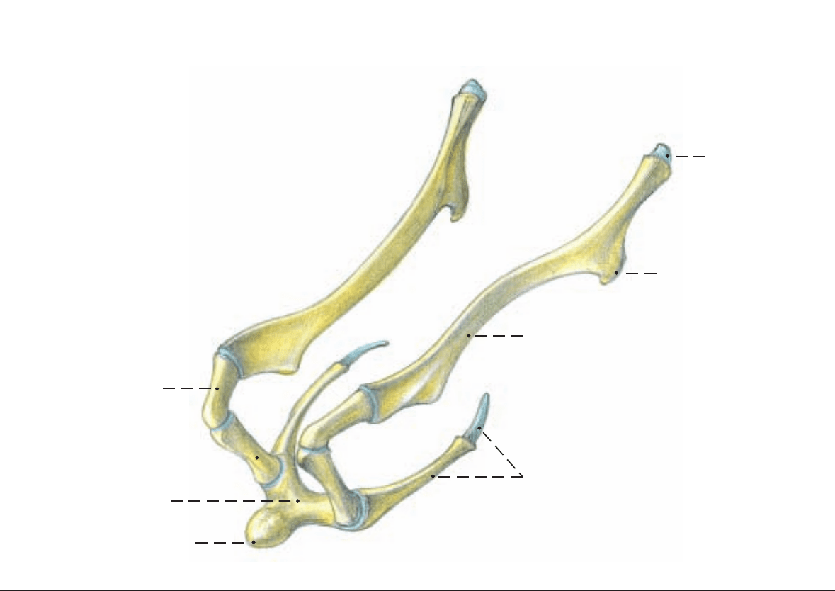
e) The HYOID APPARATUS (Text figure). The body (basihyoid)
gives off a stubby median lingual process. The thyrohyoid fuses lat-
er with the body and articulates with the rostral horn of the thyroid
cartilage of the larynx. The ceratohyoid articulates with the body
and with the rod-shaped epihyoid, which in turn articulates with
the long, flattened stylohyoid. The last three joints are synovial.
The proximal end of the stylohyoid is joined by the fibrocartilagi-
nous tympanohyoid to the styloid process. The angle of the stylo-
hyoid is drawn out in the form of a hook.
30
CHAPTER 3: HEAD
1. SKULL AND HYOID APPARATUS
Directions for the use of figures on p. 31: features marked with an asterisk (★)—upper fig.; those marked with a square (■)—lower fig.; those marked with a bullet
(
●)—p. 33 upper figure; those marked with a rhombus (◆)—p. 33 lower figure.
Lingual process
Hyoid apparatus
(rostrolateral)
Tympanohyoid
Angle of stylohyoid
Stylohyoid
Epihyoid
Ceratohyoid
Thyrohyoid
Basihyoid
Anatomie des Rindes englisch 09.09.2003 12:36 Uhr Seite 30
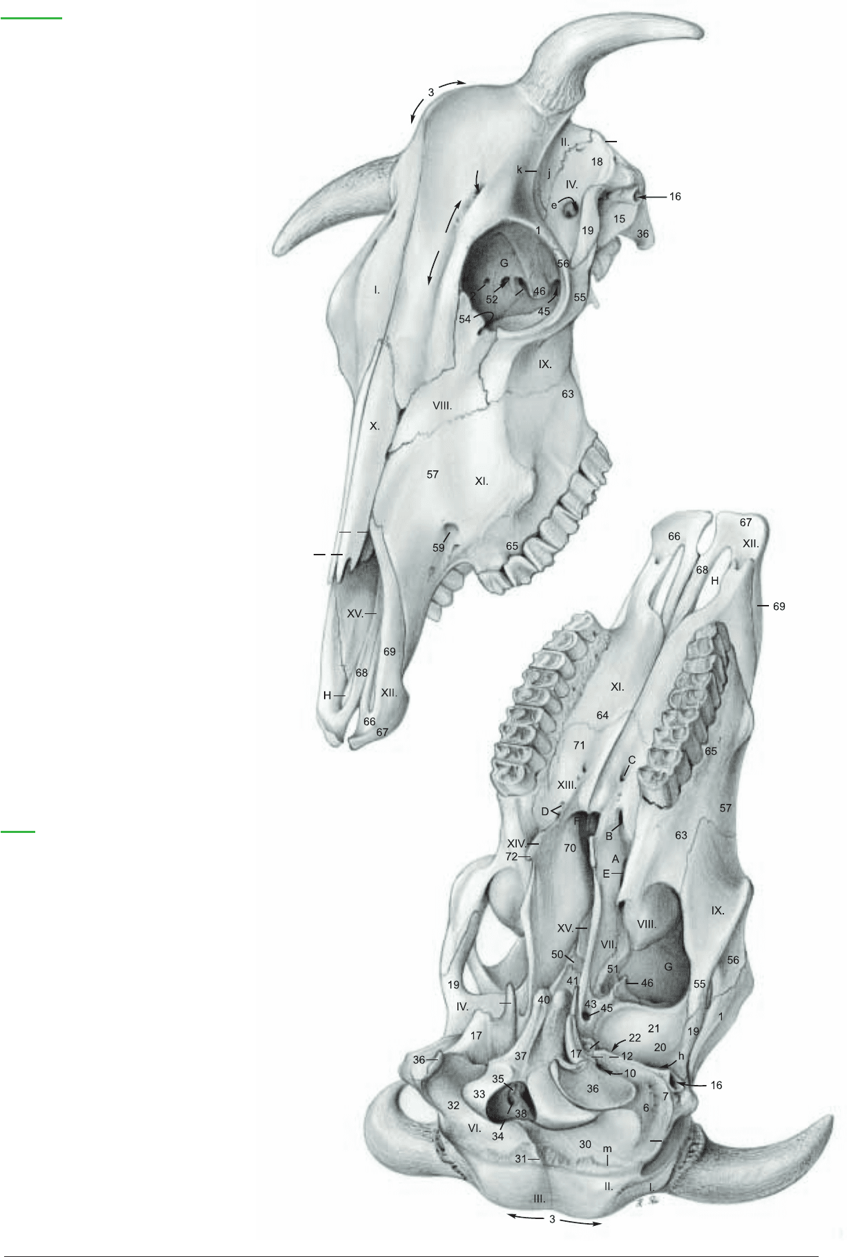
External lamina (a) ●
Diploe (b) ●
Internal lamina (c) ●
Temporal meatus (e) ★ ●
Retroarticular foramen (h) ■
Temporal fossa (j) ★
Temporal line (k) ★ [External frontal crest]
Nuchal line (m)
■
Temporal crest (m') ★ ■
Jugular foramen (q) ●
Petrooccipital fissure (q’) ●
Neurocranial bones
I. Frontal bone ★
Zygomatic process (1) ★ ■
Supraorbital groove (1') ★
Supraorbital canal (1") ★
Ethmoid foramen (2) ★
Intercornual protuberance (3) ★
■
Cornual process (3') ★ ■
II. Parietal bone ■
III. Interparietal bone ■
IV. Temporal bone ★ ■
a. Petrous part (6) ■ ●
Mastoid process (7) ■
Internal acoustic meatus
Internal acoustic pore (8)
●
Facial canal (9) ●
Stylomastoid foramen (10) ■
Styloid process (10') ■
Petrotympanic fissure (12) ■
Cerebellar fossa (13) ●
b. Tympanic part (15) ★
External acoustic meatus
External acoustic pore (16) ★
■
Tympanic bulla (17) ■
Muscular process (17") ■
c. Squamous part (18) ★
Zygomatic process (19) ★
■
Lateral opening of temporal meatus (e) ★
Mandibular fossa (20)
■
Articular surface (21) ■
Retroarticular process (22) ■
VI. Occipital bone ■ ●
Squamous part (30) ■
External occipital protuberance (31) ■ ●
Internal occipital protuberance (31') ●
Lateral part (32) ■ ●
Occipital condyle (33) ■ ●
Condylar canal (34) ■ ●
Hypoglossal nerve canal (35) ■ ●
Jugular and paracondylar process (36) ★ ■ ●
Basilar part (37) ■ ●
Foramen magnum (38) ■ ●
Muscular tubercle (40) ■ ●
VII. Sphenoid bone ■ ●
Basisphenoid bone
Body (41)
■ ●
Sella turcica (42) ●
Wing [Ala] (43) ■ ●
Groove for ophthalmic and maxillary nn. (44') ●
Foramen orbitorotundum (44") ★
Oval foramen (45) ★
■ ●
Pterygoid crest (46) ★ ■
Presphenoid bone
Body (50)
■ ●
Wing [Ala] (51) ■
Orbitosphenoid crest (51') ●
Optic canal (52) ★ ●
Face
Pterygopalatine fossa (A) ■
Major palatine canal
Caudal palatine foramen (B)
■
Major palatine foramen (C) ■
Minor palatine canals
Caudal palatine foramen (B)
■
Minor palatine foramina (D) ■
Sphenopalatine foramen (E) ■ ●
Choanae (F) ■
Orbit (G) ★ ■
Palatine fissure (H) ★ ■
Cranium
(rostrodorsal ★)
(caudobasal
■)
Facial bones
VIII. Lacrimal bone ★
Fossa for lacrimal sac (54) ★
Lacrimal bulla (54') ★
■
IX. Zygomatic bone ★ ■
Temporal process (55) ★ ■
Frontal process (56) ★ ■
X. Nasal bone ★
Rostral process (X.') ★
Nasoincisive notch (X.") ★
XI. Maxilla ★ ■
Body of maxilla (57)★ ■
Facial crest (57') ★
Facial tuber (57") ★
■
Infraorbital canal
Infraorbital foramen (59) ★
Lacrimal canal (see p. 35 D)
Zygomatic process (63) ★
■
Palatine process (64) ■
Alveolar process (65) ★ ■
XII. Incisive bone ★ ■ ●
Body of incisive bone (66) ★ ■ ●
Alveolar process (67) ★ ■ ●
Palatine process (68) ★ ■ ●
Nasal process (69) ■ ●
XIII. Palatine bone ■ ●
Perpendicular plate (70) ■ ●
Horizontal plate (71) ■ ●
XIV. Pterygoid bone ■ ●
Hamulus (72) ■ ●
XV. Vomer ●
m'
1"
1'
44"
54'
57'
57"
X."
X.'
57"
54'
17"
10'
m'
3'
31
Anatomie des Rindes englisch 09.09.2003 13:13 Uhr Seite 31
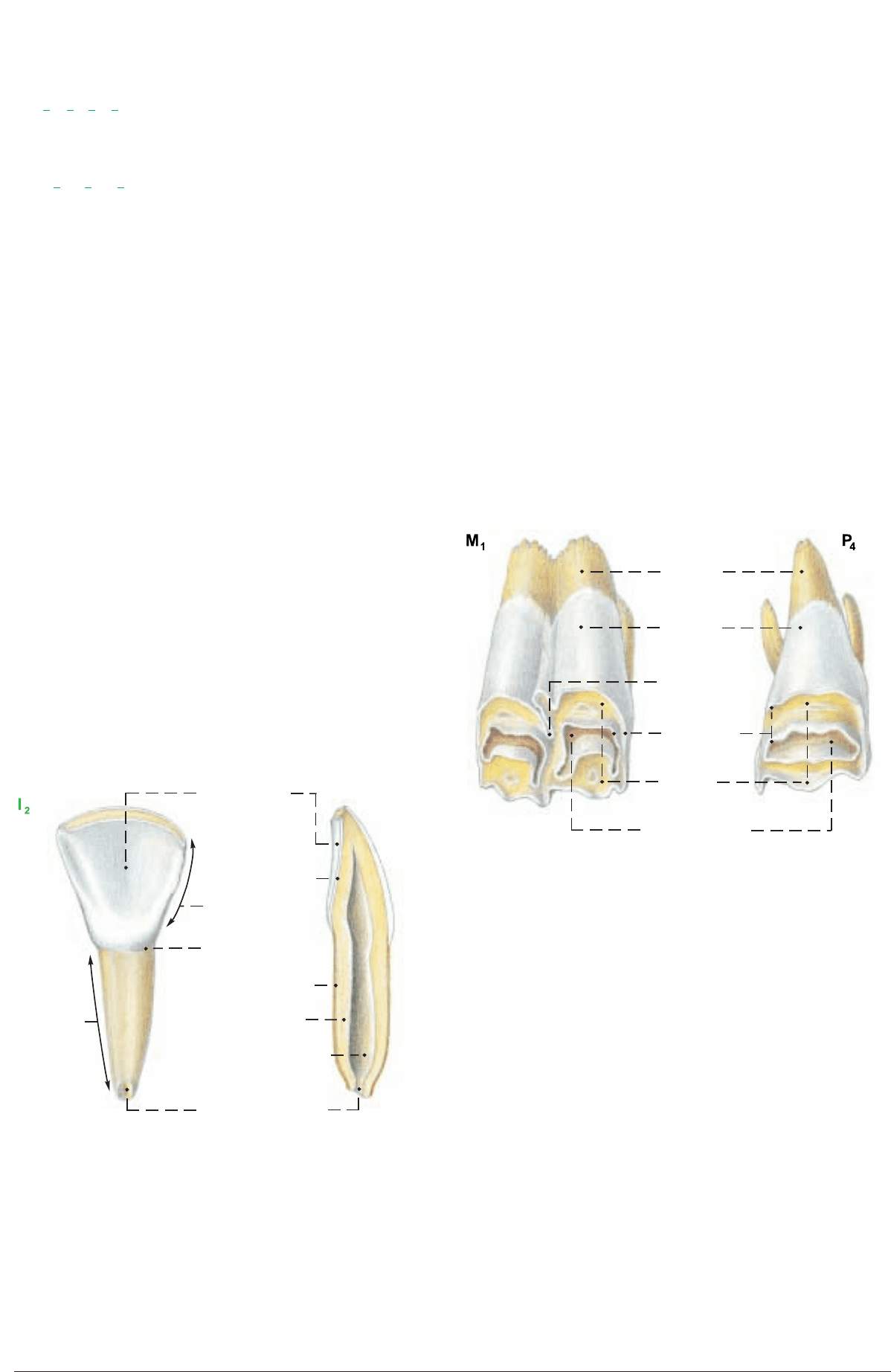
DENTITION.
The formula for the permanent teeth is:
2
ICPM
= 32
where I = incisor, C = canine, P = premolar, and M = molar.
The formula for the deciduous teeth (milk teeth) is:
2
Di Dc Dp
= 20
where Di = deciduous incisor, Dc = deciduous canine, and Dp =
deciduous premolar.
In domestic ruminants the missing upper incisors and canines are
replaced by the dental pad (p. 45, a) a plate of connective tissue
covered by cornified epithelium.
The individual TEETH have a crown, neck, and root. They consist
of dentin (ivory), enamel, and cement. The five surfaces of a tooth
are: lingual, vestibular (labial or buccal), occlusal, and two contact
surfaces. The mesial contact surface of the incisors is toward the
median plane; on all other teeth it is directed toward the incisors.
The opposite contact surface is distal. Although the upper incisors
and canines are absent after birth, the primordia are present in the
embryo.
The canine teeth (C) have the shape of incisors (I1, 2, 3) with a def-
inite neck and a shovel-shaped crown; therefore they are common-
ly counted as the fourth incisors. When these teeth erupt, the crown
is covered briefly by a thin pink layer of gingival mucosa, and
neighboring teeth overlap, but by the end of the first month they
have rotated so that they stand side by side. The permanent incisors
erupt at about the following ages: I1, 1
1
/
2
–2 yrs.; I2, 2–2
1
/
2
yrs.; I3,
3 yrs.; C, 3
1
/
2
–4 yrs. At first the crown is completely covered by
enamel; lingual and labial surfaces meet in a sharp edge. The lin-
gual surface is marked by enamel ridges extending from the
occlusal border about two thirds of the way to the neck. As the
tooth wears, the thin lingual enamel is abraded faster than the thick
labial plate, keeping the tooth beveled to a sharp edge (see text fig.).
The darker, yellowish dentin is exposed and forms most of the
occlusal surface. The dental star appears, filled with secondary
dentin. The lingual border of the occlusal surface is notched
between the ridges on the lingual surface. When the tooth wears
down to the point where the ridges disappear, the lingual border of
the occlusal surface is a smooth curve and the tooth is said to be
level. This usually occurs in sequence from I1 to C at 6, 7, 8, and 9
years. Deciduous incisors and canines are smaller than permanent
teeth and have narrower necks. The first premolar is missing, so
that the first cheek tooth is P2. Between the canines and the pre-
molars in the lower jaw there is a space, the diastema (J), with no
teeth. The size of the cheek teeth increases greatly from rostral to
caudal. The incisors and canines are brachydont teeth; they do not
grow longer after they are fully erupted, and they do not have
infundibula. The cheek teeth are hypsodont; they continue to grow
in length after eruption, but to a lesser extent than in the horse.
The infundibula of the cheek teeth develop by infolding of the
enamel organ. (See text fig.) When tooth erupts the central enamel
of each infundibulum is continuous with the external enamel in a
crest. As the crest wears off the infundibulum is separated from the
external enamel and the dentin is exposed between them. In rumi-
nants the sections of the infundibula visible on the occlusal surface
are crescentic. The infundibula are partially filled by cement and
blackened feed residue. The outside of the newly erupted tooth is
also coated with cement.
The upper premolars have one infundibulum and three roots. The
upper molars have two infundibula and three roots. The horns of
the crescents of all the infundibula of the upper cheek teeth point
toward the buccal surface. The lower premolars (P2, 3, 4) are irreg-
ular in form. P2 is small and has a simple crown, usually without
enamel folds. P3 and P4 have two vertical enamel folds on the lin-
gual surface. On P4 the caudal one may be closed to form an
infundibulum. The lower premolars have two roots. The lower
molars (M1, 2, 3) have two infundibula and two roots. The horns
of the infundibula point toward the lingual surface.
The lower jaw is narrower than the upper jaw, and the occlusal sur-
face of the upper cheek teeth slopes downward and outward to
overlap the buccal edge of the lower teeth, but the lateral motion of
the mandible in chewing, first on one side and then on the other,
wears the occlusal surfaces almost equally.
32
2. SKULL WITH TEETH
Directions for the use of figures on p. 31: features marked with an asterisk (
★
)—upper fig.; those marked with a square (
■
)—lower fig.; those marked with a bullet
(
●
)—p. 33 upper figure; those marked with a rhombus (
◆
)—p. 33 lower figure.
Lingual surface
Enamel
Crown
Neck
Cement
Root
Dentin
Pulp cavity
Apical foramen
(Upper teeth, lingual surface)
Cement
Enamel
Enamel fold
Enamel crest
Dentin
Infundibulum
0
3
0
1
3
3
3
3
0
3
0
1
3
3
Anatomie des Rindes englisch 09.09.2003 13:14 Uhr Seite 32
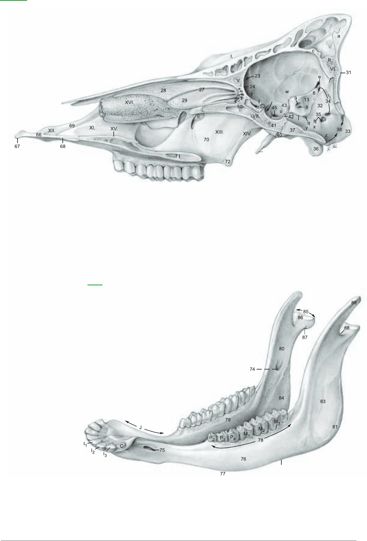
51'
44'
31'
q'
17"
77'
Cranial cavity
Rostral cran. fossa (r) ●
Middle cran. fossa (u) ●
Hypophysial fossa (v) ●
Piriform fossa (w) ●
Caudal cran. fossa (x) ●
Pontine impression (y) ●
Medullary impression (z) ●
Neurocranial bones
II. Parietal bone ★ ■
III. Interparietal bone ■
IV. Temporal bone ★ ■
a. Petrous part (6) ■ ●
Mastoid process (7) ■
Internal acoustic meatus
Internal acoustic pore (8)
●
Facial canal (9) ●
Stylomastoid foramen (10) ■
Styloid process (10') ■
Petrotympanic fissure (12) ■
Cerebellar fossa (13) ●
b. Tympanic part (15) ★
External acoustic meatus
External acoustic pore (16) ★
■
Tympanic bulla (17) ■
Muscular process (17") ■ ●
c. Squamous part (18) ★
Zygomatic process (19) ★
■
Mandibular fossa (20) ■
Articular surface (21) ■
Retroarticular process (22) ■
V. Ethmoid Bone ●
Cribriform plate (23) ●
Crista galli (24) ●
Ethmoid labyrinth (25) ●
Ethmoturbinates
Ectoturbinates (not shown)
Endoturbinates (27)
●
Dorsal nasal concha (28) ●
Middle nasal concha (29) ●
VI. Occipital bone ■ ●
Squamous part (30) ■
External occipital protuberance (31) ■ ●
Internal occipital protuberance (31') ●
Lateral part (32) ■ ●
Occipital condyle (33) ■ ●
Condylar canal (34) ■ ●
Hypoglossal nerve canal (35) ■ ●
Jugular and paracondylar process (36) ★ ■ ●
Basilar part (37) ■ ●
Foramen magnum (38) ■ ●
Muscular tubercle (40) ■ ●
VII. Sphenoid bone ■ ●
Basisphenoid bone
Body (41)
■ ●
Sella turcica (42) ●
Wing [Ala] (43) ■ ●
Groove for ophthalmic and maxillary nn. (44') ●
Foramen orbitorotundum (44") p. 31 ★
Oval foramen (45) ★
■ ●
Pterygoid crest (46) ★ ■
Presphenoid bone
Body (50)
■ ●
Wing [Ala] (51) ■
Orbitosphenoid crest (51') ●
Optic canal (52) ★ ●
Face
Facial bones
Sphenopalatine foramen (E) ■ ●
XII. Incisive bone ■ ●
Body of incisive bone (66) ★ ■ ●
Alveolar process (67) ★ ■ ●
Palatine process (68) ★ ■ ●
Nasal process (69) ★ ■ ●
XIII. Palatine bone ■ ●
Perpendicular plate (70) ■ ●
Horizontal plate (71) ■ ●
XIV. Pterygoid bone ■ ●
Hamulus (72) ■ ●
Cranium
(Paramedian section ●)
External lamina (a) ●
Diploë (b) ●
Internal lamina (c) ●
Temporal meatus (e) ★ ●
Retroarticular foramen (h) ■
Temporal fossa (j) ★
Temporal line (k) ★ [External frontal crest]
Nuchal line (m)
■
Temporal crest (m') ★ ■
Jugular foramen (q) ●
Petrooccipital fissure (q') ●
XVII. Mandible ◆
XV. Vomer ●
XVI. Ventral nasal concha ●
XVII. Mandible ◆
Mandibular canal
Mandibular foramen (74)
◆
Mental foramen (75) ◆
Body of the mandible (76) ◆
Diastema (J) ◆
Ventral border (77) ◆
Vascular groove (77') ◆
Alveolar border (78) ◆
Mylohyoid line (79) ◆
Ramus of the mandible (80) ◆
Angle of the mandible (81) ◆
Masseteric fossa (83) ◆
Pterygoid fossa (84) ◆
Condylar process (85) ◆
Head of mandible (86) ◆
Neck of mandible (87) ◆
Mandibular notch (88) ◆
Coronoid process (89) ◆
33
Anatomie des Rindes englisch 09.09.2003 13:14 Uhr Seite 33

a) The PARANASAL SINUSES (see also p. 45) may be studied
from prepared skulls, but many of the clinically important septa are
not solid bone; they are completed by membranes that do not sur-
vive maceration. The paranasal sinuses develop by evagination of
the nasal mucosa into the spongy bone (diploë, b, p. 33)
● between
the external and internal plates (a, c)
● of the cranial and facial
bones. Therefore each sinus is lined by respiratory epithelium and,
except for the lacrimal and palatine sinuses, which are diverticula
of the maxillary sinus, each has a direct opening to the nasal cavi-
ty. Unfortunately, when inflammation occurs, the mucous mem-
brane swells and closes the aperture, blocking normal drainage of
the sinus. This condition may require surgical drainage.
The paranasal Sinuses of the Ox
Group I Group II
Maxillary Frontal
Lacrimal Caudal
Palatine Rostral
Conchal Medial
Dorsal Intermediate
Ventral Lateral
Sphenoid
Ethmoid cells
Middle conchal sinus
I. The first group of sinuses open into the middle nasal meatus
(p. 45, 6)
1. The maxillary sinus (7) occupies the maxilla and extends back
under the orbit into the thin-walled lacrimal bulla (E) and into the
zygomatic bone, thereby surrounding the orbit rostrally and ven-
trally. The nasomaxillary opening is high on the medial wall just
ventral to the lacrimal canal (D) and midway between the orbit and
the facial tuber. It opens into the middle nasal meatus.
The maxillary sinus communicates with the lacrimal sinus (5) and
through the maxillopalatine opening (F) over the infraorbital canal
(G) with the palatine sinus (10). See also p. 45, j.
There is a large opening in the bony wall between the ventral nasal
meatus and the palatine sinus, but this is closed in life by the appo-
sition of their mucous membranes.
2. Also opening into the middle nasal meatus is the dorsal conchal
sinus (6) in the caudal part of the dorsal concha, and
3. the ventral conchal sinus in the caudal part of the ventral con-
cha (XVI) p. 33
●. See also p. 45.
II. The second group of sinuses open into ethmoidal meatuses in
the caudal end of the nasal cavity.
1. The frontal sinuses are variable in size and number. In the new-
born calf, they occupy only the frontal bone rostrodorsal to the
brain. In the aged ox the caudal frontal sinus is very extensive,
invading also the parietal, interparietal, occipital, and temporal
bones. Left and right frontal sinuses are separated by a median sep-
tum (B). The caudal frontal sinus (1) is bounded rostrally by an
oblique transverse septum (B') that runs from the middle of the
orbit caudomedially to join the median septum in the transverse
plane of the caudal margin of the orbit. The caudal boundary is the
occipital bone and the lateral boundary is the temporal line (k)*.
There is an extension into the zygomatic process. The supraorbital
canal (C), conducting the frontal vein, passes through the caudal
frontal sinus in a plate of bone that appears to be a septum, but is
always perforated. The caudal frontal sinus has three clinically
important diverticula: the nuchal (H), cornual (J), and postorbital
(K) diverticula. The caudal frontal sinus has only one aperture: at
its rostral extremity there is a small outlet to an ethmoid meatus.
There is no frontomaxillary opening in any domestic animal except
the Equidae. The rostral frontal sinuses (2, 3, 4) lie between the ros-
tral half of the orbit and the median plane. Each has an opening at
its rostral end to an ethmoid meatus. A part of the dorsal nasal con-
cha (6) projects caudally between two of the rostral frontal sinuses.
The lateral rostral frontal sinus is separated by a thin septum from
the lacrimal sinus.
2. The sphenoid sinus (8), when present, opens into an ethmoid
meatus.
3. The ethmoid cells (9) in the medial wall of the orbit, and
4. The sinus of the middle concha (p. 45, g) open into ethmoid
meatuses.
b) The HORNS (CORNUA) project from the caudolateral angle
of the frontal bone in both sexes, (except for hornless breeds, which
have only a knob-like thickening of the bone.) Round, and tapering
conically to a small apex, their form is not only species and breed
specific, but is also quite variable individually. In the cow they are
slender and long—in the bull, thick and short, and in the steer also
thick, but longer. We recognize a base, a body, and an apex. The
osseous core of the horn is the cornual process of the frontal bone
(p. 31, 3'), which until shortly before birth is a rounded thickening.
This elongates after birth to become a massive bony cone, and
beginning at six months is pneumatized from the caudal frontal
sinus. This is clinically important in deep wounds of the horns and
in dehorning methods.
The bony process, like the distal part of the digit, is covered by
greatly modified skin.
I. The subcutis is absent and the periosteum is fused with the der-
mis.
II. The dermis bears distinct papillae, which become longer on the
base and especially toward the body, and lie step-wise over each
other parallel to the surface. On the apex they are large free verti-
cal tapering papillae. The dermis forms the positive die on which
the living epidermis is molded.
III. The epidermis of the horn produces from its living cells the
cornified horn sheath (stratum corneum) as horn tubules corre-
sponding to the dermal papillae. The tubules are bound together by
intertubular horn. Longitudinal growth of the horns occurs under
the previously formed conical horn sheath through the production
of a new cone of horn by the living epidermal cells, pushing the
horny substance toward the apex. This can be seen on a longitudi-
nal section. The horn consists of a stack of cones, each produced
during a growth period, the horn sheath becoming thicker toward
the apex. Radial growth pressure inside the rigid sheath compress-
es and flattens the tubules so that they are not recognizable in the
body. On the apex of the cornual process additional tubular horn
is formed over the free papillae. Growth is mainly longitudinal;
growth in diameter is of lesser importance.
The formation of horn substance is steady in the bull; therefore the
horns appear smooth on the surface. In the cow, growth is period-
ical and variable in rate, causing superficial rings and grooves. The
rings are the product of regular, and the grooves the product of
irregular horn formation, which is explained primarily by repeated
pregnancies, but also by nutritional deficiencies and possibly dis-
eases.
On the base of the horn at the transition from the skin to the horn
sheath there is an epidermal zone called the epikeras that is com-
parable to the periople of the equine hoof.
The blood supply of the horns comes from the cornual aa. and vv.
from the supf. temporal a. and v.
The innervation is supplied by the cornual br. of the zygomati-
cotemporal br. (see p. 40) and also the supraorbital and
infratrochlear nn., all from the ophthalmic n.
The lymph is drained to the parotid ln.
34
3. SKULL WITH PARANASAL SINUSES AND HORNS
Anatomie des Rindes englisch 09.09.2003 13:14 Uhr Seite 34
