Budras Klaus-Dieter, Habel Robert E. Bovine Аnatomy
Подождите немного. Документ загружается.

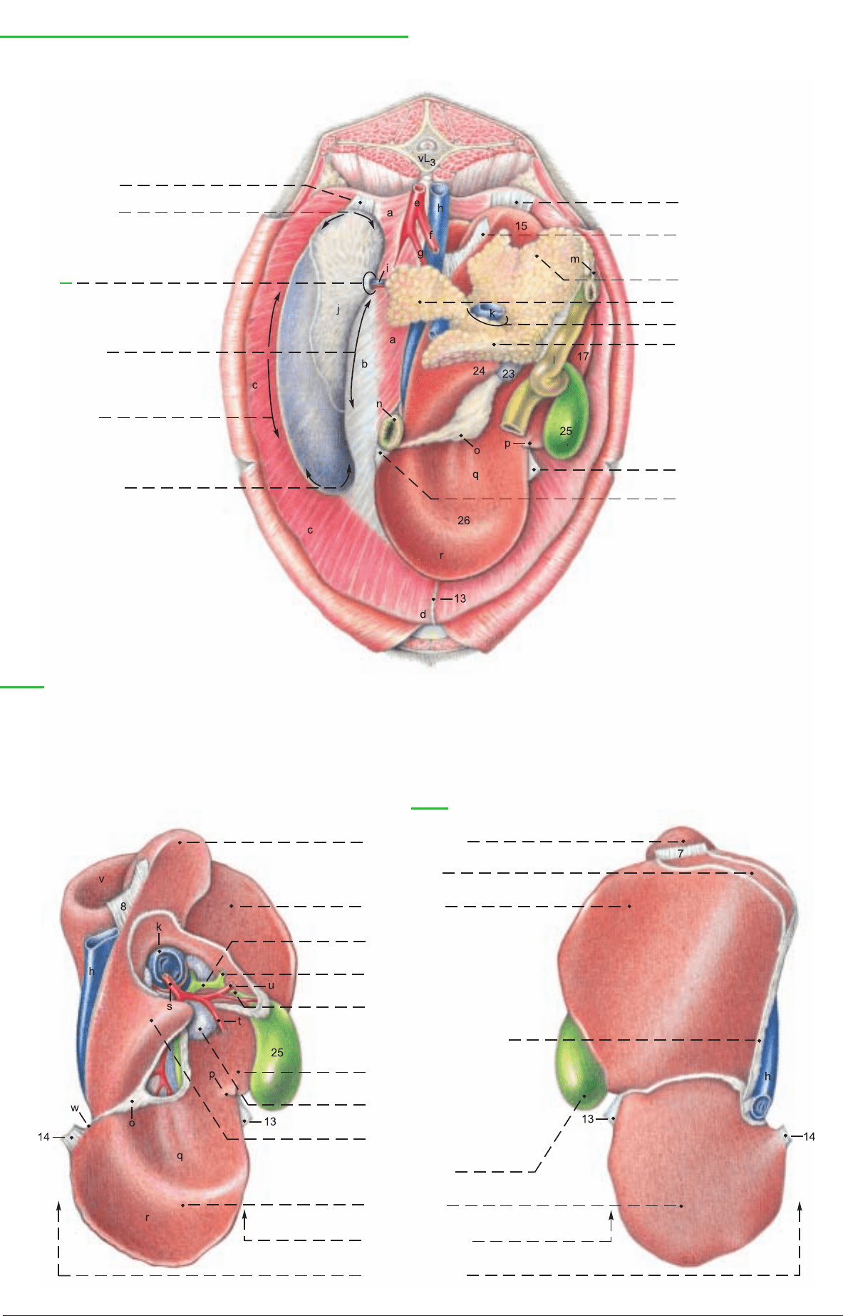
28 Dorsal border
Spleen, Liver, and Pancreas (Abdominal surface of diaphragm)
(dorsal)
Spleen:
1 Phrenicosplenic lig.
2 Dorsal end of spleen
3 Hilus of
spleen
4 Cran. border of
spleen
5 Caud. border of
spleen
6 Ventral end of spleen
7 Right triangular lig.
8 Hepatorenal lig.
Pancreas:
9 Right lobe of pancreas
10 Left lobe of pancreas
11 Pancreatic notch
12 Body of pancreas
13 Falciform and Round ligg.
14 Left triangular lig.
(See p. 69)
(ventral)
Legend:
Diaphragm:
a Lumbar part
b Tendinous center
c Costal part
d Sternal part
e Aorta
f Cran. mesenteric a.
g Celiac a.
h Caud. vena cava
i Splenic a. and v.
j Splenico-ruminal adhesion
k Portal v.
l Duodenum
m Accessory pancreatic duct
n Esophagus
o Lesser omentum
p Fissure for round lig.
q Omasal impression
r Reticular impression
s Hepatic a.
t Right gastric a.
u Gastroduodenal a.
v Renal impression
w Esophageal impression
(cut edge)
Liver
(Visceral surface)
(Diaphragmatic surface)
15 Caudate proc.
16 Bare area
(Area nuda)
17 Right lobe
18 Common hepatic duct
19 Common bile duct
(Ductus choledochus)
20 Cystic duct
21 Coronary lig.
22 Quadrate lobe
23 Hepatic lnn.
24 Papillary proc.
25 Gallbladder
26 Left lobe
27 Ventral border
75
Anatomie des Rindes englisch 09.09.2003 14:18 Uhr Seite 75
a) The INTESTINAL TRACT is displaced to the right half of the
abdominal cavity by the enormous expansion of the stomach, pri-
marily the rumen, on the left. Most of the intestines, attached by the
mesentery, lie in the supraomental recess. The intestinal tract has
considerable length – 33–59 m, whereas the lumen, especially of the
large intestine, is small compared to the horse.
The small intestine has a total length of 27–49 m. The duodenum
begins ventrally on the right at the pylorus with the cranial part (1),
which runs dorsally to the porta of the liver. Here it forms the sig-
moid flexure (1), turns caudally at the cranial flexure, and contin-
ues as the descending part of the duodenum (2) (see also p. 69).
This runs caudodorsally, accompanied at first by the right lobe of
the pancreas, to the plane of the tuber coxae. Here it turns sharply
medially around the caudal border of the mesentery at the caudal
flexure (3), and continues cranially as the ascending part of the
duodenum (4). The descending colon (17) is dorsal to the ascend-
ing duodenum and adherent to it. The caudal free border of this
adhesion is the duodenocolic fold (5). Under the left lobe of the
pancreas and on the left side of the cranial mesenteric a., the
ascending duodenum passes through the duodenojejunal flexure
into the jejunum (6). This surrounds the disc of the coiled colon like
a wreath. It begins cranially at the liver and pancreas and runs cau-
doventrally through many loops until it passes without a clear
boundary into the ileum cranial to the pelvic inlet. The caudal part,
called the “flange” is of clinical significance because of its longer
mesentery. The ileum (7) is described as the part of the small intes-
tine attached to the ileocecal fold (8), but in the ox the fold extends
on the left side of the mesentery to the apex of the flange.* There-
fore by this definition the bovine ileum has a convoluted part as
well as the 1 m long straight part near the cecum. The ileum opens
into the large intestine at the ileal orifice, on the ileal papilla (p. 77,
lower figure) which marks the boundary between the cecum and
colon at the transverse plane of the 4th lumbar vertebra.
The large intestine including the cecum, colon, and rectum has nei-
ther bands nor sacculations, unlike that of the horse. The cecum is
cylindrical, 50–70 cm long, and slightly curved. It lies in the dorsal
part of the right abdominal cavity and extends to the pelvic inlet
with a free, rounded blind apex (10). The body of the cecum (9) is
attached by the common mesentery to the proximal and distal
loops of the colon, and is continuous with the colon, with no
change in the lumen, at the cecocolic orifice (p. 77, lower figure).
The colon is about 7–9.5 m long, and consists of the ascending
colon, transverse colon, and descending colon.* The ascending
colon, the longest part of the large intestine, has three parts. The
proximal loop (11) runs cranially for a short distance to the plane
of the right kidney, where it doubles back dorsal to the first part
and the cecum. It then turns mediodorsally around the caudal bor-
der of the mesentery and runs cranially on the left side of the mesen-
tery. Near the left kidney it becomes narrower and turns ventrally
into the elliptical coil formed by the spiral loop. This is variable, but
usually consists of 1.5–2 centripetal gyri (12), the central flexure
(13), and the same number of centrifugal gyri (14). The last (outer)
centrifugal gyrus passes into the narrow distal loop (15) at the
plane of the first lumbar vertebra. The distal loop runs first dorso-
caudally on the left side of the mesentery, ventral to the ascending
duodenum and dorsal to the proximal loop. At the plane of the 5th
lumbar vertebra it turns sharply around the caudal border of the
mesentery and runs forward on the right to the short transverse
colon (16). It turns around the cranial mesenteric a. from right to
left and becomes the descending colon (17) that runs caudally ven-
tral to the vertebral column. Its fat-filled mesocolon lengthens at
the last lumbar vertebra, and the sigmoid colon (18) forms at the
pelvic inlet. The rectum (19) begins at the pelvic inlet with a short-
ened mesorectum, but no structural transition.
b) The MESENTERY. The derivatives of the primitive dorsal
mesentery that are attached to the parts of the small and large intes-
tines are fused in the intestinal mass to form a common mesentery.
Only the transverse and sigmoid colons have a free mesocolon. The
proximal and distal loops and the cranial part of the descending
colon are adherent to the cranial part of the cecum and ascending
duodenum in a fat-filled mass around the root of the mesentery.
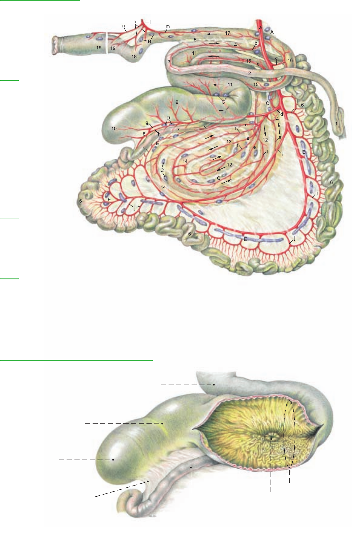
c) The BLOOD SUPPLY to the intestines comes from the cranial
and caudal mesenteric aa. The long cran. mesenteric a. (a) gives off
pancreatic brr. directly to the right lobe of the pancreas, and the
caud. pancreaticoduodenal a. (b). It also gives off the middle colic
a. (c) directly. From the proximal part of the ileocolic a. (d) the right
colic aa. (e) are given off to the distal loop of the colon and to the
centrifugal gyri. From the distal part of the ileocolic a. the colic
branches (f) go to the proximal loop of the colon and the centripetal
gyri. All of the arteries of the spiral loop may originate from the
ileocolic a. by a common trunk. They anastomose via collateral
branches. The cecal a. (g) passes to the left of the ileocolic junction
into the ileocecal fold and can give off an antimesenteric ileal
branch (h), which is constant in the dog. In addition, the cranial
mesenteric a. gives off a large collateral branch (i), peculiar to the
ox, that runs in the jejunal mesentery along the last centrifugal
gyrus, to which it gives branches, and rejoins the cranial mesenteric
a. Both give off jejunal aa. (f') and finally anastomose with the ileal
aa. (k). The mesenteric ileal branch (h') from the ileocolic a. or cecal
a. also supplies several branches to the neighboring parts of the spi-
ral colon. The caudal mesenteric a. (l) gives off the left colic a. (m)
to the descending colon, and the cranial rectal a. (n) and sigmoidal
aa. (o). The portal v. and its main branches are generally similar to
those of the horse and dog. The veins predominantly follow the
course of the corresponding arteries.
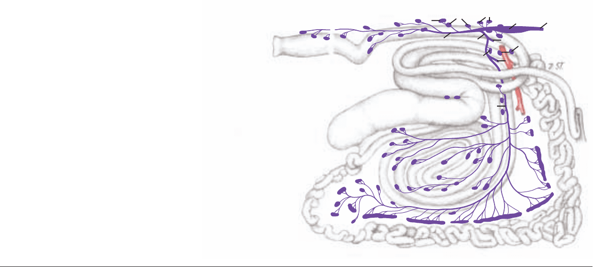
d) The LYMPH NODES. The cranial mesenteric and celiac lnn. (A)
lie at the origin of the cranial mesenteric a. The following are regu-
larly examined in meat inspection: the jejunal lnn. (E) are in the
mesentery of the jejunum and ileum near the intestinal border, unlike
the dog and horse. The cecal lnn. (D) are inconstant. Three groups
of colic lnn. (C) are most numerous on the right surface of the spiral
loop; others are present on the proximal and distal loops. The cau-
dal mesenteric lnn. (B) are on the sides of the descending colon. The
lymph drainage goes into the cisterna chyli.
76
6. INTESTINES WITH BLOOD VESSELS AND LYMPH NODES
* Smith, 1984
** see also Baum, 1912
B
G
F
O
H
R
L
M
N
A
C
C
D
E
E
K
Q
I
C
E
P
Lymph nodes and Lymphatic vessels
Legend:
A
Celiac and cran. mesenteric lnn.
B Caud. mesenteric lnn.
C Colic lnn.
D Cecal lnn.
E Jejunal lnn.
F Aortic lumbar lnn.
G Proper lumbar lnn.
H Renal lnn.
I Pancreaticoduodenal lnn.
K Anorectal lnn.
L Gastric trunk
M Hepatic trunk
N Intestinal trunk
O Cisterna chyli
P Thoracic duct
Q Lumbar trunk
R Visceral trunk
Anatomie des Rindes englisch 09.09.2003 14:18 Uhr Seite 76
h'
Intestines (Right surface)
Legend:
a Cran. mesenteric a.
b Caud. pancreaticoduodenal a.
c Middle colic a.
d Ileocolic a.
e Right colic a.
f Colic branches
g Cecal a.
h Antimesenteric ileal br.
h' Mesenteric ileal br.
i Collateral br.
j Jejunal aa.
k Ileal a.
l Caud. mesenteric a.
m Left colic a.
n Cran. rectal a.
o Sigmoidal aa.
Legend:
A Cran. mesenteric lnn.
B Caud. mesenteric lnn.
C Colic lnn.
D Cecal lnn.
E Jejunal lnn.
Legend:
Duodenum:
1 Cran. part and
Sigmoid loop
2 Descending part
3 Caud. flexure
4 Ascending part
5 Duodenocolic fold
6 Jejunum
7 Ileum
8 Ileocecal fold
Cecum:
9 Body of cecum
10 Apex of cecum
Colon:
Ascending colon:
11 Prox. loop of colon
Spiral loop of colon
12 Centripetal gyri
13 Central flexure
14 Centrifugal gyri
15 Distal loop of colon
16 Transverse colon
17 Descending colon
18 Sigmoid colon
19 Rectum
Cecum, Ileum, and Prox. loop of colon (cut open)
Body of cecum
Apex of cecum
Ileocecal fold
Cecocolic orifice
Prox. loop of colon
Ileum
Ileal papilla and Ileal orifice
77
Anatomie des Rindes englisch 09.09.2003 14:19 Uhr Seite 77
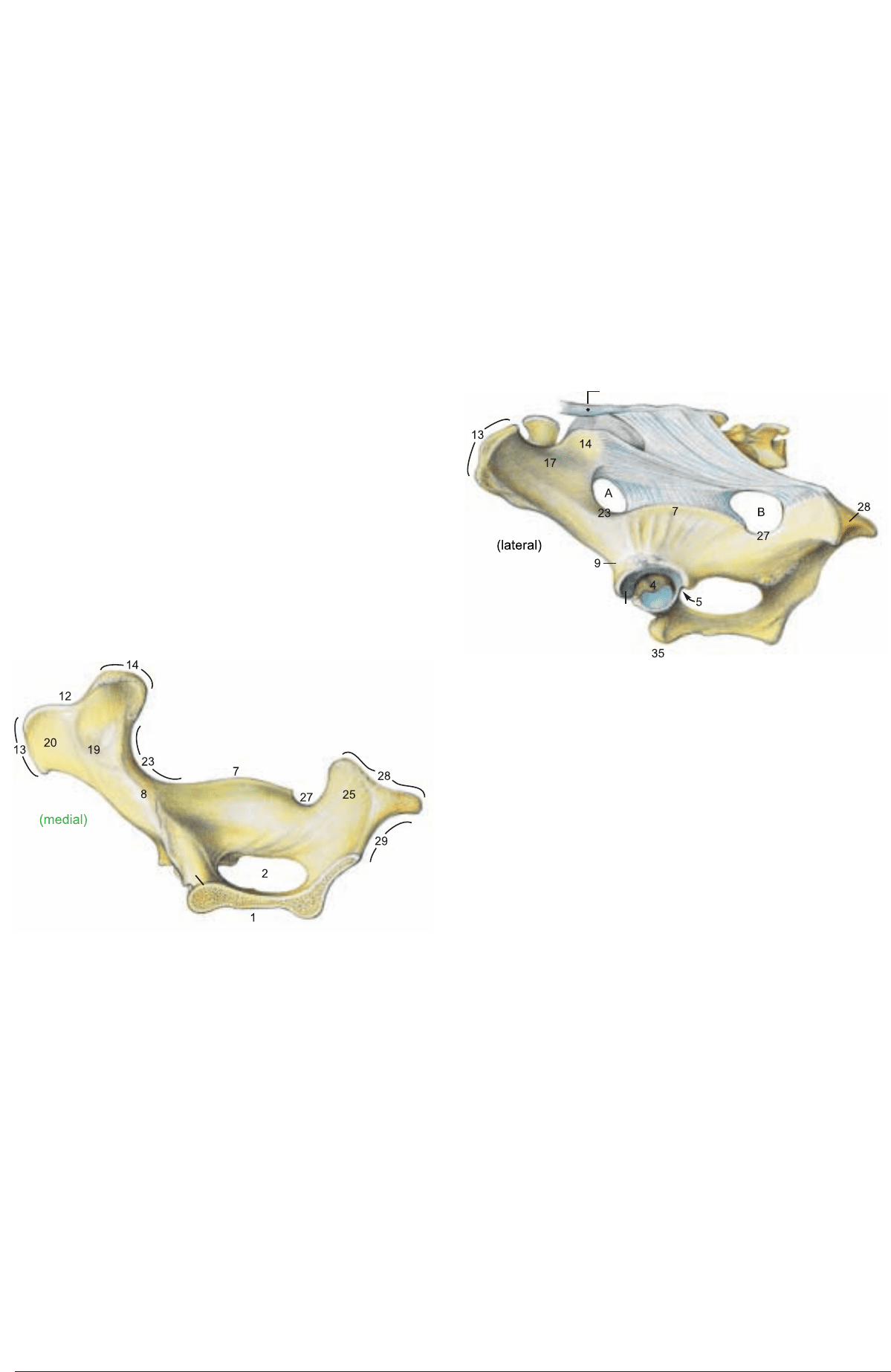
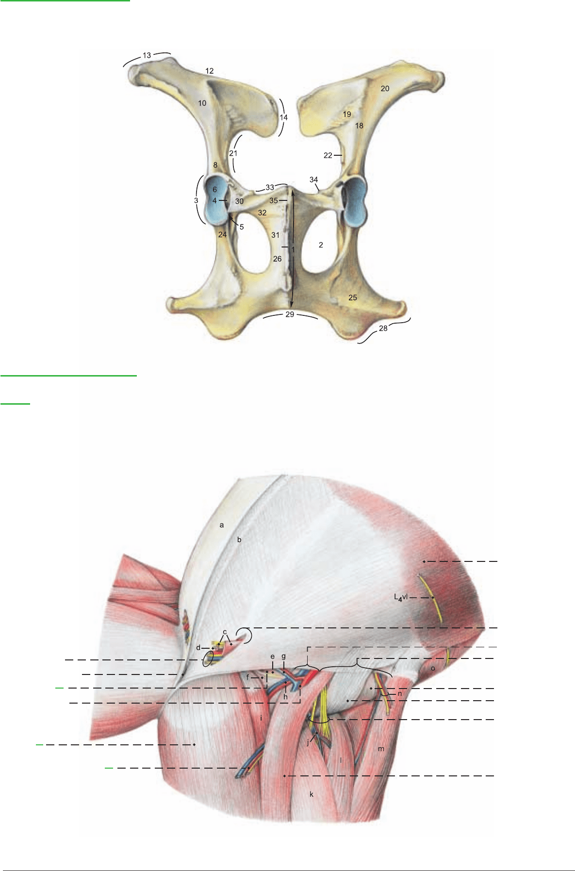
a) The PELVIC GIRDLE consists of the two hip bones (ossa
coxarum), each composed of the fused ilium, pubis, and ischium.
The two hip bones are joined in the pelvic symphysis, which ossi-
fies progressively with age.
I. On the ilium the tuber coxae (13) is thick in the middle and
undivided, and the gluteal surface (17) faces dorsolaterally. The
wing of the ilium (10) is broad, but smaller than in the horse. On
the sacropelvic surface (18) the auricular surface (19) and the iliac
surface (20) are separated by a sharp crest.
II. On the ischium the ischial tuber (28) has three processes, and
the ischial arch (29) is deep.
III. The right and left pubic bones join in the pubic symphysis to
form a ventral pubic tubercle (35) and an elongated dorsal pubic
tubercle (35'). The iliopubic eminence (34) is an imposing large
rough tubercle. The pelvic symphysis (1) is composed of the pubic
symphysis and the ischial symphysis. The latter is marked by a ven-
tral symphyseal crest (1') with a prominent caudal tubercle. The
sciatic spine (7) is high, with a sharp edge, and inclined slightly
medially. In the acetabulum (3) the lunar surface (6) is divided by
an additional cranioventral notch into a lateral greater part (6') and
a medial lesser part (6"). The oval obturator foramen (2) is espe-
cially large, with a sharp margin. The pelvic floor slopes medioven-
trally, is excavated by a deep transverse trough, and rises cau-
dodorsally. Sexual dimorphism is not as striking as in the horse.
The transverse trough is broader in the cow.
The bony pelvis is the solid framework of the birth canal which is
evaluated by measurements (pelvimetry). The transverse diameter
between the right and left psoas tubercles (22) is significant because
constriction occuring there is a hindrance to the birth process. The
vertical diameter extends from the cranial end of the pelvic sym-
physis to the dorsal wall of the pelvis. The farther caudally the verti-
cal diameter meets the dorsal wall, the more this tight passage in the
birth canal can be enlarged by drawing the pelvic floor cranially. (The
pelvic ligg. are relaxed in parturition.) On the whole, the pelvis of the
cow is not as well adapted to parturition as that of the mare.
b) The SACROSCIATIC LIGAMENT (LIG. SACROTUBERALE
LATUM) extends from the lateral part of the sacrum to the ilium and
ischium. The cranial part is attached to the sciatic spine (7) as far as
the greater sciatic notch (23). Ventral to the sacral tuber it leaves the
greater sciatic foramen (A) free for passage of the sciatic nerve and the
cranial gluteal a., v., and n. The caudal (sacrotuberous) part of the lig-
ament extends to the dorsal process of the tripartite ischial tuber (28).
Cranial to that, in the lesser sciatic notch (27), is the lesser sciatic fora-
men (B) for the passage of the caudal gluteal a. and v. Because of the
absence of vertebral heads of the caudal thigh muscles, the caudal
part of the sacrosciatic lig. is the dorsolateral boundary of the
ischiorectal fossa between the root of the tail and the ischial tuber.
The fossa is also present in the dog, but not in the horse.
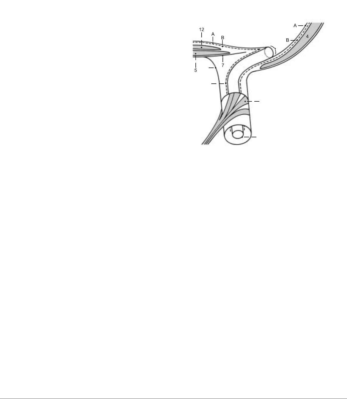
c) SUPERFICIAL STRUCTURES IN THE PUBIC AND
INGUINAL REGIONS
The intertendinous fossa (2), open ventrally, is cranial to the ven-
tral pubic tubercle and contains the terminal part of the linea alba
(b). The fossa lies between bilateral semiconical pillars converging
toward the symphyseal tendon at the apex of the prepubic tendon.
These pillars are covered by the yellow abdominal tunic (a) and are
formed by the abdominal tendons of the external oblique muscles
sheathing the ventral borders of the rectus tendons. The latter fuse
and terminate in the symphyseal tendon and on the symphyseal
crest (1').
The gracilis muscles (5) originate mainly from the symphyseal ten-
don. The external pudendal a. and v. (1) pass through the superfi-
cial inguinal ring (8) as in the dog and horse. The caudomedial
angle of the ring is close to the median plane.
The lacuna vasorum (9) is a space between the caud. border of the
pelvic tendon of the ext. oblique and the ilium. It conducts the
femoral a. and v. (4) through its lateral part and the caudal (larger)
head of the sartorius (14) through its medial part. Cranial and cau-
dal heads of the muscle embrace the femoral vessels and then unite
below them to form a single muscle belly. The femoral a. and v. and
saphenous n. pass laterally through the sartorius into the femoral
triangle (p. 18, a) and are therefore covered medially by the muscle
and not by fascia alone as in the dog and horse. (The lacuna vaso-
rum was formerly called the femoral ring, and the femoral triangle
was called the femoral canal by many veterinary anatomists, but
the terms femoral ring and femoral canal are preempted for their
meaning in human anatomy: the ring is in the medial angle of the
lacuna vasorum, covered by transversalis fascia and peritoneum,
and leads to the canal, which is only 1.25 cm long in man and con-
tains nothing but fat and a lymph node. In adult domestic mammals
the femoral ring is usually obscured by the deep femoral (h) and
pudendoepigastric (g) vessels.) The large deep femoral vessels (h)
usually originate from the external iliac vessels, give off the puden-
doepigastric trunk and vein (g) in the abdominal cavity (p. 81, s, t),
and pass out through the medial part of the lacuna vasorum, but
the origin of the deep femoral vessels is variable. They may come
from the femoral vessels in the femoral triangle, so that the puden-
doepigastric a. and v. must pass back into the abdominal cavity
through the femoral ring to reach the inguinal canal. They divide
into the caudal epigastric a. and v. (p. 81, u) and the external
pudendal a. and v. (1). The latter vessels always exit through the
inguinal canal.
Through the lacuna musculorum (10) between the inguinal lig. and
the ilium pass the iliopsoas, the smaller cranial head of the sarto-
rius (14), the femoral n. (13), divided into its branches, and the
saphenous n. (6). Ventrally the lacuna musculorum is covered by
the yellow abdominal tunic and by the tendinous femoral lamina
(12) from the external oblique (7), as in the horse.
78
CHAPTER 8: PELVIC CAVITY AND INGUINAL REGION, INCLUDING URINARY AND
GENITAL ORGANS
1. PELVIC GIRDLE WITH THE SACROSCIATIC LIG. AND SUPERFICIAL STRUCTURES IN THE PUBIC
AND INGUINAL REGIONS
35'
Hip bone
17'
17"
6"
6'
1'
Sacrosciatic ligament
Supraspinous ligament
Anatomie des Rindes englisch 09.09.2003 14:19 Uhr Seite 78
1'
(See p. 81)
(ventral)
Bones of the pelvic girdle
Hip bone (Os coxae)
Pelvic symphysis (1)
Symphysial crest (1')
Obturator foramen (2)
Acetabulum (3)
Acetabular fossa (4)
Acetabular notch (5)
Lunar surface (6)
Greater part (6')
Lesser part (6")
Sciatic spine (7)
Ilium
Body of the ilium (8)
Ventr. caud. iliac spine (9)
Wing of the ilium (10)
Iliac crest (12)
Tuber coxae (13)
Sacral tuber (14)
Gluteal surface (17)
Ventr. gluteal line (17')
Caud. gluteal line (17")
Sacropelvic surface (18)
Auricular surface (19)
Iliac surface (20)
Arcuate line (21)
Tubercle of psoas minor (22)
Greater sciatic notch (23)
Ischium
Body of the ischium (24)
Tabula of the ischium (25)
Ramus of the ischium (26)
Symphysial surface
Lesser sciatic notch (27)
Ischial tuber (28)
Ischial arch (29)
Pubis
Body of the pubis (30)
Caud. ramus of the pubis (31)
Symphysial surface
Cran. ramus of the pubis (32)
Pecten pubis (33)
Iliopubic eminence (34)
Ventr. pubic tubercle (35)
Dors. pubic tubercle (35')
Pubic and inguinal regions
Legend:
a Yellow abdominal tunic
b Linea alba
c Cremaster m. and cranial br. of genitofemoral n.
d Tunica vaginalis
e Transversalis fascia
f Transverse acetabular lig.
g Pudendoepigastric a. and v.
h Deep femoral a. and v.
i Pectineus (and long adductor)
j Cran. femoral a. and v.
k Vastus medialis
l Rectus femoris
m Tensor fasciae latae
n Deep circumflex iliac a.
and v. and lat. cut. femoral n.
o Internal oblique m.
(caudoventral)
1 Ext. pudendal a. and v.
and caudal br. of
genitofemoral n.
2 Intertendinous fossa
3 Femoral ring
4 Femoral a. and v.
5 Gracilis
6 Saphenous n. and saphenous
a. and med. saphenous v.
7 External oblique
8 Supf. inguinal ring
9 Lacuna vasorum
10 Lacuna musculorum
and ilioinguinal n.
11 Iliopsoas
12 Tendinous femoral lamina
of pelvic tendon of ext. oblique
13 Femoral n.
14 Sartorius
79
Anatomie des Rindes englisch 09.09.2003 14:19 Uhr Seite 79
a) The INGUINAL CANAL extends from the deep inguinal ring
(13) to the superficial inguinal ring (8). In the bull the vaginal tunic
(18) with its contents and the cremaster muscle (19) pass through
the canal. In the cow the vaginal tunic and the cremaster are absent.
The round lig. of the uterus, unlike that of the bitch and mare, ends
on the internal surface of the abdominal wall near the inguinal
canal without passing through it. In both sexes, the inguinal canal,
as in the dog and horse, conducts the external pudendal a. and v.,
the lymphatics, and the genital branch of the genitofemoral n. from
L2, L3, L4. The latter is divided into cranial (19) and caudal (11)
branches. In the ox the angles of the deep inguinal ring are medial
and dorsolateral; whereas those of the superficial ring are caudal
and cranial. The distance between the inguinal rings is much short-
er medially than craniolaterally.
The length of the inguinal canal, as in the horse, is about 15 cm
from the dorsolateral angle of the deep ring to the caudal angle of
the superficial ring.
I. The skin is not involved in the formation of the inguinal canal.
It is continuous with the skin of the scrotum or vulva.
II. The yellow abdominal tunic (7) is the deep elastic lamina of the
external fascia of the trunk. At the level of the superficial inguinal
ring it gives off the elastic external spermatic fascia (7'), reinforces
both angles of the ring, and ensheathes the structures that pass
through the ring. In the bull the caudal preputial muscle (see p. 66)
originates on the deep (spermatic) fascia mainly lateral to the vagi-
nal tunic. In the cow the yellow abdominal tunic forms the medial
laminae and part of the lateral laminae of the suspensory appara-
tus of the udder (see p. 88). In the bull it gives off the fundiform lig.
(from Latin: funda = sling): bilateral elastic bands that pass around
the penis and blend with the scrotal septum. From the fascia on the
lateral crus of the superficial inguinal ring, the fascial femoral lam-
ina (10)* is given off toward the thigh as in the horse. In the bull it
is thick and elastic; in the cow it is thin and collagenous. In the
inguinal groove the fascia passes to the medial surface of the thigh
as the femoral fascia. The linea alba (6) enters the prepubic tendon
and splits into a dorsal (internal) part to the pecten pubis and a ven-
tral (external) part to the symphyseal tendon and crest.
III. The aponeurosis of the external oblique abdominal m. (3) is
divided by the superficial inguinal ring (8) into an abdominal ten-
don whose border is the medial crus of the ring, and a pelvic ten-
don whose border is the lateral crus of the ring. The two tendons
overlap and join the prepubic tendon.
The aponeurosis of the internal oblique abdominal m. (12) and the
abdominal tendon of the external oblique (5) form the cranial bor-
der of the deep inguinal ring (13). The caudal border is the pelvic
tendon of the external oblique (4). The vaginal tunic with its con-
tents and the cremaster pass through the dorsolateral angle (14)
which is fixed by the origin of the internal oblique from the iliac
fascia near the external iliac vessels. The ext. pudendal vessels and
the genital branches of the genitofemoral n. go through the ring
more medially. The medial angle (15) lies close to the median line
against the prepubic tendon. The label, 2, marks only the caudal
part of the prepubic tendon, which extends to the junction of the
aponeurosis of the int. oblique (12) and the fused tendons of the
rectus abdominis mm. (17). (See c) Prepubic tendon.) The caudal
border of the transversus (16) is in the plane of the tuber coxae and
has no relation to the inguinal canal.
The cremaster (19) originates from the inguinal ligament and runs
parallel to the caudal border of the internal oblique.
IV. The fascia transversalis (B) evaginates at first as the covering of
the vaginal process of the peritoneum—the internal spermatic fas-
cia (B') and after a short course becomes loose connective tissue.
The bull lacks the annular thickening peculiar to the horse at the
beginning of the evagination.
V. The peritoneum (A) evaginates at the vaginal ring (A') as the
vaginal process of the peritoneum (A"), becoming the vaginal tunic
after descent of the testis, passing through the inguinal canal into
the scrotum, and covering the testis and epididymis.
b) The INGUINAL LIG. (20)** consists of a twisted cord of fibers
of the tendon of origin of the internal oblique that begins at the
tuber coxae, is interwoven with the iliac fascia in its course, and,
giving origin to the cremaster, ends lateral to the passage of the ext.
iliac a. and v. through the lacuna vasorum. Unlike the condition in
the dog and horse, the inguinal lig. does not join the caudal border
of the pelvic tendon of the ext. oblique at this point to form a con-
tinuous inguinal arch from the tuber coxae to the prepubic tendon.
Ligamentous fibers that radiate into the pelvic tendon as in the dog
and horse do not exist in the ox. In this region only the thickened
caudal border of the pelvic tendon is functionally important.
c) The PREPUBIC TENDON (2) is attached to the pubic bones,
primarily on the iliopubic eminences and the ventral pubic tubercle.
It is also attached to the symphyseal tendon. It extends to junction
of the aponeurosis of the int. oblique (12) and the fused tendons of
the recti (17), but is not visible interiorly, except for its attachment
on the pelvis. It consists of the crossed and uncrossed tendons of
origin of the pectineus muscles and of the cranial parts of the gra-
cilis muscles, and the pubic and symphyseal tendons of the recti and
oblique abdominal muscles. The linea alba and the yellow abdom-
inal tunic are also incorporated in it. Contrary to some authors,
transverse ligamentous fibers connecting right and left iliopubic
eminences do not exist. ***
80
2. INGUINAL REGION WITH INGUINAL CANAL, INGUINAL LIG., AND PREPUBIC TENDON
* No tendinous lamina radiates from the lateral crus (it is composed of fascia).
** Traeder, 1968 *** Habel and Budras, 1992
7'
B'
A'
A"
Caudal preputial m.
Inguinal canal (transverse section)
Anatomie des Rindes englisch 09.09.2003 14:19 Uhr Seite 80
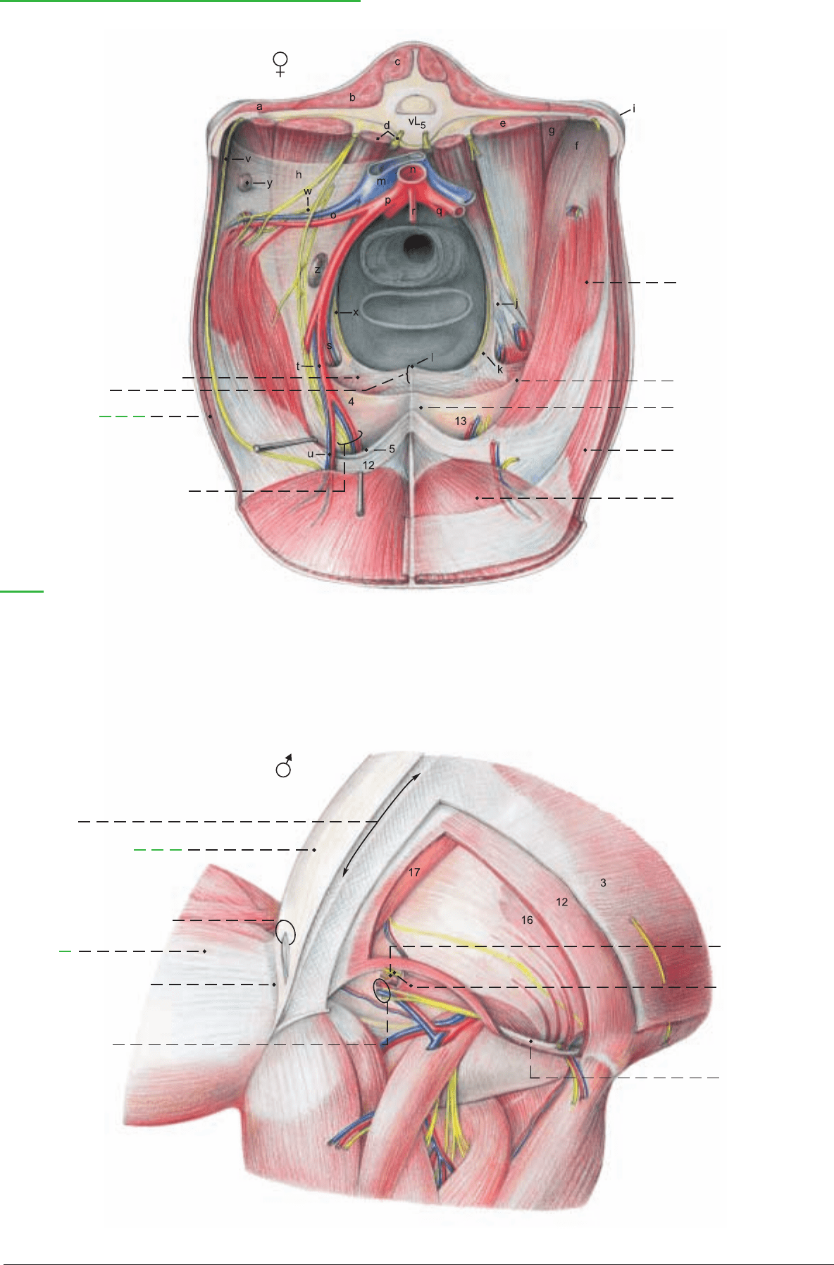
u Caudal epigastric a. and v.
v Iliohypogastric n.
w Lat. cut. femoral n.
x Obturator n.
y Lat. iliac ln.
z Iliofemoral ln.
Inguinal canal, Inguinal lig., and Prepubic tendon
(cranial)
1 Pectineus (and adductor longus)
2 Prepubic tendon
(caudal part)
3 External oblique
4 Pelvic tendon and caud. border
of deep ing. ring
5 Abdominal tendon and cran. border
of deep inguinal ring
12 Internal oblique
13 Deep inguinal ring
14 Dorsolateral angle
15 Medial angle
16 Transversus
17 Rectus
(See p. 79)
6 Linea alba
7 Yellow abdominal tunic
8 Cran. angle of supf. inguinal
ring
9 Medial
femoral fascia
10 Femoral lamina of fascia
11 Ext. pudendal vessels
and caud. br.
of genitofemoral n.
18 Vaginal tunic
19 Cremaster and cran.
br. of genitofemoral n.
20 Inguinal lig.
Legend:
a Iliocostalis
b Longissimus dorsi
c Multifidus
d Psoas minor and
sympathetic trunk
Iliopsoas
e Psoas major
f Iliacus
g Quadratus lumborum
h Internal iliac fascia
i Tuber coxae
j Psoas minor tubercle
k Iliopubic eminence
l Dorsal pubic tubercle
m Caudal vena cava
n Aorta
o Deep circumflex iliac vessels
p External iliac a.
q Internal iliac a.
r Caudal mesenteric a.
s Deep femoral a. and v.
t Pudendoepigastric vessels
(caudoventral)
(See p. 83)
Genitofemoral n. and
ext. pudendal a. and v.
81
Anatomie des Rindes englisch 09.09.2003 14:45 Uhr Seite 81
UQL|qNyNzKjvWvGT8zRmLENm5w==|1287792626
a) The LYMPHATIC SYSTEM in the dorsal abdominal and pelvic
cavities includes the following lymph nodes.
The 12–15 small lumbar aortic lnn. (8) lie dorsal and ventral to the
aorta and caudal vena cava and are examined in meat inspection in
special cases. There are also up to 5 inconstant unilateral or bilat-
eral proper lumbar lnn. between the lumbar transverse processes.
The 1–4 renal lnn. (9) are found on both sides between the renal a.
and v. (2). They are routinely examined in meat inspection. The
lymph drainage is through the lumbar trunk or directly into the cis-
terna chyli. The medial iliac lnn. (4), 1–5 in number, lie at the ori-
gin of the external iliac aa. (f). The lateral iliac ln. (12) at the bifur-
cation of the deep circumflex iliac a. (11) may be double. Both
groups are routinely examined in meat inspection. The sacral lnn.
(5), 2–8 in number, lie in the angle between the internal iliac aa. (h).
The sciatic ln. (p. 17, B) is in the lesser sciatic foramen or dorsal to
it on the outside of the sacrosciatic ligament. The anorectal lnn. (p.
77) are dorsal and lateral to the rectum and anus. The iliofemoral
ln. (6) is up to 9 cm long and located in the angle between the deep
circumflex iliac and external iliac vessels. It is clinically important
because it receives lymph from the superficial inguinal (mammary)
lnn. and can be palpated per rectum cranial to the shaft of the ili-
um. It is examined in meat inspection in cases of mastitis. The
lymph drainage from the iliac, sacral, sciatic, anorectal, and
iliofemoral lnn. passes through the medial iliac lnn., the iliofemoral
ln., or the lumbar trunk into the cisterna chyli, which is 1.5–2 cm
long and extends from the last thoracic vertebra to the 1st or 2nd
lumbar vertebra, dorsal to the vena cava and aorta.
b) The ADRENAL GLANDS (7) are 5–8 cm long, flattened, rela-
tively smooth, and reddish brown to dark gray, sometimes also
with black spots. Each weighs 15–23 g. They are retroperitoneal
and covered ventrally by fat. The right adrenal is more or less heart-
shaped and located at the 12th intercostal space craniomedial to
the right kidney. It is partly covered ventrally by the caudal vena
cava and attached to it by connective tissue. The left adrenal is com-
ma-shaped and larger and heavier than the right. It lies in the plane
of the 1st lumbar vertebra on the left side of the vena cava, to which
it also is attached by connective tissue. It is usually several cm cra-
nial to the left kidney.
c) The URINARY ORGANS
I. The kidneys differ remarkably in position as a result of the
developmental expansion of the rumen.
The flat elongated oval right kidney (1) is retroperitoneal and
extends from the 12th intercostal space to the 2nd or 3rd lumbar
vertebra. The pit-like hilus is medial. The cranial end is in contact
with the liver (see p. 75, v). The dorsal surface is applied to the right
crus of the diaphragm and the lumbar muscles. The ventral surface
lies on the pancreas, cecum, and ascending colon.
The left kidney (10) is not illustrated in its normal position. In the
live ox it is pushed to the right side by the rumen. It is almost com-
pletely surrounded by peritoneum and therefore pendulous, and
lies ventral to lumbar vertebrae 2–5, and caudal to the right kidney,
from which it is separated by the descending mesocolon. Because
the left kidney undergoes a 90-degree rotation on its long axis, its
hilus (24) is dorsal. Medially it adjoins the rumen and laterally, the
intestinal mass.
The kidneys are red-brown; their combined weight is 1200–1500 g.
They are marked on the surface by the renal lobes (26), unlike any
other domestic mammal. In the ox, two or more fetal lobes remain
distinct; others are partially or completely fused in the cortex,
resulting in 12–15 simple or compound lobes of various sizes. The
actual boundaries of the lobes can be seen only by the course of the
interlobar aa. and vv. (19). On the cut surface the reddish light
brown renal cortex (23) with its distinct renal columns (21) con-
trasts with the dark red external zone (17) and the light internal
zone (18) of the renal medulla (15). The renal pyramids (16) pro-
ject with their prominent renal papillae (20) into the urine collect-
ing renal calices (25). These open into cranial and caudal collecting
ducts which join within the irregular fat-lined renal sinus to form
the ureter. The ox lacks a renal pelvis.
II. In the standing live ox the right ureter (3) takes a course on the
ventral surface of its kidney and dorsal to the left kidney toward the
pelvic cavity. The left ureter runs along the dorsal surface of the
caudal half of its kidney, inclines to the left of the median plane and
enters the urinary bladder.*
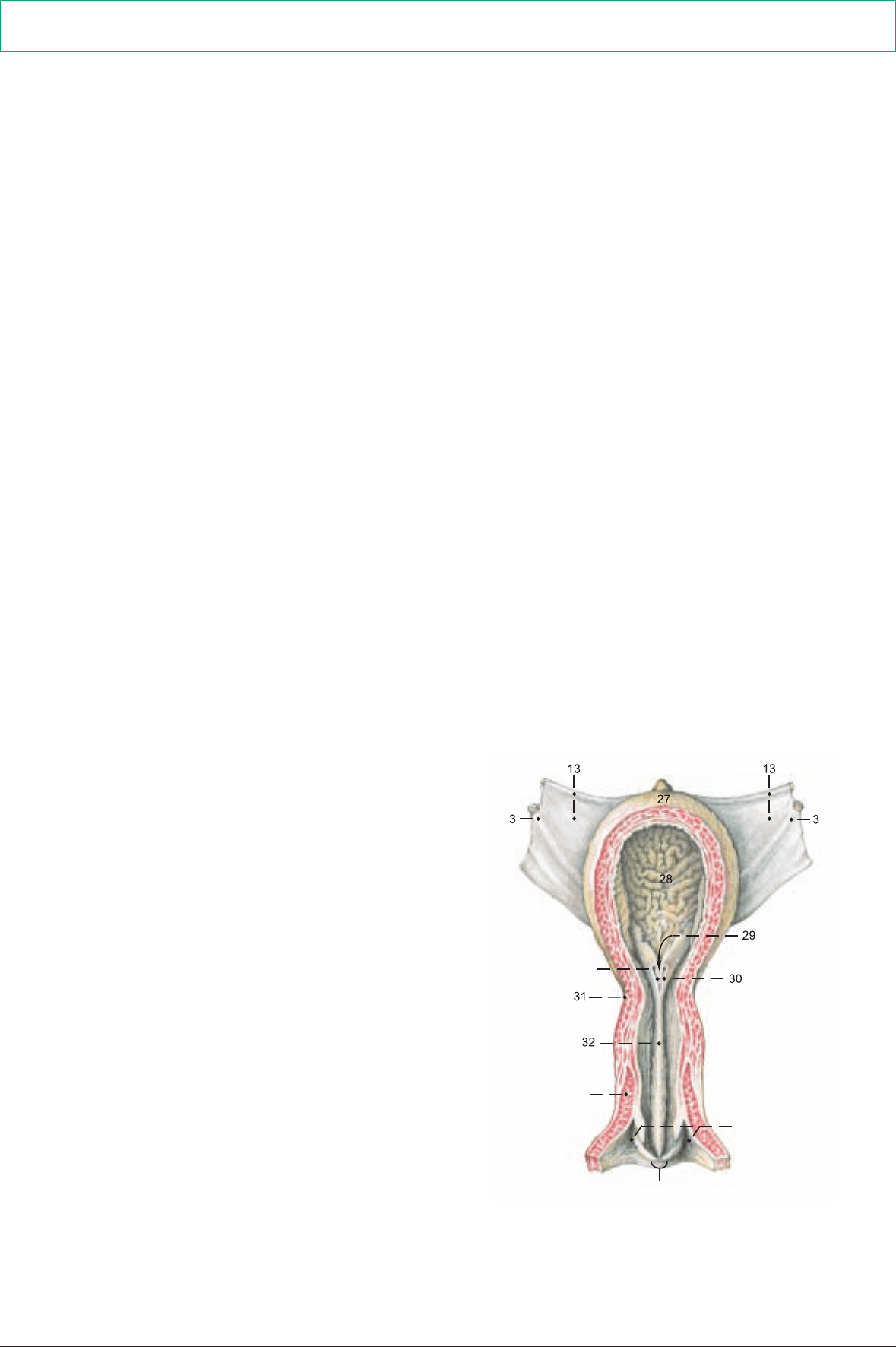
III. The urinary bladder (n) (see also text figure) is relatively large.
When moderately filled it extends into the ventral abdominal cavity
farther than in the horse. The apex (27) and body (28) are covered
with peritoneum. The neck (31) is extraperitoneal and attached to
the vagina by connective tissue. On the apex there is a distinct coni-
cal vestige of the urachus, which in the three-month-old calf can still
be as long as 4 cm. The ureters open close together in the middle of
the neck of the bladder. The ureteric folds (30) run caudally from
there inside the bladder and converge to form the narrow vesical tri-
angle (29). The lateral ligaments of the bladder (13) contain in their
free border the small, in old age almost obliterated, umbilical artery
(round lig. of the bladder; p. 87, t). The middle lig. of the bladder
(14) runs from the ventral wall of the bladder to the pelvic symph-
ysis and to the ventromedian abdominal wall.
IV. The male urethra (see p. 92, K) consists of a pelvic part sur-
rounded by a stratum spongiosum, and a penile part surrounded by
the corpus spongiosum penis. The pelvic part is also surrounded by
the disseminate prostate (see p. 92), and ventrally and laterally by
the thick striated urethral muscle (93, g). Just inside the ischial arch
is the urethral recess, present in ruminants and swine; it opens cau-
dally and practically prevents catheterization. The recess is dorsal
to the urethra and separated from it by a fold of mucosa that bifur-
cates caudally into lateral folds on which the ducts of the bul-
bourethral glands open. The lumen of the urethra passes through
the narrow slit between the folds.
V. The female urethra (see text figure) is about 12 cm long and
attached to the vagina by connective tissue and the urethral muscle.
The urethral crest (32), 0.5 cm high, passes through the urethra on
its dorsal wall to the slit-like urethral orifice, which is on the cra-
nial side of the neck of the clinically important, blind, suburethral
diverticulum (33). The latter extends cranially for 2 cm from its
common opening with the urethra on the floor of the vestibule, and
must be avoided in catheterization. (See p. 87, x.)
82
3. LYMPHATIC SYSTEM, ADRENAL GLANDS, AND URINARY ORGANS
* Fabisch, 1968
After the study of the topography of the lymph nodes, adrenals, and urinary organs, the kidneys are removed with attention to their
coverings, and their peculiarities in the ox are studied.
Ureteral orifices
(ventral)
Urethral m.
33 (Sectioned ventrally)
External urethral orifice
Anatomie des Rindes englisch 09.09.2003 14:45 Uhr Seite 82
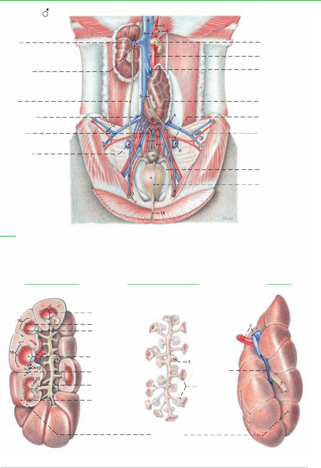
15 Renal medulla
16 Renal pyramid
17 External part
18 Internal part
Abdominal cavity and Urinary organs as seen at autopsy, in dorsal recumbency with stomach and intestines removed
(ventral)
1 Right
kidney
2 Renal a. and v.
3 Ureter
4 Medial iliac lnn.
5 Sacral lnn.
6 Iliofemoral lnn.
7 Adrenal gll.
8 Lumbar aortic lnn.
9 Renal lnn.
10 Left kidney (on the left
in dorsal recumbency
only)
11 Deep circumflex iliac a.
and v.
12 Lateral iliac lnn.
13 Lat. lig. of bladder
14 Median lig. of bladder
(See p. 81)
Legend:
a Caud. vena cava
b Aorta
c Celiac a.
d Cran. mesenteric a.
e Caud. mesenteric a.
f Ext. iliac a.
g Common iliac v.
h Int. iliac a.
i Umbilical a.
j Ductus deferens and A. ductus deferentis
k Testicular a. and v.
l Median sacral a. and v.
m Rectum
n Urinary bladder
o Vaginal ring
Right kidney (Sectioned) Ureter and Calices (Right kidney) Left kidney
26 Renal lobes
19 Interlobar a. and v.
20 Renal papilla
21 Renal columns
22 Collecting duct
in Renal sinus
23 Renal cortex
24 Renal hilus
25 Renal calices
83
Anatomie des Rindes englisch 09.09.2003 14:45 Uhr Seite 83

a) The ABDOMINAL AORTA (1) gives off the paired external
iliac aa. at the level of the 6th lumbar vertebra, and the paired inter-
nal iliac aa. and the dorsally directed unpaired median sacral a. (13)
at the level of the sacral promontory.
The external iliac a. (5), while still in the abdominal cavity, gives off
the deep circumflex iliac a. (6) and shortly before entering the
femoral triangle, it gives origin to the deep femoral a. with the
attached pudendoepigastric trunk (7), which divides into the cau-
dal epigastric a. (8) and the external pudendal a. (9). The latter
passes through the inguinal canal and gives off branches to the scro-
tum or udder (see also p. 91). The internal iliac a. (15) is, in con-
trast to that artery in the dog and horse, a long vessel that extends
to the lesser sciatic notch and ends there by dividing into the cau-
dal gluteal and internal pudendal aa. Its first branch is the umbili-
cal a. (17), which gives off the a. of the ductus deferens in the bull
and the uterine a. (18) in the cow, and in both sexes the cranial vesi-
cal a. (19) with the obliterated termination of the umbilical a. as the
round lig. of the bladder.
Also originating from the internal iliac a. are the iliolumbar a. (16)
and the cranial gluteal a. (i). The vaginal a. (23) or prostatic a. orig-
inates at the level of the hip joint. Together with the internal puden-
dal a. their branches supply most of the pelvic viscera. The vaginal
or prostatic a. supplies the uterine br. (24) or the br. to the ductus
deferens, the caudal vesical a. (25) (which can also come indirectly
from the int. pudendal a.), the urethral br. (27), the middle rectal a.
(28), and, in the cow, the dorsal perineal a. (28), which ends as the
caudal rectal a. (30). The dorsal perineal a. may give off the mam-
mary br. (37).
The int. pudendal a. (32) gives off the urethral a. (33), the vestibu-
lar a. (34), the dorsal perineal a. in most bulls, and the ventral per-
ineal a. (36) with its mammary br. (37), and ends as the a. of the
clitoris or a. of the penis (35). The obturator a. is absent.
The caudal gluteal a. (31) emerges from the pelvis through the less-
er sciatic foramen. It supplies the deep gluteal m., cran. part of the
gluteobiceps, the gemelli, and the quadratus femoris.
b) The VEINS run generally parallel to the corresponding arteries;
therefore only the important exceptions will be mentioned here.
The terminal division of the caudal vena cava (1) into paired com-
mon iliac vv. occurs at the level of the first sacral vertebra. The
median sacral v. (13) comes from the caudal vena cava, and the
deep circumflex iliac v. (6) comes from the common iliac v. The left
common iliac v. gives off the left ovarian or testicular v. (2). Medi-
al to the ilium the common iliac v. divides into the external and
internal iliac vv.
Shortly before its entry into the femoral triangle the external iliac v.
(5) gives off the pudendoepigastric v. (7) which may arise from the
deep femoral v. The internal iliac v. (15) gives off the obturator v.
(20), which runs to the obturator foramen, and the accessory vagi-
nal vein (22) neither has an accompanying artery. (See e) veins of
udder.)
The v. of the ductus deferens (24) comes from the prostatic v.; the
uterine br. (24), from the vaginal v., from which the caudal vesical
v. (25) also arises.
The blood supply of the penis, uterus, and udder follows.
c) The BLOOD SUPPLY OF THE PENIS is provided by the inter-
nal pudendal a. It ends as the a. of the penis (35) and this gives off
the a. of the bulb of the penis (38) for the corpus spongiosum and
bulb; the deep a. of the penis (39), which enters the corpus caver-
nosum at the root of the penis; and the dorsal a. of the penis (40),
which runs to the apex of the penis. The veins ramify in the same
way as the arteries of the same name.
d) The BLOOD SUPPLY OF THE UTERUS is provided mainly by
the uterine a. (18), which originates from the first part of the umbil-
ical a., near the internal iliac a. It runs on the mesometrial border of
the uterine horn in the parametrium. Its branches form anastomot-
ic arches with each other and cranially with the uterine br. (2’) of the
ovarian a. and caudally with the uterine br. (24) of the vaginal a. In
the cow the uterine a. is palpable per rectum after the third month of
pregnancy as an enlarged vessel with a characteristic thrill (fremitus)
in addition to the pulse. The uterine v. is an insignificant vessel that
comes from the internal iliac v. and accompanies the uterine a. The
main veins are the uterine br. of the ovarian v. (2), the uterine br. of
the vaginal v. (24), and the accessory vaginal v. (22), which comes
from the internal iliac v. and has no accompanying a.
e) The BLOOD SUPPLY OF THE UDDER comes mainly from the
external pudendal a. (9), and additionally from the internal puden-
dal a. (32) via the ventral perineal a. (36). The external pudendal
a., with a sigmoid flexure, enters the base of the udder dorsally and
divides into the cranial and caudal mammary aa. The cran. mam-
mary a. (caud. supf. epigastric a., 10) supplies the cranial and cau-
dal quarters, including the teats. The caud. mammary a. (11) goes
mainly to the caudal quarter. A third (middle) mammary artery may
be present, arising from the other two or from the external puden-
dal a. at its bifurcation. There are many variations in all three arter-
ies.
The cranial and caudal mammary vv. are branches of the external
pudendal. The cranial mammary v. (10) is also continous with the
caud. supf. epigastric v., which joins the cran. supf. epigastric v. to
form the large, sinous milk vein (subcutaneous abdominal v.). The
caudal mammary v. (11) joins the large ventral labial v. (37), which
is indirectly connected to the internal pudendal v. Further details of
the mammary vessels will be discussed with the udder (p. 90).
f) The SACRAL PLEXUS is joined to the lumbar plexus in the
lumbosacral plexus. The cranial gluteal n. (i) issues cranially from
the lumbosacral trunk at the greater sciatic foramen and runs with
the branches of the cran. gluteal vessels to the middle, accessory,
and deep gluteal muscles, as well as the tensor fasciae latae, fused
with the supf. gluteal muscle.
The caudal gluteal n. (j) arises caudodorsally from the lumbosacral
trunk near the greater sciatic foramen, but emerges through the
lesser sciatic foramen and innervates the parts of the gluteobiceps
that originate from the sacrosciatic ligament.
The caudal cutaneous femoral n. (k) is small in the ox. It arises from
the lumbosacral trunk just caudal to the caudal gluteal n. and runs
outside the sacrosciatic lig. to the lesser sciatic foramen, where it
divides into medial and lateral brr. The medial br. (communicating
br.) passes into the foramen and joins the pudendal n. or its deep
perineal br. In the ox the lateral br. of the caud. cut. femoral n. may
be absent; it may join the proximal cutaneous br. of the pudendal
n.; or it may contribute to the cutaneous innervation of the cau-
dolateral thigh, which is supplied mainly by the proximal and dis-
tal cutaneous brr. of the pudendal n.
The sciatic n. (f) is the direct continuation of the lumbosacral trunk.
It runs caudally over the deep gluteal m. and turns ventrally behind
the hip joint to supply the pelvic limb. It is the largest nerve in the
body.
The pudendal n. (h) originates from sacral nn. 2–4. It runs cau-
doventrally on the inside of the sacrosciatic lig., and near the lesser
sciatic foramen gives off two cutaneous brr. (p. 95): the proximal
cutaneous br. emerges through, or caudal to, the gluteobiceps, and
runs distally on the semitendinosus; the distal cutaneous br.
emerges from the ischiorectal fossa and runs distally on the semi-
membranosus. It also supplies the supf. perineal n. to the skin of the
perineum. In the bull, this provides the dorsal scrotal nn., and in the
cow, the labial nn., and branches that extend on the ventral labial
v. to the caudal surface of the udder.
The pudendal n. gives off the deep perineal n. (p. 95) to the striat-
ed and smooth perineal muscles, and continues with the internal
pudendal vessels around the ischial arch, and ends by dividing into
the dorsal n. of the clitoris and the mammary br. The latter is close-
ly associated with the loops of the ventral labial v. In the bull the
pudendal n. divides into the dorsal n. of the penis and the preputial
and scrotal br.
The caudal rectal nn. (30) are the last branches of the sacral plexus.
They have connections with the pudendal n. and supply the rectum,
skin of the anus, and parts of the perineal musculature.
g) AUTONOMIC NERVOUS SYSTEM. The sympathetic divi-
sion includes the caudal mesenteric ganglion on the caudal mesen-
teric a. cranial to the pelvic inlet. The paired hypogastric nn. leave
the ganglion and run on the dorsolateral pelvic wall to the level of
the vaginal or prostatic a. to join the pelvic plexus. The sympath-
etic trunk in the sacral region has five vertebral ganglia and in the
coccygeal region, four or five ganglia.
The parasympathetic nn. from sacral segments 2–4 leave the verte-
bral canal with the ventral roots of the pudendal n. and form the
pelvic nn., which, from a dorsal approach, join the pelvic plexus
with its contained ganglion cells. (See also p. 56.)
84
5. ARTERIES, VEINS, AND NERVES OF THE PELVIC CAVITY
Anatomie des Rindes englisch 09.09.2003 14:45 Uhr Seite 84
UQL|qNyNzKjvWvGT8zRmLENm5w==|1287792681
