Budras Klaus-Dieter, Habel Robert E. Bovine Аnatomy
Подождите немного. Документ загружается.


95
(See pp. 85, 91)
nS
3
nS
4
nS
5
a
c
b
19
18
25
d
e
f
g
j
i
k
h
k
l
15
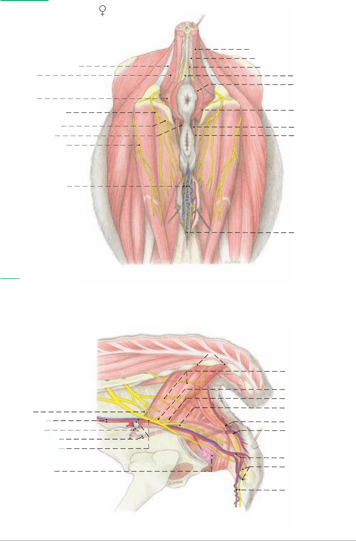
Perineal region
(Caudal aspect)
1 Sacrosciatic lig.
2 Coccygeus
3 Levator ani
4 Supf. perineal n.
5 Transversus perinei
12 Ext. anal sphincter
Anal triangle:
11 Rectocaudalis
Urogenital triangle:
13 Constrictor vestibuli
14 Constrictor vulvae
15 Retractor clitoridis
16 Vent. labial v.
Legend:
a Tuber coxae
b Tuber ischiadicum
c Gluteus medius
c Retractor clitoridis
d Biceps femoris
e Semitendinosus
f Semimembranosus
g Gracilis
h Intertransversarii
i Sacrocaudalis dors. med.
j Sacrocaudalis dors. lat.
k Sacrocaudalis vent. lat.
l Sacrocaudalis vent. med.
(Lateral aspect)
Pelvic n.
6 Int. iliac a. and v.
7 Vaginal a. and v.
8 Dors. perineal a. and v.
9 Int. pudendal a. and v.
and pudendal n.
10 Major vestibular gl.
17 Caud. rectal nn. and brr.
to coccygeus and levator ani
18 Prox. cut. br. of pudendal n.
19 Dist. cut. br. of pudendal n.
20 Deep perineal n.
21 Caud. rectal a. and v.
22 Dors. labial br. and v.
23 Vent. perineal a. and v.
24 A. and v. of clitoris and
dorsal n. of clitoris
25 Mammary brr. of vent.
perineal a. and pudendal n.;
vent. labial v.
Anatomie des Rindes englisch 09.09.2003 16:01 Uhr Seite 95

The term spongiform encephalopathy refers to spongy changes in
the brain. BSE is one of a group of diseases called transmissible
spongiform encephalopathies (TSE), of which scrapie of sheep has
been know for a long time, is widely distributed, and has been
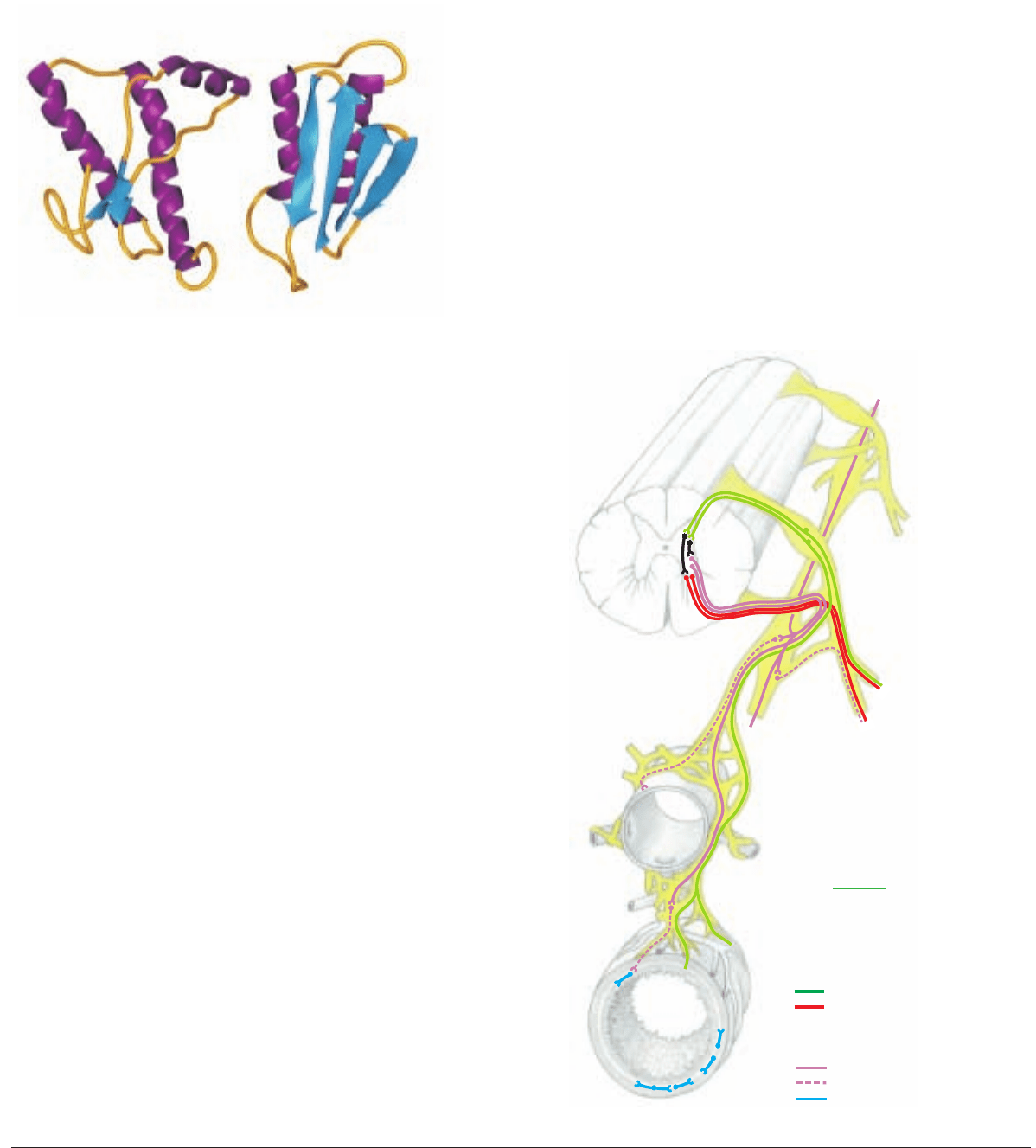
intensively investigated. The TSE are caused by prion proteins (PrP)
– minute proteinaceous infectious particles 4–6 nm in diameter.
They occur in normal and pathogenic forms on the surface of nerve
cells and various cells of lymphatic tissue. In normal PrP the amino
acid chains are predominantly wound up in alpha-helices. By
unknow processes, often by mutation in the controlling gene, path-
ogenic PrP develop, whose amino acids in some regions of the mol-
ecule are refolded from alpha helices into beta-sheets layered
antiparallel on each other.* The misfolded, pathogenic PrP cause
BSE by imposing their structure on normal PrP, thereby multiplying
the pathogen. They enter the lysosomes of nerve cells, where they
are not decomposed, but accumulate in amyloid plaques and cause
the death of the nerve cells.**
SPECIES DISTRIBUTION OF PRION DISEASES
Prion diseases have been found in sheep, goats, cattle, zoo and wild
ruminants, mink, great cats, and rhesus monkeys. Human prion
diseases are Creutzfeldt-Jakob disease, Gerstmann-Straeussler syn-
drome, fatal familial insomnia, and kuru. BSE is of great impor-
tance because:
1. Its causative agent can overcome the species barrier and become
very dangerous to man.
2. Cattle are significant sources of human food, and an undiag-
nosed BSE infection is a danger to man.
THE SIGNS OF BSE DISEASE
The average age of cattle affected with BSE is about 3 years, but the
first signs may appear at 20 months. As a result of the brain disor-
der, the following signs appear: hypersensitivity to stimuli (e. g.
noise), anxiety, aggression, and locomotor disturbance progressing
to collapse. The terminal stage is prostration until death. There is
no cure.
DIAGNOSIS OF BSE
A suspected clinical diagnosis is possible in the terminal stage, but
a certain diagnosis can be made only after death. For the rapid test,
parts of the brainstem are removed, homogenized, and digested by
proteinases. After digestion, only the pathogenic PrP remain intact,
and can be identified by a specific antibody. If the results are doubt-
ful, further tests by immunohistological or cytological (E/M) meth-
ods are required.
POSSIBLE CAUSES FOR THE APPEARANCE OF
NEW PRION DISEASES
The PrP of scrapie in sheep could have mutated in cattle to the PrP
of BSE. Scrapie was widely distributed in Great Britain, and car-
casses of affected sheep were reduced in rendering plants to fat and
tankage in large autoclaves (tanks). The tankage (meat and bone
meal) was a common source of protein in animal feed, including
cattle feed. Transmission by feed was later made highly probable by
the success of a ban on tankage in animal feed.**
In Germany BSE was probably spread by feeding calves a milk sub-
stitute made by replacing milk fat with tallow from adult bovine
mesenteric and abdominal fat.
Failure to observe proper procedures in the operation of the tank
(addition of lye and detergents and maintenance of heat at 130 °C
for 20 min.) could have led to survival of pathogenic PrP.
PATHWAYS OF INFECTION
The probable mode of infection in sheep and cattle is intestinal. Pre-
cise information on infection of cattle is not available, but infer-
ences can be drawn from experiments on rodents, which have a
much shorter incubation period. Also, possible parallels can be
drawn to scrapie in sheep.
TRANSPORT THROUGH THE AUTONOMIC SYSTEM
At least three routes to the CNS have been proposed on the basis of
experiments on rodents: ***
1. The vagus conducts parasympathetic fibers that bypass the
spinal cord. The vagal efferents have their nerve cell bodies in the
dorsal motor nucleus in the obex region of the medulla. Vagal affer-
ents have their nerve cell bodies in the proximal and distal vagal
ganglia. They send their short axons to the obex region.
2. An alternative route goes from the enteric plexuses through pre-
vertebral ganglia and the splanchnic nerves to the sympathetic
trunk, thence through the communicating branches and spinal
nerve roots to the tracts of the spinal cord leading to and from the
brain.
3. A third possibility is passage from the sympathetic trunk
through the cervicothoracic ganglion, ansa subclavia, and vago-
sympathetic trunk to the head.
96
ANATOMICAL ASPECTS OF BOVINE SPONGIFORM ENCEPHALOPATHY (BSE)
NATURE OF THE DISEASE
* Borchers, 2002 *** McBride et al., 2001
** Hoernlimann et al., 2001
Models of prion proteins (purple = alpha-helix structure, blue = beta
sheet structure
cellular (normal) pathogenic form
1
2
1
2
nd
nv
3
Afferent neurofiber
Motor neuron
(Efferent neurofiber)
Sympathetic system
Spinal cord
Aorta
Legend:
1 Spinal ganglion
2 Ganglion of sympathetic trunk
3 Prevertebral ggl.
(e. g. mesenteric)
Preganglionic neurofiber
Postganglionic neurofiber
Intramural nerve plexus
Gut
Anatomie des Rindes englisch 09.09.2003 16:02 Uhr Seite 96

97
**** Jour. AVMA, 2002 ****** USDA-APHIS, 2001
***** Sigurdson et al., 2001
AREAS OF HIGHEST CONCENTRATION IN THE BRAIN
The primary site of pathogenic prions is the region of the obex
between the medulla oblongata and the spinal cord. The dorsal
vagal nucleus and other important nuclei here show typical spongi-
form changes. Other regions of the brainstem display lesions.
Spongiform encephalopathy of the cerebellar cortex explains the
locomotor disturbances and ataxia. Insoluble amyloid forms in the
nerve cells, with high concentration of pathogenic prions and
spongiform changes. Neighboring glia cells are also affected.
Another TSE, chronic wasting disease (CWD) of North American
deer and elk, discovered in Colorado in 1967, has been found in
wild or farmed deer and elk in Wyoming, Nebraska, South Dako-
ta, Oklahoma, Montana, Wisconin, and one case in Illinois. (The
North American elk, a misnomer, is Cervus canadensis,
not Alces
alces—the European Elch and the North American moose.) There
is no evidence that other species, including man, are infected
through contact with CWD.**** In experimental deer inoculated
orally with infective deer brain, pathogenic PrP were first found in
lymphoid tissues of the alimentary system and then in autonomic
nerves leading from the gut to the brainstem, where they appeared
first in the dorsal motor nucleus of the vagus. Other peripheral
nerves, such as the brachial plexus and sciatic nerve, were tested
and found negative.*****
The U. S. government has prohibited importation of live ruminants
and most ruminant products from Europe and Canada. The U. S.
Food and Drug Administration prohibits the feeding of most mam-
malian protein to ruminants. The U. S. Dept. of Agriculture has
prohibited importation of all rendered animal products of any
species. There is no evidence of BSE in the United States after a
decade of testing for it.******
REMOVAL OF THE BRAINSTEM FOR LABORATORY TESTS
After slaughter and decapitation, brainstem tissue can be removed
with a curette through the foramen magnum. If the head is bisect-
ed, the myelencephalon and metencephalon are separated from the
more rostral parts of the brain (see p. 51, below by a transverse cut
through 13 and 14, and by cutting the roots of cranial nn. V-XII
and the cerebellar peduncles to release the sample of the brainstem.
The material of the obex region is used for the BSE rapid test. If the
results are positive, histopathologic, immunohistochemical, and
E/M investigations follow, for which more rostral parts of the
brainstem are used.
TRANSMISSION OF BSE TO MAN
Human infection with the agent of BSE and consequent illnes with
the variant of Creutzfeld-Jacob disease (vCJD) is highly probable.
In vCJD the multiplication of the agent also occurs outside the
brain and spinal cord in the lymphatic organs (e.g. tonsils); where-
as in the sporadic (classical) CJD the pathological changes remain
restricted to the CNS. The likelihood of transmission from BSE-
infected cattle to man is supported by the fact that the agents of BSE
and vCJD are biologically and biochemically identical. The con-
nection of time and place between occurences of BSE and vCJD in
Great Britain supports this probability. Apparently a genetically
determined susceptibility plays a role in transmission because, so
far, only a few people have contracted vCJD, and only a few of the
cattle in a herd contract BSE.
DANGERS OF EATING MEAT AND MEAT PRODUCTS
FROM BSE-INFECTED CATTLE OR CATTLE SUSPECTED
OF EXPOSURE TO BSE
The risks increase with the amount of infective material consumed
and its concentration of pathogenic misfolded prions. Of the com-
ponents of nervous tissue, the perikarya and therefore the ganglia
and nuclei may present a greater danger than axons, and thus more
than nerves and fiber tracts. The perikarya occupy a much larger
volume and have a concentration of prions in the lysosomes, which
are not present in the processes. The danger is increased, the near-
er the ganglia lie to valuable cuts of meat; for example, the sympa-
thetic trunk and ganglia are closely associated with the tenderloin
(iliopsoas and psoas minor (see p. 81, upper fig.) The spinal ganglia
lie in the intervertebral foramina and are included with the bone in
steaks cut from the rib and loin regions (see text fig.). Regarding the
concentration of pathogenic misfolded prions the following list
presents the opinion of the European Union on the possible risk of
infectivity in various tissues (including experiments with scrapie).
1. Highly infectious tissues: brain and spinal cord together with
surrounding membranes, eyes, spinal ganglia.
2. Tissues of intermediate infectivity: intestine, tonsils, spleen, pla-
centa, uterus, fetal tissue, cerebrospinal fluid, hypophysis, and
adrenal gl.
3. Tissues of lower infectivity: liver, thymus, bone marrow, tubu-
lar bones, nasal mucosa, peripheral nerves.
4. Infectivity was not demonstrated in the following tissues and
organs: skeletal muscle, heart, kidneys, milk, fat (exept mesen-
teric fat), cartilage, blood, salivary gll., testis, and ovary.
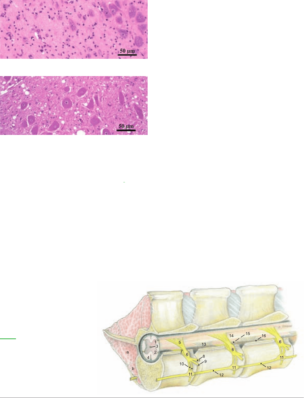
Nervous tissue of the region of the obex in BSE.
Preparation: Prof. F. Ehrensperger, Inst. of Vet. Pathology, Zürich
Normal nervous tissue in the region of the obex (dorsal vagal nucleus).
Preparation: Prof. G. Boehme, Inst. of Vet. Anatomy, FU-Berlin
Gray matter
1 Dorsal horn
2 Ventral horn
3 Central canal
4 White matter
5 Dorsal root
6 Spinal ganglion
7 Ventral root
8 Dorsal br.
9 Ventral br.
Spinal cord and sympathetic trunk after
removal of the left side of the vertebral
arches and the musculature
Legend:
10 Communicating brr.
white and gray
11 Ganglia of sympathetic trunk
12 Sympathetic trunk
13 Denticulate lig.
14 Pia mater
15 Arachnoidea
16 Dura mater
a Psoas major
b Psoas minor
3-89993-000-2.qxd 12.09.2003 8:48 Uhr Seite 97

MUSCLE / FIG. ORIGIN TERMINATION INNERVATION FUNCTION REMARKS
MEDIAL MUSCLES OF THE SHOULDER AND ARM (p. 4)
Teres major (5.2)
Caudal border Teres major tuberosity Axillary n. Flexor of shoulder joint Joined by terminal
of scapula and of humerus tendon of latissimus
subscapualis dorsi
Subscapularis (5.4) Subscapular fossa Minor tubercle of Subscapular and Mainly an extensor of 3–4 distinct parts;
of scapula humerus axillary nn. shoulder jt. tendon acts as med.
collat. lig. of shoulder
joint
Coracobrachialis Coracoid process Small part prox. and Musculocutaneous n. Extensor of shoulder joint Two bellies; synovial
(5.16) of scapula large part dist. to teres and adductor and supinator bursa under tendon of
major tuberosity of of brachium origin
humerus
Articularis humeri Inconstant in the ox.
Biceps brachii (5.26) Supraglenoid tubercle Radial tuberosity, Musculocutaneous n. Extensor of shoulder joint, Intertubercular bursa
of scapula cranial surface of flexor of elbow joint under tendon of origin;
radius, fleshy on med. thin lacertus fibrosis to
collat. lig. of elbow joint antebrachial fascia
Brachialis (5.21) Caud. surface of Radial tuberosity and Musculocutaneous n.; Flexor of elbow joint Spiral course in
humerus, close to med. collat. lig. of for distal parts, brachialis groove of
neck elbow joint radial n. humerus, added inner-
vation from radial n. in
50 %
Tensor fasciae Caud. border of Medially on olecranon Radial n. Tensor of fascia of forearm
antebrachii (5.22) scapula, latissimus and antebrachial fascia and extensor of elbow joint
dorsi
LATERAL MUSCLES OF SHOULDER AND ARM (p. 4)
Deltoideus Axillary n.
Clavicular part Clavicular intersection Crest of humerus Advances limb Part of brachiocepha-
(Cleidobrachialis) licus; see p. 60
(5.23)
Scapular part (5.6) Caud. border of Deltoid tuberosity of Flexor of shoulder joint Small flat muscle
scapula, aponeurosis humerus, fascia of
from scapular spine triceps
Acromial part (5.7) Acromion Deltoid tuberosity of Flexor of shoulder joint Interspersed with
humerus tendinous strands
Teres minor (5.12) Distal half of cd. Prox. to deltoid tube- Axillary n. Flexor of shoulder joint
border of scapula rosity of humerus on
teres minor tuberosity
Supraspinatus (5.1) Supraspinous fossa, Major and minor Suprascapular n. Extensor and stabilizer of Tendon of origin of
cran. border of tubercles of humerus shoulder jt.; also flexor biceps passes between
scapula dependent on state of joint the terminal tendons
Infraspinatus (5.11) Infraspinous fossa Deep part on prox. Suprascapular n. Abductor and lateral Largely tendinous, flat;
and spine of scapula border and med. surface rotator of arm; acts as lat. supf. tendon passes
of major tubercle; supf. collateral lig. over infraspinatus
part distal to tubercle bursa
Triceps brachii All heads together on Radial n. Extensor of elbow joint; Relatively flat
olecranon long head also flexes
shoulder joint; stabilizer
of elbow
Long head (5.18) Caud. border of scapula
Lat. head (5.17) Lateral on humerus
Med. head (5.19) Medial on humerus
Accessory head Caudal on humerus Partially separable from
med. head
Anconeus (5.25) Borders of olecranon Lateral on olecranon Radial n. Extensor of elbow joint Separable with diffi-
fossa culty from lat. head of
triceps
98
SPECIAL ANATOMY, TABULAR PART
1. MYOLOGY
Anatomie des Rindes englisch 09.09.2003 16:11 Uhr Seite 98

MUSCLE / FIG. ORIGIN TERMINATION INNERVATION FUNCTION REMARKS
CRANIOLATERAL MUSCLES OF THE FOREARM
Generally extensors, which originate predominantly on the lateral epicondyle of the humerus (p. 4)
Common digital
Radial n. Extensor of the digits
extensor (5.40) and carpus
Medial head Lateral epicondyle Middle and distal Extensor of fetlock and Receives extensor
(Proper extensor of humerus phalanges of pastern joints of digit III branches of interosseus
of digit III, Med. digit III III
digital extensor)
Lateral head Lateral epicondyle Branches to extensor Extensor of coffin joints Narrow muscle;
(Common extensor of humerus, head processes of dist. humeral and ulnar
of digits III and IV) of ulna phalanges of digits heads unite in a
III & IV common tendon
Lateral digital exten- Proximal on radius Middle and distal Radial n. Extensor of fetlock and Unified; corresponds to
sor (Proper extensor and ulna phalanges of digit IV pastern joints of digit IV medial dig. extensor,
of digit IV) (5.41) with extensor branches
from interosseus IV
Extensor carpi Lat. supracondylar Tuberosity of Mc III Radial n. Extensor and stabilizer Has synovial bursae on
radialis (5.35) crest and radial fossa of carpus carpus and at termina-
of humerus tion; may have rudi-
mentary extensor
digiti I
Ulnaris lateralis Lateral epicondyle Accessory carpal bone Radial n. Flexor (!) of the carpus
(Extensor carpi of humerus and Mc V
ulnaris) (5.38)
Ext. carpi obliquus
Craniolat. in middle Mc III Radial n. Extensor of the carpus Terminal tendon has
(Abductor pollicis third of radius synovial bursa
longus) (5.39)
CAUDOMEDIAL MUSCLES OF THE FOREARM
Generally FLEXORS, which originate predominantly on the medial epicondyle of the humerus (p. 4)
Superficial digital
Med. epicondyle of Flexor tuberosities Ulnar n. Flexor of the carpus Larger supf. belly supf.
flexor (5.36 and 5.37) humerus of middle phalanges and digits to flexor retinaculum;
deep belly within carpal
canal
99
Anatomie des Rindes englisch 09.09.2003 16:11 Uhr Seite 99

MUSCLE / FIG. ORIGIN TERMINATION INNERVATION FUNCTION REMARKS
Deep digital
Flexor tubercles of Flexor of coffin jts.; The single deep flexor
flexor (5.34) distal phalanges support of fetlock jts. tendon is surrounded
by a synovial bursa in
the carpal canal
Humeral head Med. epicondyle Ulnar and median nn. The humeral head is
of humerus tripartite and inter-
spersed with many
tendinous strands
Ulnar head Olecranon Ulnar n. Ulnar head is small
Radial head Caudomedial on Median n.
prox. third of radius
Flexor carpi Accessory carpal bone Ulnar n. Flexor of carpus
ulnaris (5.29)
Humeral head Medial epicondyle
of humerus
Ulnar head Medially on olecranon Ulnar head is small
Flexor carpi Medial epicondyle Proximopalmar on Median n. Flexor of carpus Surrounded by a
radialis (5.28) of humerus Mc III tendon sheath in carpal
canal
Pronator teres Medial epicondyle Craniomedial on Median n. Pronator of forearm Weakly muscular
(5.27) of humerus radius and manus
METACARPUS (p. 4 and 18)
Interflexorii
Muscle fibers connecting the supf. and deep Median n. Auxiliary flexors of
digital flexors as well as their tendons, in and the digits
near the carpal canal
Interosseus III and Prox. end of mtc. Prox. sesamoid bones; Palmar branch Support fetlock joints; Predominantly
Interosseus IV (p. 18) bone; deep palmar branches to proper of ulnar n. oppose tension of deep tendinous in older
carpal lig. extensor tendons; flexor on distal phalanx cattle
accessory lig. to supf.
flexor
MUSCLES OF THE HIP JOINT (p. 16)
Tensor fasciae latae
Tuber coxae By the fascia lata on the Cran. gluteal n. Flexor of hip joint, advances Includes cran. parts of
(17.5) patella, lat. patellar lig., limb; extensor of stifle; gluteus supf.; especially
and cran. border of tibia tensor of fascia lata robust in cattle
Gluteus superficialis Tuber coxae Cran. part of biceps Cran. and caud. Extensor of hip joint; Not separable from the
(gluteal fascia) femoris and fascia lata gluteal nn. retractor of limb total mass of the glu-
teobiceps
Gluteus medius Gluteal surface Major trochanter of Cran. gluteal n. Extensor of hip joint; Has a lumbar process
(17.1) of ilium femur abductor of limb on the longissimus lum-
borum
Gluteus accessorius Gluteal surface Craniolat. on femur Cran. gluteal n. Same as gluteus medius Clearly separable from
(17.3) of ilium just distal to maj. gluteus medius;
trochanter trochanteric bursa
under terminal tendon
Gluteus profundus Sciatic spine, lat. Craniolat. on femur, Cran. gluteal n. Abductor of limb Synovial bursa under
(17.4) on body of ischium, distal to gluteus terminal tendon
sacrosciatic lig. accessorius
100
Anatomie des Rindes englisch 09.09.2003 16:11 Uhr Seite 100

MUSCLE / FIG. ORIGIN TERMINATION INNERVATION FUNCTION REMARKS
CAUDAL THIGH MUSCLES (p. 16)
Gluteobiceps
(Biceps Vertebral head: caud. Patella; lat. patellar Vert. head: caud. Extensor of hip and stifle; Not clearly separable
femoris)
(17.7) part of median sacral lig.; cran. border of gluteal n. with caud. part, flexor of into cran. and caud.
crest and last trans- tibia (by fascia cruris Pelvic head: tibial n. stifle; abductor of limb; parts; almost complete
verse processes; and fascia lata); extensor of hock fusion with gluteus
sacrosciatic lig.; and common calcanean supf.
tuber ischiadicum. tendon
Pelvic head: tuber
ischiadicum
Semitendinosus Tuber ischiadicum Cran. border of tibia, Tibial n. In supporting limb: extensor No vertebral head;
(17.20) terminal aponeurosis of hip, stifle and hock; in transverse intersection
of gracilis, common swinging limb: flexor of between prox. and
calcanean tendon stifle; also adductor and middle thirds.
retractor of limb
Semimembranosus Tuber ischiadicum Med. condyles of Tibial n. In supporting limb: extensor No vertebral head.
(17.18) femur and tibia of hip and stifle; in swinging Belly divides into two
limb: retractor, adductor, branches
and pronator of limb
DEEP MUSCLES OF THE HIP JOINT (p. 16, 18)
Gemelli (17.25)
Lesser sciatic notch Trochanteric fossa Muscular brr. of Rotate thigh laterally Thick, unified muscle
of femur sciatic n. plate
Internal obturator is absent in the ox.
Quadratus femoris
Ventral surface of Lat. surface of body Muscular brr. of Supinator of thigh,
(17.26) ischium of femur sciatic n. auxiliary extensor of
hip joint
External obturator Outer and inner Trochanteric fossa Obturator n. Supinator of thigh; The intrapelvic part is
(19.7) surface of ischium of femur adductor of limb small and not homo-
around obturator for. logous to internal
obturator
MEDIAL THIGH MUSCLES: Adductors (p. 18)
Gracilis (19.10)
Prepubic tendon; Fascia cruris Obturator and Adductor (and extensor Right and left tendons
by symphysial tendon saphenous nn. of stifle jt.) of origin fused to form
from pelvic symphysis symphyseal tendon; ter-
minal tendon fused
with that of sartorius
Adductor magnus Symphyseal tendon; Facies aspera Obturator n. Adductor and retractor Joined by connective
(et brevis) (19.9) ventrally on pelvis of femur of the limb tissue with semimem-
branosus; on split car-
cass cut surface of
adductor in bull is tri-
angular; in cow it is
bean-shaped
Pectineus (et adduc- Contralateral pubis: Caudomedial on Adductor part: Adductor of limb, More robust than in
tor longus) (19.8) iliopubic eminence; femur obturator n.; flexor of hip horse; crossed tendons
ilium up to tubercle pectineus part: of origin form the bulk
of psoas minor saphenous n. of the prepubic tendon
EXTENSORS OF THE STIFLE (p. 18)
Sartorius (19.3)
Cranial: iliac fascia Fascia cruris Saphenous n. Flexor of hip joint; Lacuna vasorum for
and tendon of psoas protractor and adductor femoral vessels lies
minor; caudal: of limb; extensor of stifle between the two
iliopubic eminence tendinous heads
and adjacent ilium
Quadriceps femoris By middle patellar Femoral n. Flexor of the hip joint Very large and clearly
lig. on the tibial (rectus); extensor and four heads
tuberosity stabilizer of the stifle
Rectus femoris Ilium: main tendon
(19.1) from med. fossa cran.
to acetabulum; small
tendon from lat. area
near acetab.
Vastus lateralis Proximolateral on femur
(17.29)
Vastus medialis Proximomedial on femur
(19.2)
Vastus intermedius Proximocranial on femur
101
Anatomie des Rindes englisch 09.09.2003 16:11 Uhr Seite 101

MUSCLE / FIG. ORIGIN TERMINATION INNERVATION FUNCTION REMARKS
SPECIAL FLEXOR OF THE STIFLE: Caudal to the stifle (p. 18)
Popliteus (29.4)
Lateral femoral Proximomedial on Tibial n. Flexor of stifle
condyle caud. surface of tibia
EXTENSORS OF THE HOCK AND FLEXORS OF THE DIGITS: Caudal on the crus (p. 18)
Gastrocnemius
On both sides of By the common Tibial n. Extensor of the hock, Very tendinous;
(19.11) supracondylar fossa calcanean tendon flexor of the stifle intermediate fleshy
Lateral head of the femur on calcanean tuber tract connects origin of
Medial head lat. head to tendon of
med. head
Soleus (17.31) Prox. rudiment of Joins common Tibial n. Auxiliary extensor of Fused with the lat. head
the fibula calcanean tendon the hock of gastrocnemius
Supf. digital flexor Supracondylar fossa Flexor tuberosities of Tibial n. Extensor of hock; digital Very tendinous, fused
(19.22) of femur middle phalanges flexor; and flexor of the stifle proximally with lat.
head of gastroc.; ten-
don caps calcaneus
Deep digital flexors Distal phalanges Tibial n. Flexors of coffin joints; Tendons join to form
support of hock and fetlock the common deep
joints flexor tendon in the
metatarsus
Lat. digital flexor Lat. condyle and Passes over
(17.32) caud. surface of tibia sustentaculum tali
Caudal tibial m. Lat. condyle of tibia Passes over
(17.33) sustentaculum tali
Med. digital flexor Lat. condyle of tibia Crosses hock separately
(19.5)
FLEXORS OF THE HOCK AND EXTENSORS OF THE DIGITS: Craniolateral on the crus (p. 16)
Tibialis cranialis
Cran. border and T I; proximomedial Deep peroneal n. Flexor of hock Smaller than in horse;
(17.8) proximolat. surface of on Mt III and Mt IV perforates terminal
tibia; prox. rudiment tendon of peroneus
of fibula and replace- tertius; smaller head
ment ligament corresponds to extensor
digiti I
Peroneus tertius Extensor fossa Prox. on Mt III and Deep peroneal n. Flexor of hock Large and fleshy;
(17.10) of femur Mt IV; T II and T III completely fused at
origin with long digital
extensor
Long digital Extensor fossa Deep peroneal n. Extensor of digits and Mostly covered by
extensor (17.13) of femur flexor of hock peroneus tertius
Medial head Middle and distal Receives extensor
(Proper extensor phalanges of digit III branches of interosseus
of digit III, Med. III
digital extensor)
Lateral head Branches to extensor
(Extensor of processes of distal
digits III and IV) phalanges of digits III
and IV
102
Anatomie des Rindes englisch 09.09.2003 16:11 Uhr Seite 102

MUSCLE / FIG. ORIGIN TERMINATION INNERVATION FUNCTION REMARKS
Lateral digital
Lat. collateral Middle and distal Deep peroneal n. Extensor of digit IV and Relatively large and
extensor (Proper lig. of stifle; phalanges of digit IV flexor of hock pennate; receives
extensor of digit IV) lat. condyle of tibia extensor branches from
(17.12) interosseus IV
Extensor digitalis Ligamentous mass Joins tendon of long Deep peroneal n. Digital extensor Small
brevis (17.15) on dorsal surface digital extensor
of tarsus
Peroneus longus Lat. condyle of tibia, Tendon crosses lat. sur- Deep peroneal n. Flexor of hock Small, with long thin
(17.11) rudiment of fibula face of hock and tendon tendon
of lat. dig. ext. and
plantar surface of hock
to T I
METATARSUS:
Interossei III and IV: (see Muscle tables, p. 100 and p. 18)
MUSCLES INNERVATED BY THE FACIAL NERVE (p. 36 and 37)
Cervicoauricularis
Nuchal lig. Dorsolat. surface of Caud. auricular n. Raises auricle
superficialis auricle from facial nerve
Cervicoauricularis Nuchal lig. and Caudolat. and caud. Caud. auricular n. Turn intertragic notch
profundus and cervical fascia surface of auricle from facial nerve laterally
medius
Cervicoscutularis
Nuchal lig., parietal Caud. border of Caud. auricular n. Raises auricle and tenses Broad muscle plate
(37.2) bone caud. to inter- scutiform cartilage from facial nerve scutiform cartilage
cornual protuberance
Interscutularis (37.3) Cornual proc., Medially on scutiform Rostral auric. brr. of Tensor of scutiform Has no connection to
temporal line cartilage auriculopalpebral n. cartilage contralateral muscle
from facial n.
Frontoscutularis Temporal line and Scutiform cartilage Rostral auric. brr. of Tensor of scutiform Two distinct parts
zygomatic proc. of auriculopalpebral n. cartilage according to origin
frontal bone from facial n.
Zygomaticoscutularis Zygomatic arch Rostrally on scutiform Rostral auric. brr. of Tensor of scutiform
(37.B) cartilage auriculopalpebral n. cartilage
from facial n.
Scutuloauricularis Scutiform cartilage Rostromedial on Rostral auric. brr. of Levator and protractor Two muscles crossed on
superficialis et auricle auriculopalpebral n. of auricle scutiform cartilage
profundus from facial n.
(37.D, E)
Zygomaticoauricularis
Zygomatic arch Auricular concha, at Rostral auric. brr. of Turns intertragic notch
(37.12) intertragic notch auriculopalpebral n. rostrally
from facial n.
Parotidoauricularis Parotid fascia Auricular concha, at Auriculopalpebral n. Depressor and retractor
(37.13) intertragic notch from facial nerve of auricle
Styloauricularis Cartilage of Rostromedial border Caud. auricular n. Muscle of the acoustic May be absent
acoustic meatus of auricle from facial nerve meatus
MUSCLES OF THE LIPS AND CHEEKS (p. 36)
Orbicularis oris
Surrounds the opening of the mouth, except Buccal brr. of facial Closes rima oris Contralat. fibers do not
(37.10) the middle of the upper lip nerve join in the upper lip
Buccinator (37.26) Between coronoid process of mandible and Buccal brr. of facial Muscular substance of Separable into a molar
angle of the mouth nerve cheek; presses food from part with rostroventral
vestibule into oral cavity fiber course, and buccal
proper part with dorsoventral
fiber course
Zygomaticus (37.11) Parotidomasseteric In orbicularis oris Auriculopalpebral n. Retractor of angle of mouth Well developed
fascia at angle of mouth from facial n.
Caninus (37.23) Rostrally on facial With 3 tendons on Buccal brr. of facial Dilates nostril and raises Passes through levator
tuber lat. rim of nostil nerve upper lip nasolabialis; lies
between levator and
depressor of upper lip
103
Anatomie des Rindes englisch 09.09.2003 16:11 Uhr Seite 103

MUSCLE / FIG. ORIGIN TERMINATION INNERVATION FUNCTION REMARKS
Levator labii
Facial tuber Planum nasolabiale Buccal brr. of facial Levator and retractor of Passes through levator
superioris (37.22) dors. and med. to nostril nerve upper lip and planum nasolabialis; right and
nasolabiale left tendons may join
between nostrils
Depressor labii Rostrally on facial Upper lip and planum Buccal brr. of facial Depressor of upper lip Lies ventral to caninus
superioris (37.24) tuber nasolabiale nerve and planum nasolabiale
Depressor labii Caudal alveolar Lower lip Buccal brr. of facial Depressor and retractor
inferioris (37.25) border of mandible nerve of lower lip
MUSCLES OF THE EYELIDS AND NOSE (p. 36)
Orbicularis oculi
The muscular ring around the eye in the eyelids Auriculopalpebral n. Narrowing and closure of
(37.4) from facial n. the palpebral fissure
Levator nasolabialis Frontal bone Deep part on nasal Auriculopalpebral n. Levator of upper lip, dilator Broad and thin; levator
(37.5) proc. of incisive bone from facial n. of nostril labii superioris and
and lat. nasal cartilages; caninus pass between
supf. part between supf. and deep parts
nostril and upper lip
Malaris (37.20) Lacrimal bone and Cheek; orbicularis oculi Buccal brr. of facial n. Levator of the cheek Can be divided into
parotidomasseteric near medial angle of rostral and caudal parts
fascia eye
Frontalis (37.1) Base of horn and Upper eyelid and Auriculopalpebral n. Levator of upper eyelid Much reduced in other
intercornual frontal region from facial n. and medial angle of eye domestic mammals
protuberance
The retractor anguli oculi lat. is absent and the levator anguli oculi med. is replaced in the ox by the frontalis.
MUSCLES INNERVATED BY THE MANDIBULAR NERVE (p. 38)
SUPERFICIAL MUSCLES OF THE INTERMANDIBULAR REGION
Digastricus (39.31)
Tendinous on Medially on vent. Caud. belly: digastric Opens the mouth Two bellies not
paracondylar process border of mandible br. of facial n.; rostral distinctly divided;
rostral to vascular belly: mylohyoid n. connected to contralat.
groove from mandib. n. m. by fibers on lingual
proc. of hyoid bone
Mylohyoideus (39.25) Rostral part from Lingual proc. of Mylohyoid n. from Raises the floor of the The two parts have
angel of chin to first hyoid bone mandib. nerve mouth and elevates the different fiber
cheek tooth; caud. part tongue against the palate directions
from 3rd to beyond
last cheek tooth
LATERAL MUSCLES OF MASTICATION
Temporalis (39.17)
Temporal fossa Coronoid proc. of Deep temporal nn. Masticatory m.: raises and Relatively poorly
mandible from masticatory n. presses mandible to maxilla, developed
from mandibular n. closing the mouth
Masseter (39.13) Masseteric n. from Masticatory m.: raises and Very tendinous
masticatory n. from presses mandible to maxilla;
Supf. Part Facial tuber Angle and caud. mandibular n. closes the mouth; unilat.
border of mandible contraction pulls mandible
Deep part Facial crest; laterally
zygomatic arch Lat. surface of ramus
of mandible
MEDIAL MUSCLES OF MASTICATION
Pterygoideus (39.22)
Pterygoid bone and Pterygoid fossa medial Pterygoid nn. from Synergists of masseter; Brr. of mandibular n.
surroundings on ramus of mandible; mandibular n. unilateral contraction pulls pass between pterygoid
—medialis condylar proc. of mandible laterally mm.
mandible
—lateralis
104
Anatomie des Rindes englisch 09.09.2003 16:11 Uhr Seite 104
