Budras Klaus-Dieter, Habel Robert E. Bovine Аnatomy
Подождите немного. Документ загружается.

f
o
8
10
12
2
11
9
7
40
37
39
38
35
36
28
31
c
b
a
1
e
d
33
32
14
l
h
13
j
21
27
k
4
6
3
m
15
16
5
n
g
24
25
23
20
i
17
26
19
18
30
p
29
14'
c
b
a
14''
f
o
8
10
12
2'
11
9
7
37
35
36
14''
28
31
1
e
d
33
32
14
l
h
13
j
21
27
k
4
6
3
m
15
16
n
g
24
23
20
i
17
26
19
18
5
30
22
2
18
29
25
34
p
14'
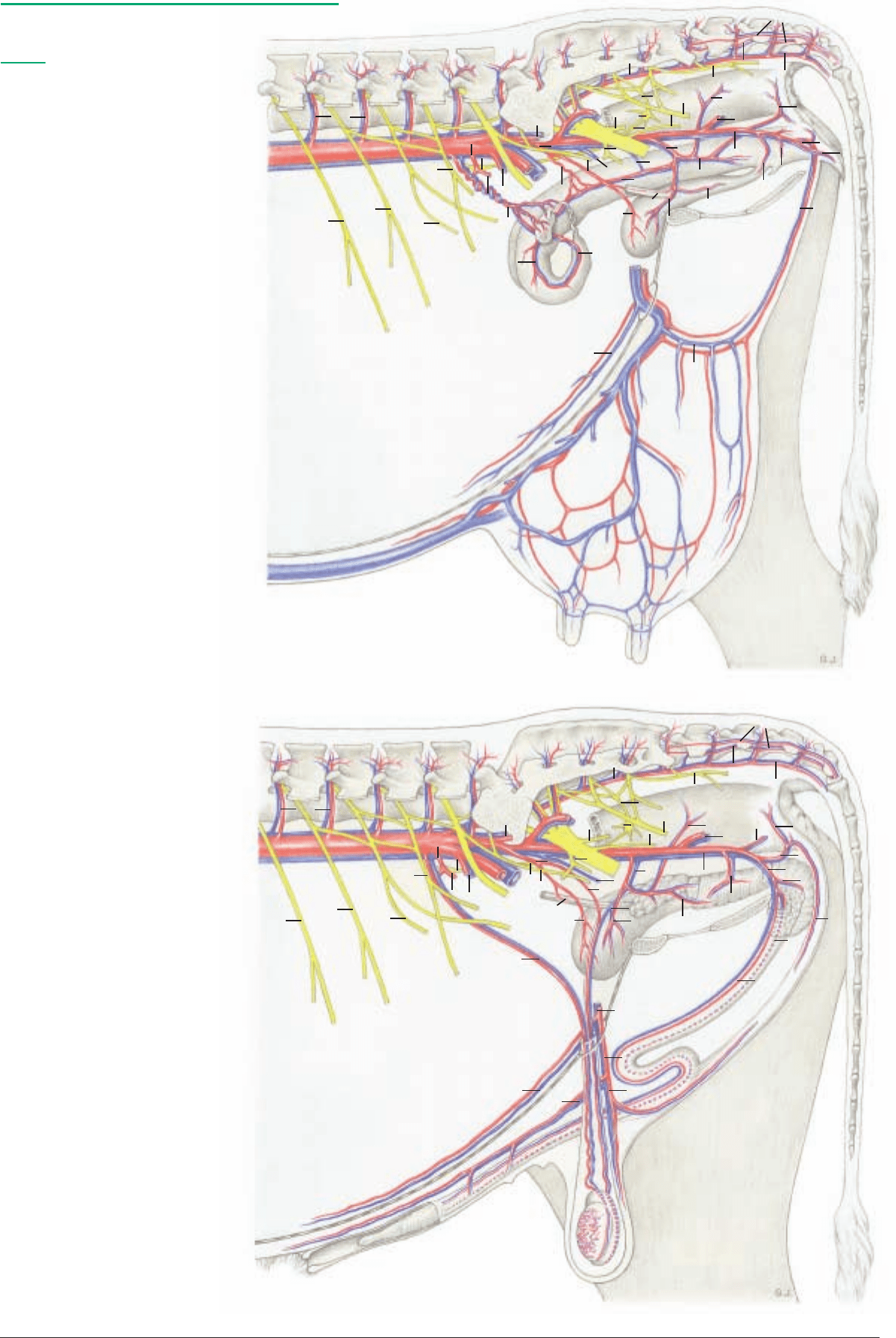
Pelvic arteries, veins, and nerves (left side)
(See pp. 17, 19, 21, 91)
Legend:
A
rteries, veins:
1 Abd. aorta and caud. vena cava
2 Ovarian or testicular a. and v.
2' Uterine br.
3 Caud. mesenteric a. and v.
4 Cran. rectal a. and v.
5 Ext. iliac a. and v.
6 Deep circumflex iliac a. and v.
7 Pudendoepigastric trunk and v.
8 Caud. epigastric a. and v.
9 Ext. pudendal a. and v.
10 Caud. supf. epigastric a. and v.
(Cran. mammary a. and v.)
11 Caud. mammary a. and v. or
Ventr. scrotal br. and v.
12 Lumbar aa. and vv.
13 Median sacral a. and v.
14 Median caud. a. and v.
14' Ventrolat. caudal a. and v.
14'' Dorsolat. caudal a. and v.
15 Int. iliac a. and v.
16 Iliolumbar a. and v.
17 Umbilical a.
18 Uterine a. or a. of ductus deferens
19 Cran. vesical a.
20 Obturator v.
21 Cran. gluteal a. and v.
22 Accessory vaginal v.
23 Vaginal or prostatic a. and v.
24 Uterine br. and v. or v. of ductus deferens
25 Caud. vesical a. and v.
26 Ureteric br.
27 Urethral br.
28 Middle rectal a. and v.
29 Dors. perineal a. and v.
30 Caud. rectal a. and v.
31 Caud. gluteal a. and v.
32 Int. pudendal a. and v.
33 Urethral a. and v.
34 Vestibular a. and v.
35 A. and v. of clitoris or penis
36 Ventr. perineal a. and v.
37 Ventr. labial v. and mammary br.
of ventr. or dors. perineal a.
In bull, br. and v. from ventr. perineal vessels
38 A. and v. of bulb of penis
39 Deep a. and v. of penis
40 Dors. a. and v. of penis
Nerves:
a Iliohypogastric n.
b Ilioinguinal n.
c Genitofemoral n.
d Lat. cut. femoral n.
e Femoral n.
f Sciatic n.
g Obturator n.
h Pudendal n.
i Cran. gluteal n.
j
Caud. gluteal n.
k Caud. cut. femoral n.
l Caud. rectal nn.
m Caudal mesenteric plexus
n Hypogastric n.
o Pelvic plexus
p Pelvic n.
85
Anatomie des Rindes englisch 09.09.2003 14:45 Uhr Seite 85
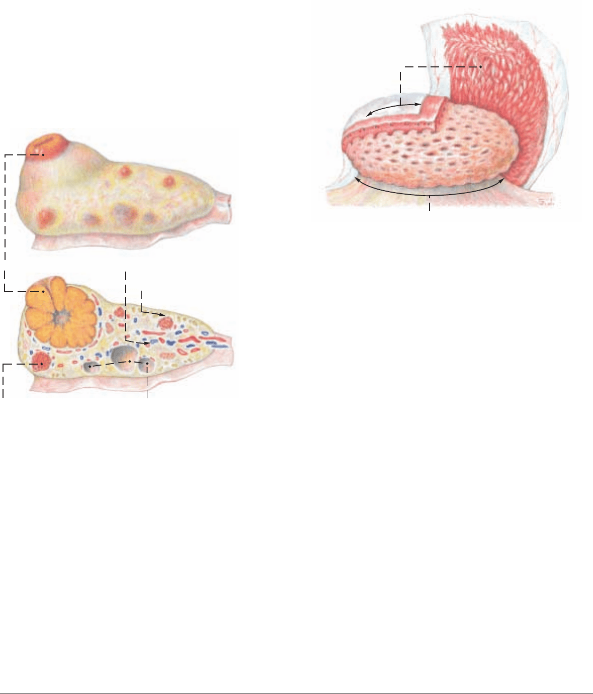
a) The OVARY (6) has a different position from that of the bitch
and mare because of the longer developmental “descent” of the
ovary and parietal attachment of the mesovarium (2) toward the
pelvis. This results in the spiral of the uterine horn and gives the
long axis of the ovary an obliquely transverse direction. The tubal
end of the ovary is dorsolateral and the uterine end is ventromedi-
al. The ovary lies near the lateroventral part of the pelvic inlet, cra-
nial to the external iliac a. In the pregnant cow it is drawn cra-
nioventrally. The mesovarium contains the ovarian a., coming from
the aorta, and gives off laterally the thin mesosalpinx (3) for the
uterine tube. The cranial border of the mesovarium is the suspen-
sory lig. of the ovary (1). Caudally the mesovarium is continuous
with the mesometrium (4). The mesovarium, mesosalpinx, and
mesometrium together form the broad ligament (lig. latum uteri)
which contains smooth muscle.
The ovary measures 3.5 x 2.5 x 1.5 cm, about the size of the distal
segment of the human thumb. Compared to that of the mare it is
relatively small. It is covered by peritoneum on the mesovarian
margin only, and by the superficial epithelium elsewhere. There is
no ovarian fossa, which is a peculiarity of the mare. The cortex and
medulla are arranged as in the bitch. On the irregularly tubercu-
lated surface there are always follicles and corpora lutea of various
stages of the estrous cycle which can be palpated per rectum. A fol-
licle matures to about 2 cm; a corpus luteum can reach the size of
a walnut. The single corpus luteum changes color during the cycle
from yellow or ocher-yellow to dark red, red-brown, gray-white,
and black. This can be seen on a section through the ovary.
b) The UTERINE TUBE (14) is somewhat tortuous and at 28 cm,
relatively long. The mesosalpinx (3) with the uterine tube sur-
rounds the ovary cranially and laterally like a mantle and forms
with the mesovarium the flat voluminous ovarian bursa (13) with
a wide cranioventromedial opening. The infundibulum of the tube
(16) with its fimbriae surrounds the ovary. It funnels into the
abdominal orifice of the tube (15). The ampulla and isthmus of the
tube do not show any great difference in the size of the lumen. The
uterine tube ends, unlike that of the bitch and mare, without a uter-
ine papilla at the uterine orifice of the tube (7) in the apex of the
uterine horn. Here the proper ligament of the ovary (8) ends and
the round lig. of the uterus begins. The latter is attached by a seros-
al fold to the lateral surface of the mesometrium and extends to the
region of the inguinal canal. Both ligaments develop from the
gubernaculum of the ovary. (The mammalian uterine tube differs in
form and function from the oviduct of lower animals.)
c) The UTERUS, as in all carnivores and ungulates, is a uterus
bicornis. The horns of the uterus (cornua uteri, 9) are 30–40 cm
long, rolled through cranioventral to caudodorsal, and fused cau-
dally into a 10–15 cm long double cylinder. Cranial to the union the
horns are connected by the dorsal and ventral intercornual ligg.
(11). Internally the true, undivided body of the uterus (12) is only
2–4 cm long. The neck of the uterus (cervix uteri, 26) with the cer-
vical canal (26) begins at the internal uterine orifice (27) and ends
at the external uterine orifice (25) on the vaginal part of the cervix
(portio vaginalis, 25). The cervix is 8–10 cm long and can be dis-
tinguished from the body of the uterus and the vagina by its firm
consistency.
The three layers of the wall of the uterus are formed by the peri-
toneum (perimetrium), the muscular coat (myometrium), and the
mucosa (endometrium). The mucosa of the uterus forms longitudi-
nal and transverse folds and in each uterine horn four rows of
10–15 round or oval caruncles (10)* of various sizes. These project
dome-like on the internal surface, and in the pregnant uterus can
reach the size of a fist. The total number of caruncles in the uterus,
including the body, is about 100.
During pregnancy they form, together with the cotyledons,** the
placentomes. Cotyledons are bunches of villi on the fetal amnio-
chorion and allantochorion that invade the caruncles. (See text fig-
ure.) The cervical mucosa presents longitudinal folds and, with the
support of the musculature, bulges into the lumen, usually in four
characteristic circular folds, and closes the cervical canal. This is
clinically important. The last circular fold projects into the vagina
as the portio vaginalis cervicis (25).
d) The Vagina (18), 30 cm long, is longer than in the mare, hollow
and its fornix (17) arches over the portio vaginalis cervicis dorsal-
ly. The cranial part of the vagina is covered by peritoneum in the
area of the rectogenital excavation (5) which extends caudally to
the middle of the pelvic cavity or to the first caudal vertebra. Cau-
dally the vagina joins the vestibule (23), sometimes without a dis-
tinct boundary, sometimes with only a faint transverse fold, the
hymen (19). The external urethral orifice (20) opens into the cra-
nial end of the vestibule 7–11 cm from the ventral commissure of
the labia. The suburethral diverticulum (x) lies ventral to the ure-
thral orifice.
The openings of the vestigial deferent ducts (remnants of the cau-
dal parts of the mesonephric ducts) are found on each side of the
urethral orifice. The ducts run between the mucosa and the muscu-
lature and can reach a considerable length. They end blindly and
can become cystic. The major vestibular gland (w) is cranial to the
constrictor vestibuli (m). It is 3 cm long and 1.5 cm wide, and has
2–3 ducts that open in a small pouch (24) lateral to the urethral ori-
fice. The microscopic minor vestibular glands open on the floor of
the vestibule cranial to the clitoris.
e) The VULVA surrounds with its thick labia (22) the labial fissure
(rima pudendi). The dorsal commissure of the labia is, in contrast
to the mare, more rounded, and the ventral commissure is pointed,
with a tuft of long coarse hairs.
The clitoris (21) is smaller than in the mare, although 12 cm long
and tortuous. The end is tapered to a cone. The glans is indistinct.
The prepuce is partially adherent to the apex of the clitoris so that
an (open) fossa clitoridis is almost absent.
86
5. FEMALE GENITAL ORGANS
* Caruncula -ae, L. = papilla
** Cotyledo(n) -onis L., Gr. = cup
Ovary
Corpus luteum
Corpus luteum in regression
Medulla [Zona vasculosa]
Cortex [Zona parenchymatosa]
Vesicular follicles
Caruncle
Placentome
Cotyledon
Anatomie des Rindes englisch 09.09.2003 14:45 Uhr Seite 86
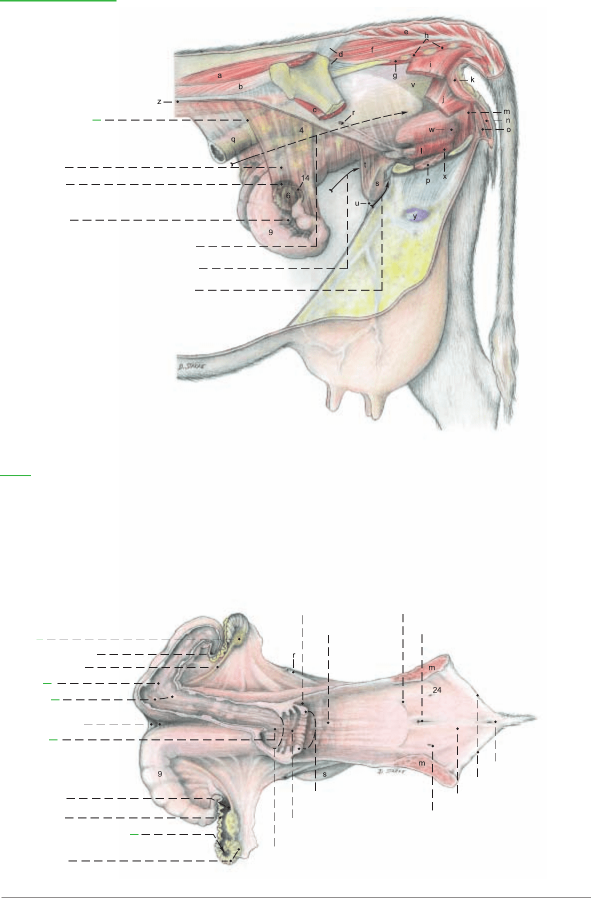
27 Int. orifice of uterus
Female genital organs
(Left side)
1 Suspensory lig. of the
ovary
Broad lig. of the uterus:
2 Mesovarium
3 Mesosalpinx
4 Mesometrium
5 Rectogenital excavation
Vesicogenital excavation
Pubovesical excavation
(See pp. 17, 19, 93)
Legend:
a Middle gluteal m.
b Longissimus lumborum
c Iliacus
d Sacroiliac ligg.
e Sacrocaudalis dorsalis
medialis
f Sacrocaudalis dorsalis lateralis
g Sacrocaudalis ventralis lateralis
h Intertransversarii
i Coccygeus
j Levator ani
k Ext. anal sphincter
l Urethralis
Bulbospongiosus:
m Constrictor vestibuli
n Constrictor vulvae
o Retractor clitoridis
p Intrapelvic part of
ext. obturator
q Descending colon
r Ureter
s Urinary bladder
t Lat. lig. and round
lig. of bladder
u Median lig. of bladder
v Rectum
w Major vestibular gl.
x Suburethral diverticulum
y Supf. inguinal lnn.
z Peritonum (Sectio)
(dorsal)
6 Ovary
7 Uterine orifice of uterine
tube
8 Proper lig. of ovary
9 Horn of
uterus
10 Caruncles
11 Intercornual ligg.
12 Body of
uterus
13 Ovarian bursa
14 Uterine tube
15 Abdominal orifice of uterine tube
16 Infundibulum and fimbriae
of uterine tube
17 Fornix of vagina
18 Vagina
19 Hymen
20 Ext. urethral orifice
21 Clitoris
22 Labia of vulva
23 Vestibule of vagina
24 Orifice of major vestibular gl.
25 Ext. orifice of uterus and
portio vaginalis of cervix
26 Cervix and
cervical canal
87
Anatomie des Rindes englisch 09.09.2003 14:45 Uhr Seite 87
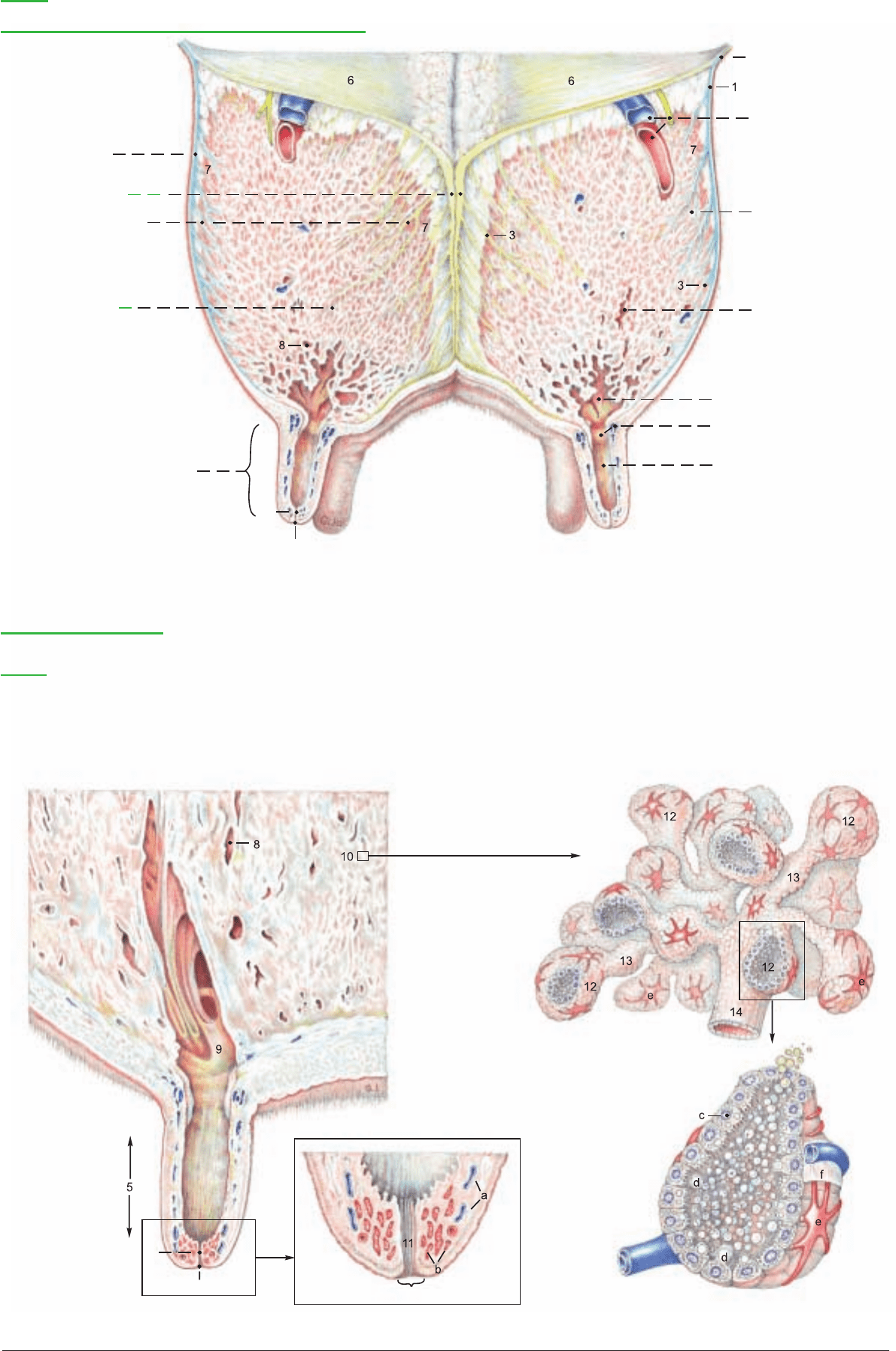
The udder is composed of four mammary glands—modified skin
glands that occur in this form only in true mammals (Eutheria). The
mammary secretion is milk (lac). The first milk secreted after par-
turition is colostrum, containing a high concentration of antibod-
ies, which give the newborn passive immunity. Cow’s milk and also
milk from sheep and goats, is a valuable human foodstuff. It con-
tains proteins, fats, sugar, and minerals (for example, calcium and
phosphorus). Therefore milk production is of great economic sig-
nificance in agriculture. Diseases of the udder lead directly to
reduced milk production that persists throughout the lactation
period. For that reason early treatment of udder diseases is espe-
cially important in veterinary practice. The diagnosis of udder dis-
eases and the possible need for surgery, such as removal of half of
the udder (mastectomy) or the amputation of a teat, require
anatomical knowledge of the structure of the udder, its suspensory
apparatus, blood vessels, lymph drainiage, and innervation.
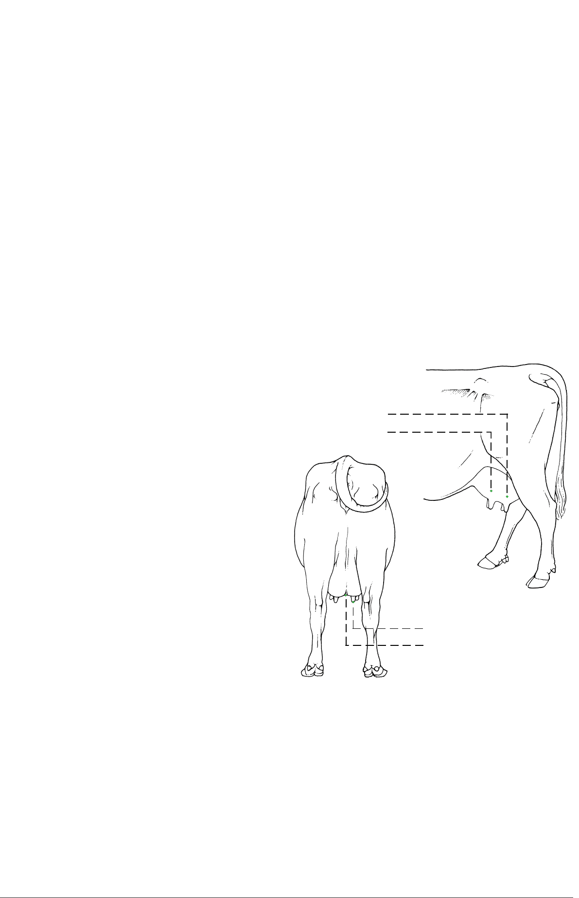
The four mammary glands of the bovine udder are attached to the
body in the inguinal region and are commonly called quarters. At
the height of lactation each quarter may reach enormous size.
Each mammary gland consists of a teat (papilla mammae, 5) and a
body (corpus mammae, 4). The size of the body and the length of
the teat vary with the individual cow, functional status, and form.
The teats are about as thick as the thumb and as long as the index
finger. The teat canal, with its orifice on the end of the teat, may be
incompletely closed, permitting ascending bacterial inflammation
of the udder (mastitis). A narrow, partly blocked teat canal will
restrict the flow of milk. Rudimentary accessory glands and teats
occur and are not rare. They are usually caudal to the normal teats,
but may be between them or cranial to them. Rudimentary teats
occur in the bull cranial to the scrotum. The right and left halves of
the udder are divided by a median intermammary groove. The
udder is covered by modified skin that is hairless and without skin
glands on the teat, and sparsely haired elsewhere. The skin of the
healthy udder is easily slipped on the subcutis, but this mobility is
lost in inflammation, and together with pain, edema, and heat
serves to diagnose mastitis. Suspensory apparatus: lateral laminae
(1) of fascia pass over the surface of the udder from the symphyseal
tendon and the lateral crus of the superficial inguinal ring in a
mainly cranioventral direction. The medial laminae (2) separate the
right and left halves of the udder. (This median separation can be
demonstrated by blunt dissection between the medial laminae from
their caudal borders.) Composed mostly of elastic tissue, they orig-
inate as a paired paramedian suspensory lig. (2). This comes from
the yellow abdominal tunic on the exterior surface of the prepubic
tendon (p. 66) at its junction with the symphysial tendon.
*
From
both the lateral and the medial laminae, thin suspensory lamellae
(3) penetrate the mammary gland, separating the parenchyma into
curved, overlapping lobes (7).
**
When filled with milk the udder
has considerable weight, which stretches the suspensory apparatus,
especially the medial laminae. Therefore the teats of the tightly
filled udder project laterally and cranially because the elastic medi-
al laminae are stretched more than the lateral laminae, which con-
sist mainly of regular dense collagenous tissue.
In contrast to the bitch and mare, each mammary gland of the cow
contains only one duct system and the associated glandular tissue.
In addition the gland contains interstitial connective tissue with
nerves, blood vessels, and lymphatics. The duct system ends on the
apex of the teat with the orifice (5") of the narrow teat canal (pap-
illary duct, 5'), surrounded by the teat sphincter (b).
The teat canal drains the lactiferous sinus with its papillary part
(teat sinus, 9") and glandular part (gland sinus, 9). The boundary
between the parts is marked by the annular fold (9') of mucosa,
containing a venous circle (of Fuerstenberg). A venous plexus (a) in
the wall of the teat forms an erectile tissue that makes hemostasis
difficult in injuries or surgery. The mucosa of the teat canal bears
longitudinal folds (11), and the proximal ends of the folds form a
radial structure called Fuerstenberg’s rosette at the boundary
between the teat sinus and the teat canal.
In the gland sinus are the openings of several large collecting ducts
(ductus lactiferi colligentes, 8). Each of these receives milk from one
of the numerous lobes through small lactiferous ducts (14) and
alveolar lactiferous ducts (13), which drain the lobules (10). A lob-
ule resembles a bunch of grapes, measures 1.5 x 1.0 x 0.5 mm, and
consists of about 200 alveoli.
***
Many alveoli are connected direct-
ly, and this construction has led to the term, “storage gland”. The
alveoli are surrounded by septa containing nerves and vessels. The
duct systems are separate for each quarter, as demonstrated by
injections of different colored dyes, even though quarters on the
same side have no septum between them. Therefore ascending
infections may be limited to one quarter. The separate medial lam-
inae make it possible to amputate one lateral half of the udder. The
teat canal has a defensive mechanism in its lining of stratified
squamous epithelium that produces a plug of fatty desquamated
cells in the canal between milkings. This is an important factor in
resistance to infection.
****
88
6. THE UDDER
* Habel and Budras, 1992 *** Weber et al., 1955
** Ziegler and Mosimann, 1960 **** Adams and Rickard, 1963
(lateral)
Caudal mammary gl.
(hindquarter)
Cranial mammary gl.
(forequarter)
(caudal)
Papilla (teat)
Intermammary groove
Anatomie des Rindes englisch 09.09.2003 14:46 Uhr Seite 88
5'
5"
5'
5"
9'
9"
5"
a Venous plexus of the teat
b Teat sphincter muscle
c Lactocyte
Udder
Transverse section through forequarters, cranial surface
(cranial)
Suspensory apparatus:
1 Lateral lamina
2 Medial laminae
(Suspensory lig.)
3 Suspensory lamellae
4 Body (Corpus)
of right forequarter
5 Teat (Papilla)
5' Teat canal
(Ductus papillaris)
5" Teat orifice
6 Yellow abdominal tunic
Cran. mammary a. and
v. and cran. br. of
genitofemoral n.
7 Lobe of
mammary gl.
8 Collecting duct
Lactiferous sinus:
9 Glandular part (Gland sinus)
9' Annular fold and
Venous circle
9" Papillary part (Teat sinus)
Mammary gland and Teat
Legend:
10 Lobule
11 Longitudinal folds
12 Alveoli
13 Alveolar lactiferous ducts
14 Lactiferous duct
d Fat droplet
e Myoepithelial cell
f Basement membrane
89
Anatomie des Rindes englisch 09.09.2003 14:46 Uhr Seite 89

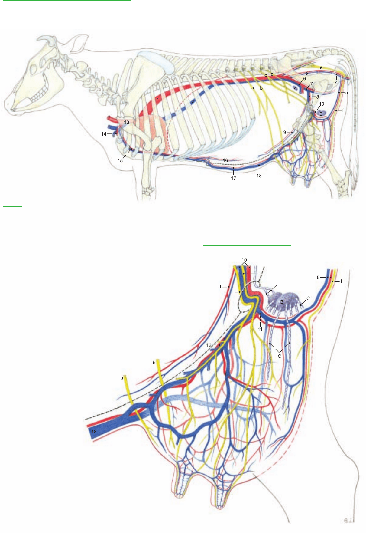
I. The blood vascular system is adapted to the high milk produc-
tion of the udder. Up to 600 liters of blood must flow through the
udder to produce one liter of milk. Therefore the blood vessels are
remarkable for their large calibre, and they have received addition-
al names. The ext. pudendal a. and v. bifurcate into the cran. (12)
and caud. (11) mammary a. and v. The cran. mammary vessels are
also known as the caud. supf. epigastric vessels. The caud. mam-
mary a. and v. are continuous with the mammary br. and ventral
labial v. (5), which usually come from the ventral perineal vessels,
but in some cows they come from the dorsal perineal vessels (see p.
95, 16).
The cran. supf. epigastric vein in milk cows can be seen bulging
under the skin of the ventral abdominal wall. It is therefore called
the subcutaneous abdominal v. (18). The place where it perforates
the abdominal wall in the xiphoid region from the int. thoracic v. is
the “milk well” [anulus venae subcutaneae abdominis]. The caud.
supf. epigastric v. is also called the cran. mammary v. (12). The cau-
dal and cranial supf. epigastric vv. anastomose end-to-end and form
the “milk vein”. This is enlarged during the first lactation and its
valves become incompetent, making blood flow possible in either
direction. The right and left cran. mammary vv. anastomose on the
cran. border of the udder. This connection, with that of the caudal
mammary vv., completes the venous ring around the base of the
udder. Many veins of the udder join this ring. The vent. labial v. is
large and tortuous in the dairy cow (see p. 95, 16). In most of its
extent the valves indicate that blood flows toward the caud. mam-
mary v.
II. The lymph from the udder is conducted to 1–3 supf. inguinal
lnn. (mammary lnn., B). They lie caudally on the base of the udder
(the surface applied to the body wall) and can be palpated between
the thighs about 6 cm from the skin at the caudal attachment of the
udder. Small intramammary lnn. may be present. The lymph flows
to the iliofemoral ln. (deep inguinal ln., A). These lnn. are routine-
ly incised in meat inspection.
III. The innervation of the udder is sensory and also autonomic
(sympathetic). The skin and teats of the forequarters and the cra-
nial part of the base of the udder are supplied by the iliohypogas-
tric n. (a), ilioinguinal n. (b), and the cran. br. of the genitofemoral
n. (c'). The skin and teats of the hindquarters are innervated by the
caud. br. of the genitofemoral n. (c") and the mammary br. of the
pudendal n. (f). The cran. and caud. brr. of the genitofemoral n.
pass through the inguinal canal into the body of the udder. The sen-
sory innervation of the teats and skin of the udder is the afferent
pathway of the neurohormonal reflex arc, which is essential for the
initiation and maintenance of milk expulsion from the mammary
glands. The stimulus produced by sucking the teats and massaging
the mammary gll. is conducted by the afferent nerves to the CNS,
where, in the nuclei of the hypothalamus, the hormone oxytocin is
produced. The afferent nervous stimulus causes the hormone to be
released through the neurohypophysis into the blood, which carries
it into the mammary gll. Here oxytocin causes contraction of the
myoepithelial cells on the alveoli, by which milk is pressed into the
lactiferous ducts and sinus. This expulsion of milk is disturbed
under stress by secretion of the hormone adrenalin, which sup-
presses the action of oxytocin on the myoepithelial cells. (For
details, see textbooks of histology and physiology.)
IV. The prenatal development of the udder begins in the embryo in
both sexes on the ventrolateral body wall between the primordia of
the thoracic and pelvic limbs. This linear epidermal thickening is
the mammary ridge. It is shifted ventrally by faster growth of the
dorsal part of the body wall. Local epithelial sprouts grow down
into the underlying mesenchyme from the ridge, forming the mam-
mary buds in the location and number of mammary glands of each
species. The mesenchyme surrounding the epithelial sprout is called
the areolar tissue. Each mammary bud is bordered by a slightly
raised ridge of skin. The teat develops in ruminants, as in the horse,
by the growth of this areolar tissue, as a proliferation teat. The sur-
rounding skin ridge is completely included in the formation of the
teat. (For details see the textbooks of embryology.)
Postnatally the mammary glands are inconspicuous in calves of
both sexes because the teats are short and the mammary glands are
hardly developed. The duct system consists only of the teat canal,
the sinus, and the primordia of the collecting ducts, which are short
solid epithelial cords. Normally, the male udder remains in this
stage throughout life. During puberty some bull calves can under-
go a further temporary growth of the mammary glands under the
influence of an elevated level of estrogen, as is natural in females.
In young heifers during pubertal development ovarian follicles
ripen and cause the level of estrogen in the blood to rise. In the
udder this results in an increase of connective and adipose tisssue,
and also further proliferation of the epithelial buds as primordia of
the lactiferous ducts, which divide repeatedly, producing the small
collecting ducts. The mammary gland primordia rest in this stage of
proliferation until the first pregnancy.
During the first pregnancy further generations of lactiferous ducts
develop by growth and division of the epithelial cords. In the sec-
ond half of pregnancy the still partially solid glandular end-pieces
are formed, while space-occupying adipose tissue is displaced.
Toward the end of gestation (about 280 days) under the influence
of progesterone and estrogen, a lumen develops in these glandular
end-pieces, and under the influence of prolactin the lactocytes begin
the secretion of milk (lactogenesis ). In the first five days after par-
turition the milk secreted is colostrum. This is rich in proteins; it
contains immunoglobulins, and it may be reddish due to an admix-
ture of erythrocytes. In addition to the passive immunization of the
newborn, colostrum has another function: it has laxative properties
that aid in the elimination of meconium (fetal feces). Lactation can
begin a few days or a few hours before parturition, and the first
drops of milk on the end of a teat are taken as an indication of
impending birth.
After birth milk secretion is maintained only in the quarters that the
suckling uses. The unused quarters rapidly undergo involution.
This occurs naturally when the calf is weaned by the dam, but in
U.S. dairy practice the calf is removed from the dam and fed artifi-
cially, beginning with colostrum from the dam. Milk secretion is
maintained by milking twice a day. After about ten months, lacta-
tion is stopped by decreasing the ration and reducing the milking to
provide a dry period of about 60 days before calving.* During invo-
lution the secretory cells in the alveoli and in the alveolar lactifer-
ous ducts degenerate. The glandular tissue is replaced by fat and
connective tissue. This is important for the clinical evaluation of the
consistency of individual quarters. The size of the udder decreases,
but never returns to the small size of an udder that has not yet pro-
duced milk.
Accessory (supernumerary) mammary gll. may be present on the
udder, a condition called hypermastia. The presence of supernu-
merary teats is called hyperthelia (Gk. thele, nipple). They may be
located before, between, or behind the main teats. If they occur on
a main teat they interfere with milking and must be removed.
90
7. THE UDDER WITH BLOOD VESSELS, LYMPHATIC SYSTEM, NERVES, AND DEVELOPMENT
* Ensminger, 1977
Anatomie des Rindes englisch 09.09.2003 14:46 Uhr Seite 90
c'
c"
c'
c"
C'
1 Aorta
2 Caud. vena cava
3 Int. iliac a. and v.
4 Int. pudendal a. and v.
5 Vent. labial v. and mammary br.
of vent. perineal a.
6 Ext. iliac a. and v.
7 Deep femoral a. and v.
8 Pudendoepigastric vessels
9 Caud. epigastric a. and v.
10 Ext. pudendal a. and v.
11 Caud. mammary a. and v.
12 Cran. mammary a. and v.
[Caud. supf. epigastric a. and v.]
13 Brachiocephalic trunk and
cran. vena cava
14 Left subclavian a. and v.
15 Int. thoracic a. and v.
16 Cran. epigastric a. and v.
17 Cran. supf. epigastric a.
18 Subcutaneous abdominal v.
[Cran. supf. epigastric v.]
A Iliofemoral ln.
[Deep inguinal ln.]
B Mammary lnn.
[Supf. inguinal lnn.]
C Afferent lymphatic vessels
C' Efferent lymphatic vessels
Arteries, Veins, and Nerves of the Udder
Left side
Legend:
a Iliohypogastric n.
b Ilioinguinal n.
c Genitofemoral n.
c' Cran. branch
c" Caud. branch
d Lat. cut. femoral n.
e Pudendal n.
f Mammary br. of pudendal n.
Left cran. and caud. mammary gll.
91
Anatomie des Rindes englisch 09.09.2003 16:01 Uhr Seite 91
a) The SCROTUM (5) is attached in the cranial pubic region. It is
elongated dorsoventrally and bottle-shaped. It is generally flesh-
colored and fine-haired, and bears two rudimentary teats on each
side of the cranial surface of the neck.
b) The elongated oval TESTES (4 and 16) hang vertically in the scro-
tum and weigh about 300 g each. The capital end is proximal and the
caudate end is distal.(Names derived from the head and tail of the epi-
didymis.) In ruminants the epididymal border of the testis is medial
or caudomedial and the free border is lateral. The part of the mesor-
chium (p) between the vaginal ring and the testis contains the testic-
ular vessels and nerves. It is covered by the visceral lamina of the vagi-
nal tunic, which is attached to the parietal lamina along the caudo-
medial surface. The ductus deferens runs in the mesoductus deferens
(q), a narrow fold attached to the cranial surface of the mesorchium.
This location is important for vasectomy. The spermatic cord (10)
extends from the vaginal ring (d) to the testis and consists of the
mesorchium and its contents, the ductus deferens, and the mesoduc-
tus deferens. The mesorchium continues distally along the epididy-
mal border of the testis. At the tail of the epididymis the mesorchium
ends in a short free fold, the lig. of the tail of the epididymis (o), the
vestige of the distal part of the gubernaculum testis. Between the testis
and the tail of the epididymis is the very short proper lig. of the testis
(n), the vestige of the proximal part of the gubernaculum.
c) The EPIDIDYMIS begins with a long head (caput, 12) on the
capital end and adjacent free boder of the testis. The head consists
of a descending limb, and an ascending limb that crosses the mesor-
chium to the slender body of the epididymis (14). This descends
medial to the testis along the caudal side of the mesorchium to the
prominent tail of the epididymis (19). Between the body of the epi-
didymis and the testis is the testicular bursa (17), often obliterated
by adhesion.
d) The DUCTUS DEFERENS (e) ascends in its mesoductus on the
medial side of the testis, cranial to the mesorchium, to the spermatic
cord (10), which is longer and narrower than in the horse. After it
enters the abdominal cavity the duct crosses the lateral lig. of the
bladder and the ureter (f) and enters the genital fold. It ends in the
urethra on the colliculus seminalis in a common orifice with the
duct of the vesicular gland.
e) The ACCESSORY GENITAL GLANDS are all present as in the
horse, but fully developed only in the bull—not in the steer. The
bilateral vesicular gland (11) is the largest accessory genital gland in
the bull. It is a lobated gland of firm consistency—not vesicular. It is
10–20 cm long and lies dorsal to the bladder and lateral to the ureter
and the ampulla of the ductus deferens (13). The ductus deferens
narrows again caudal to the ampulla and, with the duct of the vesic-
ular gland, passes under the body of the prostate. The two ducts
open on the colliculus seminalis (see above). The body of the
prostate (15) projects on the dorsal surface of the urethra between
the vesicular glands and the urethral muscle. The disseminate part of
the prostate, 12–14 cm long, is concealed in the wall of the urethra
and covered ventrally and laterally by the urethral muscle. The bilat-
eral bulbourethral gland (18) is the size of a walnut. It lies on each
side of the median plane dorsal to the urethra in the transverse plane
of the ischial arch. It is mostly covered by the bulbospongiosus mus-
cle. Its duct opens on the lateral fold that extends caudally from the
septum between the urethra and the urethral recess (see p. 82).
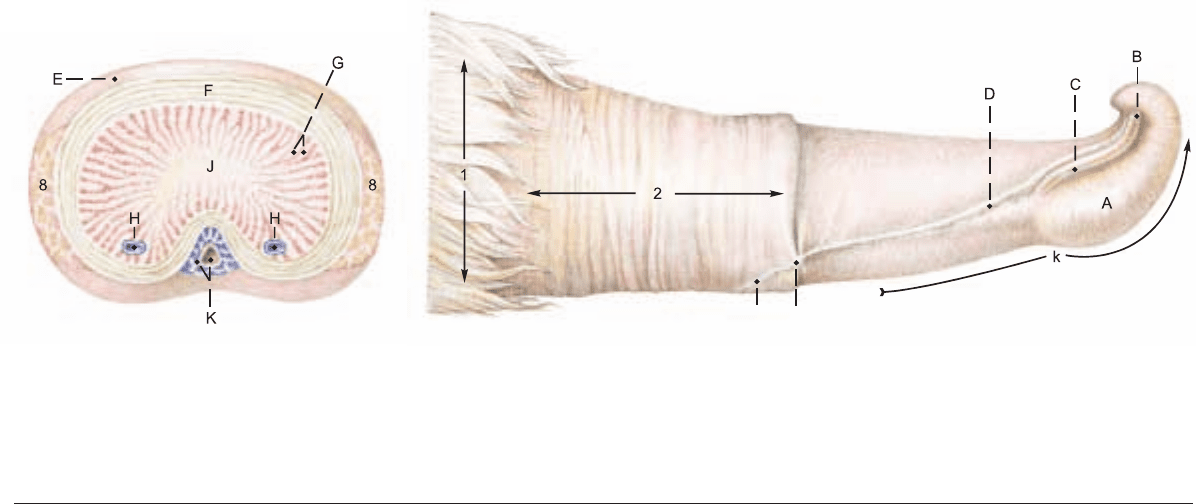
f) The PENIS of the bull belongs to the fibroelastic type. It extends
from its root (h) at the ischial arch to the glans penis (A) in the
umbilical region. It is covered by skin, is about one meter long, and
in the body [corpus penis (i)], has a sigmoid flexure (j) that is cau-
dal to the scrotum. The proximal bend is open caudally and the dis-
tal bend, open cranially, can be grasped through the skin caudal to
the thighs. The penis is sheathed by telescoping fascia. The short
collagenous suspensory ligg. of the penis (l) are attached close
together on the ischial arch, and the dorsal nn. and vessels of the
penis pass out between them. They should not be confused with the
fundiform lig. of the penis (p. 80). The penis consists of the dense
corpus cavernosum penis, which begins at the junction of the crura
penis (7), attached to the ischial arch. It is surrounded by a thick
tunica albuginea (F) containing cartilage cells. The cavernae are
mainly peripheral, and axially there is a dense connective tissue
strand (J). The free part of the penis (k), 8 cm long, is distal to the
attachment of the internal lamina of the prepuce (2). It is twisted to
the left as indicated by the oblique course of the raphe of the penis
(D) from the midventral raphe of the prepuce (D") to the external
urethral orifice (B) on the right side. Just before ejaculation an
added left-hand spiral of the free part of the penis is caused by the
internal pressure acting against the right-hand spiral of the collage-
nous fibers of the subcutaneous tissue and tunica albuginea, and
against the apical lig. The latter originates dorsally from the tunica
albuginea, beginning distal to the sigmoid flexure.* Midventral on
the penis is the penile urethra, surrounded by the corpus spongio-
sum penis (K). The urethral process (C) lies in a shallow groove
between the raphe and the cap-like glans penis (A), which is con-
nected to the corpus spongiosum, but contains little erectile tissue.
The prepuce consists, as in the dog, of an external lamina (1) and
an internal lamina (2), and has bristle-like hairs at the preputial ori-
fice (3). The frenulum of the prepuce (D') connects the raphe of the
prepuce to the raphe of the penis. The muscles of the penis: The
ischiocavernosus (7) extends from the medial surface of the ischial
tuber to the body of the penis, covering the crus penis. The bul-
bospongiosus (6) covers the bulb of the penis and a large part of the
bulbourethral gland and extends to the beginning of the body of the
penis. During erection both muscles regulate the inflow and out-
flow of blood. The paired smooth muscle retractor penis (8) origi-
nates from the caudal vertebrae, receives reinforcing fibers from the
internal anal sphincter, extends across the first bend of the sigmoid
flexure and is attached to the second bend. The two muscles then
approach each other on the ventral surface and terminate on the
tunica albuginea 15–20 cm proximal to the glans. In erection these
muscles relax, permitting the extension of the sigmoid flexure and
elongation of the penis.
The lymphatic vessels of the scrotum, penis, and prepuce drain to
the superficial inguinal lnn. (9) which lie dorsolaterally on the penis
at the transverse plane of the pecten pubis, just caudal to the sper-
matic cord. The lymph vessels of the testes go to the medial iliac
lnn. (p. 82).
92
8. MALE GENITAL ORGANS AND SCROTUM
* Ashdown, 1958; Ashdown 1969; Seidel and Foote, 1967
D"
D'
Penis
(Right surface)
Legend:
A Glans penis
B Ext. urethral orifice
C Urethral process
(Cross section cranial to sigmoid flexure)
D Raphe of penis
D' Frenulum of prepuce
D" Raphe of prepuce
E Fascia of penis
F Tunica albuginea
G Trabeculae of J
H Deep veins of penis
J Corpus cavernosum
K Corpus spongiosum and urethra
k Free part of penis
Anatomie des Rindes englisch 09.09.2003 16:01 Uhr Seite 92

n
19 Tail of epididymis
Male genital organs
(Left side)
Prepuce:
1 Ext. lamina
2 Int. lamina
3 Preputial orifice
4 Left testis
5 Scrotum
6 Bulbospongiosus and
Bulb of penis
7 Ischiocavernosus and
Crus penis
8 Retractor penis
9 Supf. inguinal lnn.
(See pp. 17, 19, 87)
Legend:
a Rectus abdominis
b Cremaster
c Testicular a. and v.
d Vaginal ring
e Ductus deferens
f Ureter
g Urethralis
Penis:
h Root of the penis
i Body of the penis
j Sigmoid flexure
k Free part of penis
l Suspensory ligg. of penis
m Male mammary gl.
n Proper lig. of testis
o Lig. of the tail of the epididymis
p Mesorchium
q Mesoductus deferens
r Mesofuniculus
s Pampiniform plexus
Testis and Epididymis
(caudal)
Accessory genital gll.
(dorsal)
10 Spermatic cord
11 Vesicular gl.
12 Head of epididymis
13 Ampulla of ductus deferens
14 Body of epididymis
15 Body of prostate
16 Left testis
17 Testicular bursa
18 Bulbourethral gl.
93
Anatomie des Rindes englisch 09.09.2003 16:01 Uhr Seite 93
94
9. PERINEUM, PELVIC DIAPHRAGM, ISCHIORECTAL FOSSA, AND TAIL
* Larson, 1953
** Erasha, 1987
vCy3
Tail (Cauda)
Sacrocaudalis dors. medialis
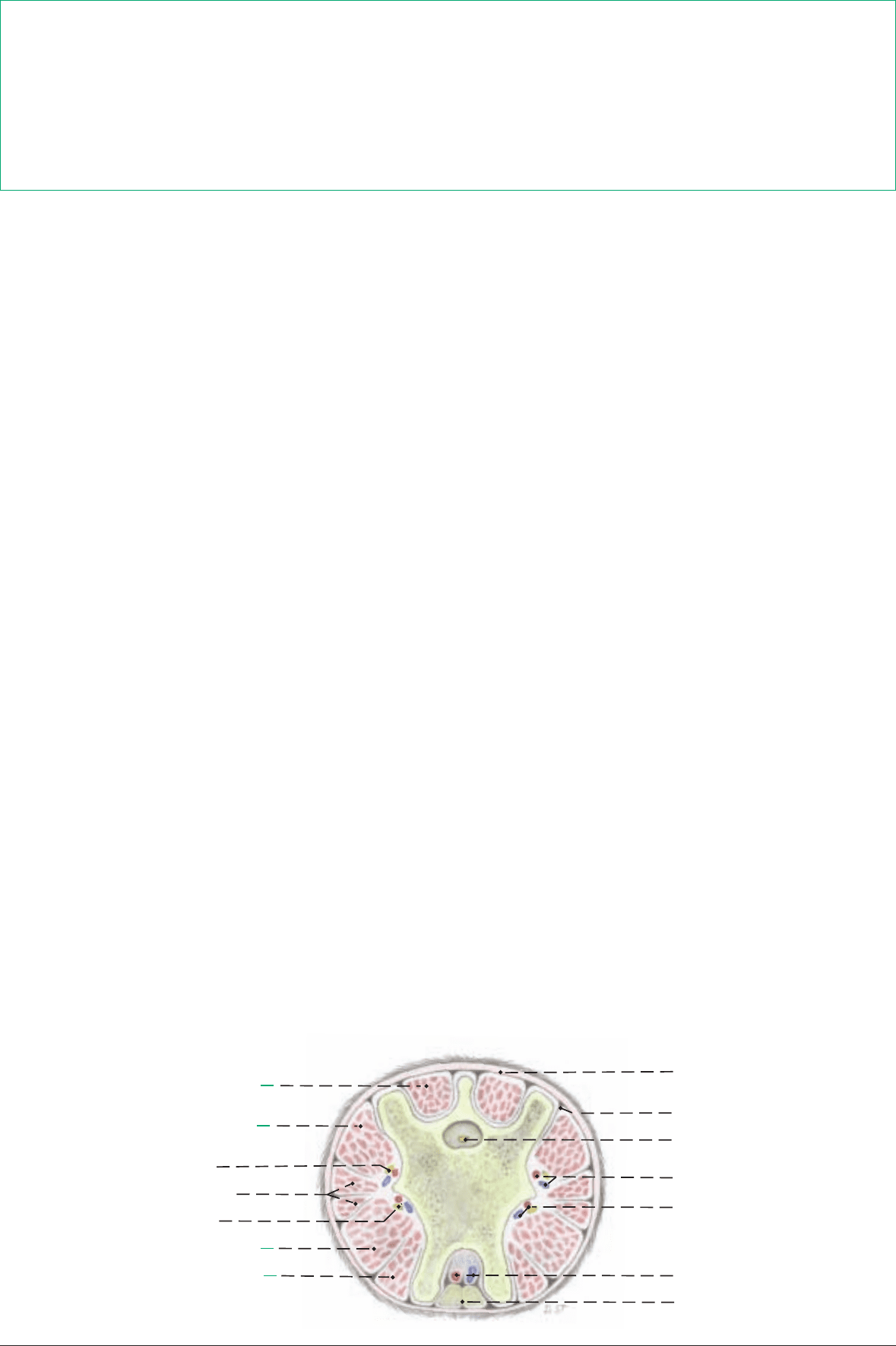
Sacrocaudalis dors. lateralis
Dors. caudal plexus
Intertansversarii caudae
Ventr. caudal plexus
Sacrocaudalis ventr. lateralis
Sacrocaudalis ventr. medialis
Skin
Caudal fascia
Filum of spinal dura mater
Dorsolat. caudal a. and v.
Ventrolat. caudal a. and v.
Median caudal a. and v.
Rectocaudalis
(transverse)
a) The PERINEUM and PERINEAL REGION. The perineum is
the part of the body wall that closes the pelvic outlet, bounded by
the first caudal vertebra, the sacrosciatic ligg. (1), the tubera
ischiadica (b), and the ischial arch. The part of the perineum dorsal
to a line connecting the tubera ischiadica is the anal triangle, sur-
rounding the anal canal and closed by the pelvic diaphragm. The
part of the perineum ventral to the line is the urogenital triangle,
surrounding the urogenital tract and closed by the perineal mem-
brane. A more restricted definition includes only the perineal body
between the anus and the urogenital tract. The perineal region is the
surface area over the perineum and adjacent parts. In the ox it is
bounded dorsally by the root of the tail and ventrally by the attach-
ment of the scrotum or udder. The lateral border is formed by the
sacrosciatic ligament, tuber ischiadicum, and a line from the tuber
to the scrotum or udder. The perineal region is divided into anal
and urogenital regions by a line connecting the medial processes of
the tubers. The urogenital region is greatly elongated in ruminants
by the ventral position of the scrotum and udder.
b) The ANAL TRIANGLE. The pelvic diaphragm is composed of
right and left coccygeus (2) and levator ani (3) muscles and the exter-
nal anal sphincter (12), together with the deep fascia on their exter-
nal and internal surfaces. Each half of the diaphragm is oblique,
extending caudomedially from the origin of the muscles on the medi-
al surface of the sciatic spine, to the termination of the coccygeus on
the caudal vertebrae and of the levator ani on the external anal
sphincter. The perineal body [centrum tendineum perinei], is the
fibromuscular mass between the anus and the urogenital tract.
c) The UROGENITAL TRIANGLE. The perineal membrane in
the cow is a strong sheet of deep perineal fascia extending from the
ischial arch to the ventral and lateral walls of the vestibule, cranial
to the constrictor vestibuli (13) and caudal to the major vestibular
gland (10). Together with the urogenital muscles it closes the uro-
genital triangle, joining the pelvic diaphragm at the the level of the
perineal body and anchoring the genital tract to the ischial arch.
d) The ISCHIORECTAL FOSSA is a fat-filled, wedge-shaped
space lateral to the anus. The laterodorsal wall is the sacrosciatic
lig., the caudal border of which, the sacrotuberous lig (1), is easily
palpable. The lateroventral wall is the tuber ischiadicum and the
obturator fascia. The medial wall is the deep fascia covering the
coccygeus, levator ani, and constrictor vestibuli. In the ox, unlike
the horse, the sacrotuberous lig. and tuber ischiadicum are subcu-
taneous (see p. 16).
e) NERVES AND VESSELS. For the intrapelvic origins of the per-
ineal nerves and vessels, see pp. 84–85. The pudendal n. (9) gives
off the proximal and distal cutaneous branches and the deep per-
ineal n. (20), and continues caudally on the pelvic floor with the
internal pudendal a. and v. (9), supplying the vestibule and the
mammary br. (25) and terminating in the clitoris. In the bull, the
pudendal n. gives off the preputial and scrotal brr. and continues as
the dorsal n. of the penis. The deep perineal n. supplies the vagina,
major vestibular gland, and perineal muscles, and ends in the labi-
um and the skin lateral to the perineal body. The caudal rectal n.
(17), which may be double, supplies branches to the rectum, coc-
cygeus, levator ani, ext. anal sphincter, retractor clitoridis (penis),
perineal body, constrictor vestibuli, roof of the vestibule, and labi-
um. Anesthesia of the penis and paralysis of the retractor penis, or
anesthesia of the vestibule and vulva can be produced by blocking
bilaterally the pudendal and caudal rectal nn. and the communi-
cating br. of the caud. cutaneous femoral n. (p. 84) inside the
sacrosciatic lig.* The internal iliac a. (6), at the level of the sciatic
spine, gives off the vaginal or prostatic a. (These arteries may orig-
inate from the internal pudendal a.) The internal iliac ends by divid-
ing at the lesser sciatic foramen into the caud. gluteal a. and inter-
nal pudendal a. (9). The latter supplies the coccygeus, levator ani,
ischiorectal fossa, vagina, urethra, vestibule, and major vestibular
gl. The internal pudendal a. ends by dividing into the ventral per-
ineal a. (23) and the a. of the clitoris (24). The ventral perineal a.
usually gives off the mammary branch (25). In some cows the ven-
tral perineal a. and mammary br. are supplied by the dorsal labial
br. of the dorsal perineal a. The vaginal a. (7), after giving off the
uterine br., divides into the middle rectal a. and the dorsal perineal
a. (8). The latter divides into the caud. rectal a. (21) and the dorsal
labial br. (22), which gives off the perineal br. seen on the tuber
ischiadicum, and runs ventrally in the labium. It may also supply
the mammary br. and the ventral part of the perineum. The dorsal
labial br. may be cut in episiotomy. In the male, the prostatic a.
gives branches to the urethra, prostate, and bulbourethral gl., and
may terminate as the dorsal perineal a., but the latter usually comes
from the internal pudendal a.**
f) The TAIL contains 16–21 caudal vertebrae. The rectocaudalis is
longitudinal smooth muscle from the wall of the rectum, attached to
caud. vertebrae 2 and 3. The smooth muscle retractor clitoridis
(penis) originates from caud. vertebrae 2 and 3 or 3 and 4. The cau-
dal nerves in the cauda equina run in the vertebral canal. The medi-
an caudal a. and v. on the ventral surface are convenient for the vet-
erinarian working behind stanchioned cows. The pulse is best pal-
pated between the vertebrae or about 18 cm from the root of the tail
to avoid the hemal processes. Tail bleeding is done by raising the tail
and puncturing the median caudal v. between hemal processes.
The clinically important perineum is studied by first removing the skin from the perineal region to see the superficial muscles, nerves,
and vessels. The fat is removed from the ischiorectal fossa, exposing the distal cutaneous br. of the pudendal n. (19) where it emerges
on the medial surface of the tuber ischiadicum and supplies the superficial perineal nn. (4). The caudal rectal a. (21) is exposed in its
course along the lateral border of the ext. anal sphincter, and branches of the dorsal and ventral perineal aa. are seen. The superficial
fascia is incised from the labia to the udder to expose the large, convoluted, and often double ventral labial v. (16), draining blood from
the perineum to the caudal mammary v. The mammary brr. of the pudendal nn. are traced on the lateral borders of the vein. The cor-
responding nerve in the bull is the preputial and scrotal br., and the vein is the ventral scrotal. In deeper dissections the fascia is removed
from the terminations of the coccygeus (2) and levator ani (3) and from the constrictor vestibuli (13) and constrictor vulvae (14). The
smooth muscle retractor clitoridis (15) is seen between the constrictor vestibuli and constrictor vulvae in the cow, and the retractor penis
between the bulbospongiosus and ischiocavernosus in the bull.
Anatomie des Rindes englisch 09.09.2003 16:01 Uhr Seite 94
