Bhushan B. Nanotribology and Nanomechanics: An Introduction
Подождите немного. Документ загружается.

262 A. Schirmeisen et al.
conditions have been reported so far. Interestingly, a low-budget construction set
(employing a tuning-fork force sensor) for a CE-mode dynamic AFM set-up has
been published on the internet (http://www.sxm4.uni-muenster.de).
If it is possible to measure atomic scale forces with the NC-AFM, it should
vice versa also be possible to exert forces with a similar precision. In fact, the new
and exciting field of nanomanipulation could be driven to a whole new dimension,
if defined forces can be reliably applied to single atoms or molecules. In this re-
spect, Loppacher et al. [47] were able to push on different parts of an isolated Cu-
tetrabromobisphenol (TBBP) molecule, which is known to possess four rotatable
legs. They measured the force–distance curves while pushing one of the legs with
the AFM tip. From the force curves they we able to determine the energy which
was dissipated during the switching process of the molecule. The manipulation of
single silicon atoms with NC-AFM was demonstrated by Oyabu et al. [48], who
removed single atoms from a Si(111)-7×7 surface with an AFM tip and could sub-
sequently deposit atoms from the tip onto the surface again. The possibility to exert
and measure forces simultaneously during single-atom or molecule manipulation is
an exciting new application of high-resolution NC-AFM experiments.
6.4 Q-Control
We have already discussed the virtues of a high Q value for high-sensitivity meas-
urements: the minimum detectable force gradient was inversely proportional to the
square root of Q. In vacuum, Q mainly represents the internal dissipation of the can-
tilever during oscillation, an internal damping factor. Low damping is obtained by
using high-quality cantilevers, which are cut (or etched) from defect-free single-
crystal silicon wafers. Under ambient or liquid conditions, the quality factor is
dominated by dissipative interactions between the cantilever and the surrounding
medium, and Q values can be as low as 100 for air or even 5 in liquid. Still, we
ask if it is somehow possible to compensate for the damping effect by exciting the
cantilever in a sophisticated way?
It turns out that the shape of the resonance curves in Fig. 6.5 can be influenced
towards higher (or lower) Q values by an amplitude feedback loop. In principle,
there are several mechanisms to couple the amplitude signal back to the cantilever,
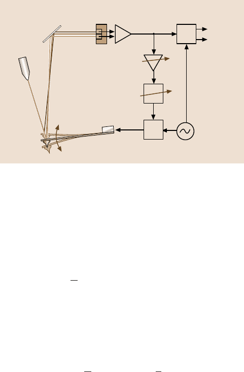
e.g., by the photothermal effect [49] or capacitive forces [50]. Figure 6.19 shows
a method in which the amplitude feedback is mediated directly by the excitation
piezo [51]. This has the advantage that no additional mechanical setups are neces-
sary.
The working principle of the feedback loop can be understood by analyzing the
equation of motion of the modified dynamic system:
m
∗
¨z(t)+ α˙z(t)+ kz(t)−F
ts
(z
0
+ z(t)) = F
ext
cos(ωt)+ Ge
iφ
z(t). (6.26)
This ansatz takes into account the feedback of the detector signal through a phase
shifter, amplifier and adder as an additional force, which is linked to the cantilever
6 Dynamic Modes of Atomic Force Microscopy 263
G
Φ
Laser
Detector
Mirror
Lock-in
amplifier
Piezo
Adder
Cantilever
+ tip
Detector signal
Variable
phase shifter
Function
generator
Variable
gain amplifier
Phase
Amplitude
+
Fig. 6.19. Schematic diagram of operating a Q-control feedback circuit with an externally
driven dynamic AFM. The tapping-mode set-up is in effect extended by an additional feed-
back loop
deflection z(t) through the gain G and the phase shift e
iφ
. We assume that the oscil-
lation can be described by a harmonic oscillation trajectory. With a phase shift of
φ = ±π/2 we find:
e
±iπ/2
z(t) = ±
1
ω
˙z(t). (6.27)
This means, that the additional feedback force signal Ge
iφ
z(t) is proportional to the
velocity of the cantilever, just like the damping term in the equation of motion. We
can define an effective damping constant α
eff
, which combines the two terms:
m
∗
¨z(t)+ α
eff
˙z(t)+ kz(t)−F
ts
(z
0
+ z(t)) = F
ext
cos(ωt), (6.28)
with α
eff
= α ±
1
ω
G for φ = ±
π
2
.
Equation (6.28) shows that the damping of the oscillator can be enhanced or weak-
ened by choosing φ =+π/2orφ = −π/2, respectively. The feedback loop there-
fore allows us to vary the effective quality factor Q
eff
= mω
0
/α
eff
of the complete
dynamic system. Hence, this system was termed c-Control. Figure 6.20 shows ex-
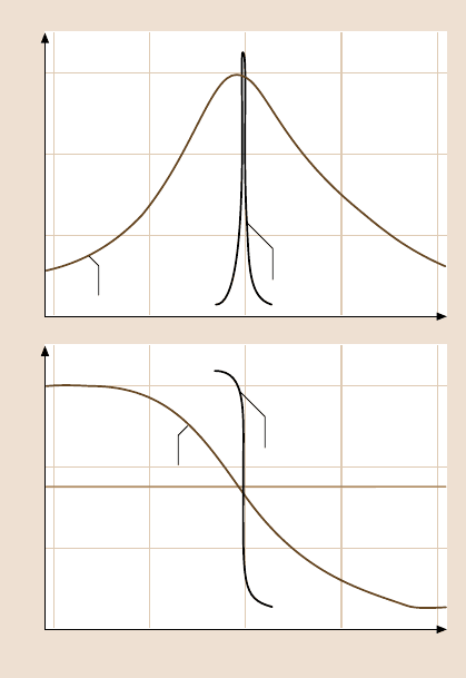
perimental data regarding the effect of Q-control on the amplitude and phase as
a function of the external excitation frequency [51]. In this example, Q-control was
able to increase the Q-value by a factor of over 40.
The effectof improvedimage contrastis demonstratedin Fig. 6.21.Here, a com-
puter hard disk was analyzed with a magnetic tip in tapping mode, where the mag-
netic contrast is observed in the phase image. The upper part shows the recorded
magnetic data structures in standard mode, whereas in the lower part of the image
Q-control feedback was activated, giving rise to an improved signal, i.e., magnetic
contrast. A more detailed analysis of measurements on a magnetic tape shows that
264 A. Schirmeisen et al.
259.5
0.6
0.4
0.2
0.0
Amplitude (arb.units)
0
90
180
Frequency (kHz)
Phase (deg)
260.0 260.5 261.5261.0
Q-control off
Q = 499
Q-control on
Q = 19.379
Q-control off
Q = 499
Q-control on
Q = 19.379
Fig. 6.20. Amplitude and
phase diagrams measured
in air with a Si cantilever
far away from the sample.
The quality factor can be in-
creased from 450 to 20,000
by using the Q-control feed-
back method
the signal amplitude (upper diagrams in Fig. 6.22) was increased by a factor of 12.4
by the Q-control feedback. The lower image shows a noise analysis of the signal,
indicating an improvement of the signal-to-noise ratio by a factor of 2.3.
Note that the diagrams represent measurements in air with an AFM operated in
AM mode. Only then can we make a distinction between excitation and vibration
frequency, since in the FM mode these two frequencies are equal by definition. Al-
though the relation between sensitivity and Q-factor in (6.17) is the same for AM
and FM mode, it must be critically investigated whether the enhanced quality factor
by Q-control can be inserted in the equation for FM-mode AFM. In vacuum appli-
cations, Q is already very high, which makes it unnecessary to operate an additional
Q-control module.
As stated before, we can also use Q-control to enhance the damping in the os-
cillating system. This would decrease the sensitivity of the system. But on the other
hand, the response time of the amplitude change is decreased as well. For tapping-
mode applications, where high-speed scanning is the goal, Q-control was able to
reduce the scan speed that limits relaxation time [52].
6 Dynamic Modes of Atomic Force Microscopy 265
Norm. standard deviation (%)
MFM phase (deg)
X-position (nm)
42
41
40
39
38
X-position (nm)
3
2
1
0
70
60
50
40
30
20
3
2
1
0
0 200 400 600 800 1.000 0 200 400 600 800 1.000
MFM phase (deg)
Norm. standard deviation (%)
Q-control off Q-control on
0.25
0
0 0.25 0.50 0.75 1.00
3.7
46.0
2.1 %
0.9 %
Fig. 6.21. Signal-to-noise analysis with a magnetic tip in tapping mode AFM on a magnetic
tape sample with Q-control
Q-control off
Q-control on
Fig. 6.22. Enhancement of the contrast in the phase
channel due to Q-control on a magnetic hard disk
measured with a magnetic tip in tapping-mode
AFM in air. Scan size 5×5 µm, phase range 10deg
(www.nanoanalytics.com)
266 A. Schirmeisen et al.
A large quality factor Q does not only have the virtue of increasing the force
sensitivity of the instrument. It also has the advantage of increasing the parameter
space of stable AFM operation in AM-mode AFM. Consider the resonance curve
of Fig. 6.5. When approaching the tip towards the surface there are two competing
mechanisms: on one hand, we bring the tip closer to the sample, which results in an
increase in attractive forces (see Fig. 6.2). On the other hand, for the case ω>ω
0
,
the resonant frequency of the cantilever is shifted towards smaller values due to
the attractive forces, which causes the amplitude to become smaller. This is the de-
sirable regime, where stable operation of the AFM is possible in the net-attractive
regime. But as explained before, below a certain tip–sample separation d
app
,the
system switches suddenly into the intermittent contact mode, where surface mod-
ifications are more likely due to the onset of strong repulsive forces. The steeper
the amplitude curve the larger the regime of stable, net-attractive AFM operation.
Looking at Fig. 6.20 we find thatthe slope of the amplitudecurve is governedby the
quality factor Q. A high Q, therefore, facilitates stable operation of the AM-AFM
in the net-attractive regime.
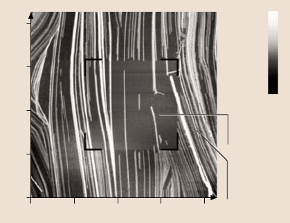
An example can be found in Fig. 6.23. Here, a surface scan of an ultrathin or-
ganic film is acquired in tapping mode under ambient conditions. First, the inner
square is scanned without the Q enhancement, and then a wider surface area was
scanned with Q-control applied. The high quality factor provides a larger parameter
space for operating the AFM in the net-attractive regime, allowing good resolution
of the delicate organic surface structure. Without the Q-control the surface struc-
8.00
6.00
4.00
2.00
0
0 2.00 4.00 6.00 8.00
25.0 nm
12.5 nm
0.0 nm
μm
Tapping
mode
with active
Q-control
Standard
tapping
mode
Fig. 6.23. Imaging of a delicate organic surface with Q-Control. Sample was a Langmuir–
Blodgett film (ethyl-2,3-dihydroxyoctadecanoate) on a mica substrate. The topographical
image clearly shows that the highly sensitive sample surface can only be imaged non-
destructively with active Q-control, whereas the periodic repulsive contact with the probe
in standard operation without Q-control leads to a significant modification or destruction of
the surface structure (data courtesy of Lifeng Chi and coworkers, University of Münster, Ger-
many)
6 Dynamic Modes of Atomic Force Microscopy 267
tures are deformed and even destroyed due to the strong repulsive tip–sample inter-
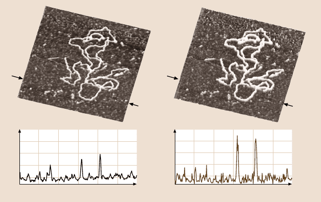
actions [53–55]. The Q-control feedback also allowed imaging of DNA structures
without predominantlydepressing the soft material duringimaging. It was then pos-
sible to observe a DNA diameter close to the theoretical value [56].
The same technique has been successfully employed to minimize the interaction
forces during scanning in liquids. This is of special relevance for imaging delicate
biologicalsamples in environmentssuch as water or buffer solution. When the AFM
probe is submerged in a liquid medium, the oscillation of the AFM cantilever is
strongly affected by hydrodynamic damping. This typically leads to quality factors
below 10 and, accordingly, to a loss in force sensitivity. However, the Q-control
technique allows one to increase the effective quality factor by about three orders
of magnitude in liquids. Figure 6.24 shows results of scanning DNA structures on
a mica substrate under a buffer solution. Comparison of the topographic data ob-
tained in standard tapping mode and under Q-Control, in particular the difference
in the observed DNA height, indicates that the imaging forces were successfully
reduced by employing Q-control.
Height (nm)
x-Position (nm)
0
600
2.0
1.5
1.0
0.5
0
500400300200100
Height (nm)
x-Position (nm)
0
600
2.0
1.5
1.0
0.5
0
500400300200100
Q-control onQ-control off
Fig. 6.24. AFM images of DNA on mica scanned in buffer solution (600 nm×600 nm). Each
scan line was scanned twice: in standard tapping mode during the first scan of the line (left
data) and with Q-control activated by a trigger signal during the subsequent scan of the same
line (right data). Thisinterleave technique allows a direct comparison of the resultsof thetwo
modes obtained on the same surface area while minimizing drift effects. Cross sections of the
topographic data reveal that the observed DNA height is significantly higher in the case of
imaging under Q-control (data courtesy of Daniel Ebeling and Hendrik Hölscher, University
of Münster, Germany)
268 A. Schirmeisen et al.
In conclusion,we have shown that, by applyingan additional feedbackcircuit to
the dynamic AFM system, it is possible to influence the quality factor Q of the os-
cillator system. High-resolution, high-speed, or low-force scanning is then possible.
6.5 Dissipation Processes Measured with Dynamic AFM
Dynamic AFM methods have proventheir great potential for imaging surface struc-
tures at the nanoscale, and we have also discussed methods that allow the assess-
ment of forcesbetween distinct singlemolecules.However, thereis another physical
mechanism that can be analyzed with the dynamic mode and has been mentioned in
some previous paragraphs: energy dissipation.
In Fig. 6.12, we have already shown an example, where the phase signal in tap-
ping mode cannot be explained by conservative forces alone; dissipative processes
must also play a role. In constant-amplitude FM mode, where the quantitative in-
terpretation of experiments has proven to be less difficult, an intuitive distinction
between conservative and dissipative tip–sample interaction is possible. We have
shown the correlation between forces and frequency shifts of the oscillating system,
but we have neglected one experimental input channel; the excitation amplitude,
which is necessary to keep the oscillation amplitude constant, is a direct indication
of the energy dissipated during one oscillation cycle. Dürig [57] has shown that
in self-excitation mode (with an excitation–oscillation phase difference of 90 deg),
conservativeand dissipative interactions can be strictly separated. Part of this energy
is dissipated in the cantilever itself, another part is due to external viscous forces in
the surrounding medium. But more interestingly, some energy is dissipated at the
tip–sample junction. This is the focus of the following paragraphs.
In contrast to conservative forces acting at the tip–sample junction, which
at least in vacuum can be understood in terms of van der Waals, electrostatic,
and chemical interactions, the dissipative processes are poorly understood. Stowe
et al. [58] have shown that, if a voltage potential is applied between tip and sam-
ple, charges are induced in the sample surface, which will follow the tip motion (in
their set-up the oscillation was parallel to the surface). Due to the finite resistance
of the sample material, energy will be dissipated during the charge movement. This
effect has been exploited to image the doping level of semiconductors. Energy dis-
sipation has also been observedin imaging magnetic materials. Liu et al. [59] found
that energy dissipation due to magnetic interactions was enhanced at the boundaries
of magnetic domains, which was attributed to domain-wall oscillations, and even
a simple system such as two clean metal surfaces which are moved in close prox-
imity can give rise to frictional forces. Stipe et al. [60] have measured the energy
dissipation due to fluctuating electromagnetic fields between two closely spaced
gold surfaces, which was later interpreted by Volokitin and Persson [61] in terms of
van der Waals friction.
Energy dissipation was also observed, in the absence of external electromag-
netic fields, when the tip and sample were in close proximity, within 1 nm. Clearly,
mechanical surfacerelaxations must give rise to energy losses. One could model the
6 Dynamic Modes of Atomic Force Microscopy 269
AFM tip as a small hammer,hitting the surface at high frequency, possibly resulting
in phonon excitations. From a continuum mechanics point of view, we assume that
the mechanical relaxation of the surface is not only governed by elastic responses.
Viscoelastic effects of soft surfaces will also render a significant contribution to en-
ergy dissipation. The whole area of phase imaging in tapping mode is concerned
with those effects [62–64].
In the atomistic view, the last tip atom can be envisaged as changing position
while experiencing the tip–sample force field. A strictly reversible change of po-
sition would not result in a loss of energy. Still, it has been pointed out by Sasaki
et al. [65] that a change in atom position would result in a change in the force inter-
action itself. Therefore, it is possible that the tip atom changes position at different
tip–surface distances during approach and retraction, effectively causing an atomic-
scale hysteresis to develop. Hoffmann et al. [13] and Hembacher et al. [66] have
measured the short-range energy dissipation for different combinations of tip and
surface materials in UHV. For atomic-resolution experiments at low temperatures
on graphite [66] it was found that the energy dissipation is a step-like function.
A similar shape of dissipation curves was found in a theoretical analysis by Kan-
torovich and Trevethan [67], where the energy dissipation was directly associated
with atomic instabilities at the sample surface.
Thedissipation channelhas also beenused to image surfaceswith atomic resolu-
tion [68]. Instead of feeding back the distance on the frequency shift, the excitation
amplitude in FM mode has been used as the control signal. The Si(111)-(7×7) re-
construction was successfully imaged in this mode. The step edges of mono-atomic
NaCl islands on single-crystalline copper have also rendered atomic-resolution con-
trast in the dissipation channel [69]. The dissipation processes discussed so far are
mostly in the configuration in which the tip is oscillated perpendicular to the sur-
face. Friction is usually referred to as the energy loss due to lateral movement of
solid bodies in contact. It is interesting to note in this contextthat Israelachivili [70]
has pointed out a quantitative relationship between lateral and vertical (with respect
to the surface) dissipation. He states that the hysteresis in vertical force–distance
curves should equal the energy loss in lateral friction. An experimental confirma-
tion of this conjecture at the molecular level is still missing.
Physical interpretation of energy dissipation processes at the atomic scale seems
to be a daunting task at this point. Nevertheless, we can find a quantitative relation
between the energy loss per oscillation cycle and the experimental parameters in
dynamic AFM, as will be shown in the following section.
InstaticAFMitwasfoundthatpermanentchangesofthesamplesurfacebyinden-
tations cancause a hysteresisbetweenapproachand retraction.The area between the
approachand retraction curvesin a force–distancediagram representsthe lostor dis-
sipatedenergycausedby the irreversiblechangeof thesurfacestructure.In dynamic-
mode AFM, the oscillation parameterssuch as amplitude,frequency,and phase must
containtheinformationaboutthedissipatedenergypercycle.Sofar,wehaveresorted
toatreatmentoftheequationofmotionofthecantilevervibrationinordertofindaquan-
270 A. Schirmeisen et al.
titativecorrelationbetweenforcesandtheexperimentalparameters.Forthedissipation
it is useful to treat the system from thepoint of viewof energyconservation.
Assuming that a dynamic system is in equilibrium, the average energy input
must equal the average energy output or dissipation. Applying this rule to an AFM
running in a dynamic mode means that the average power fed into the cantilever os-
cillation by an external driver, denoted by
P
in
, must equal the average power dissi-
pated by the motion of the cantileverbeam
P
0
andby thetip–sampleinteraction P
tip
:
P
in
= P
0
+ P
tip
. (6.29)
The term
P
tip
is what we are interested in, since it gives us a direct physical quan-
tity to characterize the tip–sample interaction. Therefore, we first have to calculate
and then measure the two other terms in (6.29) in order to determine the power
dissipated when the tip periodically probes the sample surface. This requires an
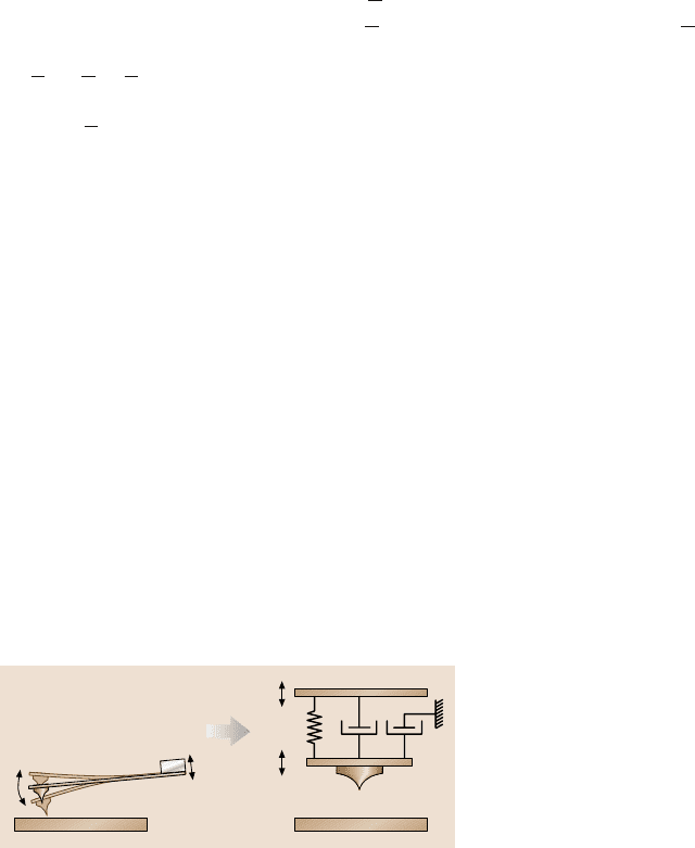
appropriate rheological model to describe the dynamic system. Although there are
investigations in which the complete flexural motion of the cantilever beam has
been considered [71], a simplified model, comprising a spring and two dashpots
(Fig. 6.25), represents a good approximation in this case [72].
The spring, characterized by the constant k according to Hooke’s law, represents
the only channel through which power P
in
can be delivered to the oscillating tip z(t)
by the external driverz
d
(t). Therefore,the instantaneouspower fed into the dynamic
system is equal to the force exerted by the driver times the velocity of the driver (the
force that is necessary to move the base side of the dashpot can be neglected, since
this power is directly dissipated and, therefore, does not contribute to the power
delivered to the oscillating tip):
P
in
(t) = F
d
(t)˙z
d
(t) = k[z(t)−z
d
(t)]˙z
d
(t). (6.30)
Assuming a sinusoidal steady-state response and that the base of the cantilever is
driven sinusoidally (see (6.6)) with amplitude A
d
and frequency ω, the deflection
z
d
(t)
z
d
(t)
z(t)
z(t)
k
α
2
α
1
?
Fig. 6.25. Rheological models applied to describe the dynamic AFM system, comprising
the oscillating cantilever and tip interacting with the sample surface. The movement of the
cantilever base and the tip is denoted by z
d
(t)andz(t), respectively. The cantilever is char-
acterized by the spring constant k and the damping constant α. In a first approach damping
is broken into two pieces α
1
and α
2
: firstly, intrinsic damping caused by the movement of
the cantilever’s tip relative to its base, and secondly, damping related to the movement of the
cantilever body in a surrounding medium, e.g. air damping

6 Dynamic Modes of Atomic Force Microscopy 271
from equilibrium of the end of the cantilever follows (6.9), where A and 0 ≤ ϕ ≤ π
are the oscillation amplitudeand phase shift, respectively. This allowsus to calculate
the average power input per oscillation cycle by integrating (6.30) over one period
T = 2π/ω:
P
in
=
1
T
T
0
P
in
(t)dt =
1
2
kωA
d
Asinϕ. (6.31)
This contains the familiarresultthat the maximum poweris deliveredto an oscillator
when the response is 90deg out of phase with the drive.
The simplified rheological model, as depicted in Fig. 6.25, exhibits two major
contributions to the damping term
P
0
. Both are related to the motion of the can-
tilever body and assumed to be well modeled by viscous damping with coefficients
α
1
and α
2
. The dominant dampingmechanism in UHV conditionsis intrinsic damp-
ing, caused by the deflection of the cantilever beam, i.e., the motion of the tip rel-
ative to the cantilever base. Therefore the instantaneous power dissipated by such
a mechanism is given by
P
01
(t) = |F
01
(t)˙z(t)| = |α
1
[˙z(t)−˙z
d
(t)]˙z(t)|. (6.32)
Note that the absolute value has to be calculated, since all dissipated power is lost
and, therefore, cannot be returned to the dynamic system.
However, when running an AFM in ambient conditions an additional damping
mechanism has to be considered; damping due to the motion of the cantilever body
in the surrounding medium, e.g., air damping, is in most cases the dominant effect.
The corresponding instantaneous power dissipation is given by
P
02
(t) = |F
02
(t)˙z(t)| = α
2
˙z
2
(t). (6.33)
In order to calculate the average power dissipation, (6.32) and (6.33) have to be
integrated over one complete oscillation cycle. This yields
P
01
=
1
T
T
0
P
01
(t)dt
=
1
π
α
1
ω
2
A
⎡
⎢
⎢
⎢
⎢
⎢
⎢
⎢
⎢
⎢
⎢
⎣
(A−A
d
cosϕ)arcsin
⎛
⎜
⎜
⎜
⎜
⎜
⎜
⎜
⎜
⎜
⎜
⎝
A−A
d
cosϕ
!
A
2
+A
2
d
−2AA
d
cosϕ
⎞
⎟
⎟
⎟
⎟
⎟
⎟
⎟
⎟
⎟
⎟
⎠
+ A
d
sinϕ
⎤
⎥
⎥
⎥
⎥
⎥
⎥
⎥
⎥
⎥
⎥
⎦
(6.34)
and
P
02
=
1
T
T
0
P
02
(t)dt =
1
2
α
2
ω
2
A
2
. (6.35)
Considering the fact that commonly used cantilevers exhibit a quality factor of at
least several hundreds (in UHV even several ten thousands), we can assume that
the oscillation amplitude is significantly larger than the drive amplitude when the
dynamic system is driven at or near its resonance frequency: A A
d
. Therefore
(6.34) can be simplified in a first-order approximation to an expression similar to