ASM Metals HandBook Vol. 17 - Nondestructive Evaluation and Quality Control
Подождите немного. Документ загружается.

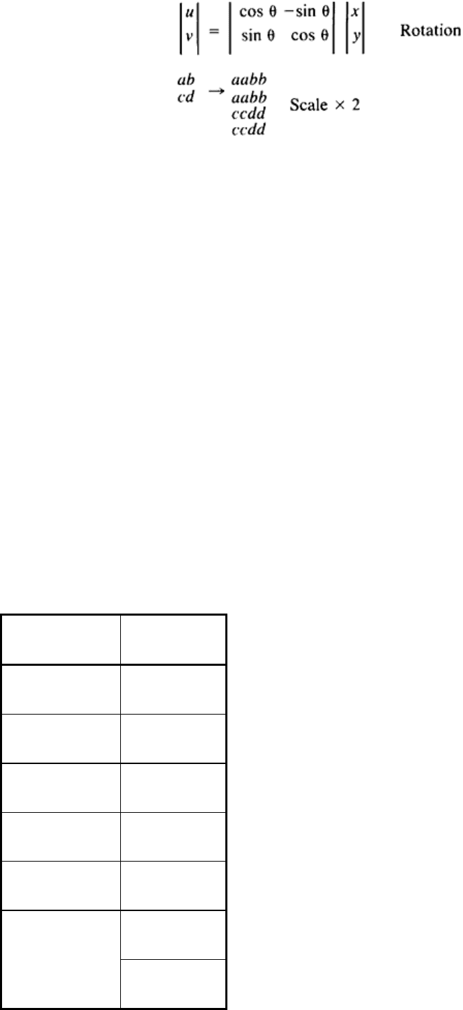
(Eq 3)
Warping is a nonlinear scaling process. The equation for a sixth-order warp is:
u = a
1
+ a
2
x + a
3
y + a
4
xy + a
5
x
2
+ a
6
y
2
v = b
1
+ b
2
x + b
3
y + b
4
xy + b
5
x
2
+ b
6
y
2
(Eq 4)
This transform can be of use if an object is at an angle to the viewing plane and it is necessary to measure a feature of
interest or to correct the image for the viewing angle.
Image registration can be performed by trial and error with the rotation, translation, and scaling functions. The
goodness of fit can be calculated with the correlation function or by subtraction. Registration is usually a prior step to
image combination.
Image Combination. Image combination operations generally include the following: AND, NAND, OR, NOR, XOR,
difference, subtract, add, multiply, divide, and average. All these operations require two operands. One will be the data in
the primary image, the other can be either a constant or data in a secondary image. The image combination operations are
used to mask certain areas of images, to outline features, to eliminate backgrounds, to search for commonality, and/or to
combine images. For example, a video of a part can be taken, edge enhanced, boosted, ANDed with 128 added to 127 and
scaled and registered with a radiograph or other image, and then added to the other image.
Filtering. There are many types of spatial filters. The most useful are low pass, high pass, edge, noise, and
morphological, as listed in Table 4.
Table 4 Commonly used image filters
Convolution filters
Other filters
High pass
Median
Low pass
Erosion
Horizontal edge
Dilation
Vertical edge
Unsharp mask
Laplacian
Roberts
Sobel or Prewitt
Compass
Wiener
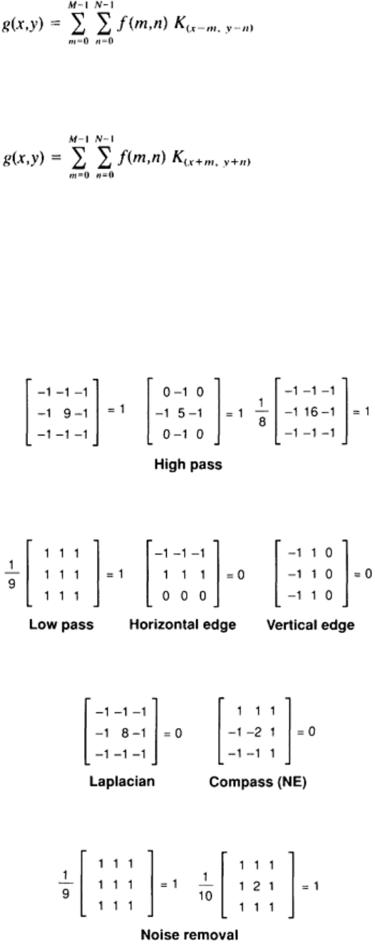
Many filters are classified as convolution filters. The convolution filter uses the discrete form of the two-dimensional
convolution integral to compute new pixel values. In Eq 5, the image matrix is defined as f(x,y), and the filter is a small 3
× 3 or (in general, m × n) matrix called a kernel, K. In practice, the kernel is limited to a size of less than 20 × 20 because
the FFT filtering procedure becomes faster than direct convolution for large kernels. The discrete form of the convolution
integral is:
(Eq 5)
Because kernels are often symmetric about a 180° rotation about the center pixel, algorithms that use the correlation
integral will give identical results to the convolution operation. The discrete form of the correlation integral is:
(Eq 6)
In this representation, one can visualize the kernel sliding over the image, with each pixel under the kernel being
multiplied by the kernel value and then all values being summed to produce the new center pixel. It is customary to
perform the convolution such that the initial and final image sizes are identical, with the outer row and column of the
image not convolved with the kernel. Some of the most useful convolution kernels are shown in Fig. 8. Those kernels that
add to 0, such as the Laplacian and the horizontal and vertical edge kernels, will remove most of the image except the
edge components. The low-pass and noise reduction filters will blur the image and remove point noise, while the high-
pass filters tend to sharpen the images and accentuate noise.
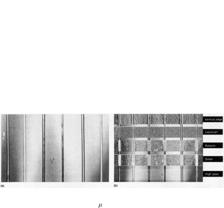
Fig. 8 Various convolution kernels and their use
Other filters that are useful in image processing are the median, erosion, dilation, and unsharp mask filters. The median
filter selects the median value in an image neighborhood (usually 3 × 3) and replaces the center pixel with the median
value. This filter can be very effective in removing isolated pixel noise without blurring the image as severely as the low-
pass filter. The effect of a low-pass and a median filter is shown on a noisy ultrasonic image of a hot-rolled plate in Fig.
3(a), (b), and (c) in the article "Use of Color for NDE" in this Volume. The erosion and dilation filters work by replacing
the center pixel value in a neighborhood with the smallest or largest index in the neighborhood, respectively. They can be
used to thicken or to thin boundaries. The unsharp mask filter algorithm is:
g(x,y) = 2 · f(x,y) - L P F (f)
xy
(Eq 7)
where LPF is the low-pass filter. The new image is the difference between the original image multiplied by a factor and
the low-pass filtered image. This operation tends to enhance the contrast and to sharpen the edges slightly.
The Roberts and Sobel filters are similar types of edge detectors. The Roberts filter algorithm is:
g(x,y) = {[f(x,y) - f(x + 1, y + 1)]
2
+ [(f(x,y + 1)
- f(x + 1, y)]
2
}
1/2
(Eq 8a)
This algorithm operates on a 2 × 2 neighborhood and will enhance high frequencies, while the Sobel filter operates with a
3 × 3 kernel and will produce good (but thick) outlines of images. The Sobel filter algorithm is:
g(x,y) = ({[(f(x + 1, y - 1) + 2 · f(x + 1, y)
+ f(x + 1, y + 1)] - [(f(x - 1, y - 1)
+ 2 · f(x - 1, y) + f(x - 1, y + 1)]}
2
+ {[f(x - 1, y - 1) + 2 · f(x, y - 1)
+ f(x + 1, y - 1)] - [f(x - 1, y + 1)
+ 2 · f(x, y + 1) + f(x + 1, y + 1)]}
2
)
1/2
(Eq 8b)
A summary of the effect of various filters and edge detectors is shown in Fig. 9.
Fig. 9 Ultrasonic image of an adhesive bond (250
m, or 0.01 in., thick) between two opaque plastic parts. (a)
Unfiltered. (b) Filtered with numerous filter types (vertical edge, high pass, Laplacian, Roberts, and Sobel)
There is a class of filters that restore image quality by considering a priori knowledge of the noise or the degradation
mechanism. An example is the inverse filter. In this case, the image has been degraded and is reestimated by the use of

the inverted degrading transfer function and a noise model to minimize the least square error. This type of filter can be
useful if a transfer function model is derived for the imaging chain.
For noise cleaning, the filters listed in Table 4 and the median filter can be used. Fast Fourier transform techniques are
particularly effective for the removal of periodic noise, especially filtering in the frequency domain.
Trend removal, or field flattening, is an important processing function to perform before contrast enhancement. In
most cases, an x-ray film will not have a uniform density even if the part is uniform. In addition, variations in density may
occur when the part is digitized with a camera. When the technique of contrast enhancement is attempted, parts of the
image will saturate, and defects will be difficult to detect, as in the ceramic disk shown in Fig. 6(a) of the article "Use of
Color for NDE" in this Volume. Listed below are techniques used to reduce the effect:
1. High-pass filter using an FFT
2. Low-pass filter using a large kernel or FFT, and subtracted from the original image
3. Polynominal fitted to image line and fitted curve subtracted from original
Most of these algorithms produce artifacts and are not entirely satisfactory, especially if there is a high spatial frequency
superimposed on a low-frequency trend. Figure 6(b) of the article "Use of Color for NDE" in this Volume shows the
results of using technique 2 (similar to unsharp masking) on a noisy image with a top-to-bottom trend.
Digital Image Enhancement
T.N. Claytor and M.H. Jones, Los Alamos National Laboratory
Information Extraction
Listed in the third column of Table 3 are image-processing functions that extract quantitative information concerning
images.
Image Statistics and Measurement. The most useful image statistic needed for subsequent processing is the
histogram or the probability density function. This will define the dynamic range of the image and determine subsequent
processing. Also of use is the line profile and frequency analysis of the line or small area of interest. The line profile is of
use when quantitative analysis needs to be performed on an image--for example, measurement of the MTF of the imaging
system or the variation in pixel value across a particular defect (such as a void). The line profile can also be useful in
determining the amount of trend present in an image.
Simple measurement functions that are often used are:
• Determination of the pixel value at a particular point (such as on a defect)
• Conversion of the pixel value to film density or part density
• The distance from one pixel to another in terms of pixel units or engineering units
• The angle of one line with respect to another
It is very desirable to be able to measure both the perimeter and the area of a defect. In many cases, such as a part with
many voids, it may be desirable to obtain the total area of all defects above (or below) a threshold and their radius in
terms of pixel values or engineering units and then to produce a histogram of the defect radii. This type of quantitative
analysis is useful for material evaluation as a function of processing variables.
Fast Fourier Transform. The FFT algorithm is central to many filtering and information extraction schemes. The two-
dimensional FFT is analogous to the one-dimensional FFT in that the function extracts frequency-dependent information
from the waveform or image. A discrete form of the two-dimensional FFT algorithm is:
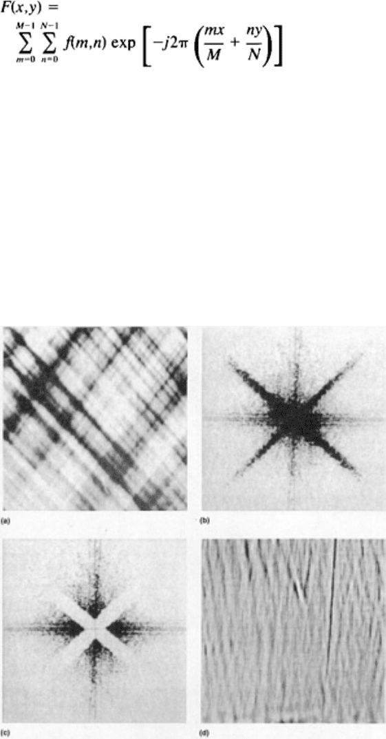
(Eq 9)
Specific methods for calculating Eq 9 are given in Ref 6 and 17.
The most direct uses of the FFT and inverse FFT are as filters to eliminate periodic noise from an image. The noise may
be a periodic pattern, as in the case of the filament-wound vessel shown in Fig. 10(a), or it could be 60-cycle noise. The
general filter procedure is shown in Fig. 10. The image is simply transformed, edited, and inverse transformed. In Fig.
10(a), the original radiograph shows a section of a filament-wound vessel with a cut in the windings. After the transform,
the signal from the filament-wound structure is seen to radiate from the center at 45° (Fig. 10b). The filament-wound
structure is removed from the image by removing the frequency components of the structure as shown in Fig. 10(c). As
shown in Fig. 10(c) the zero frequency component (center pixel) is left intact to restore the average gray-level value. The
image is then inverse transformed to recreate the original image minus the 45° filament pattern. Figure 10(d) shows the
pattern after the inverse transform; the cut section is readily apparent.
Fig. 10
General procedure used to filter an image with the FFT. (a) Original image. (b) Image transformed to
the frequency domain. (c) Image edited. (d) Image inverse transformed into the filtered image
A correlation function can be used directly to detect objects or features of an image that are of interest or to align
images for combination. If two images that are generated from different modalities are to be combined and a registration
mark is not present, the two images can be correlated over a region of interest and moved until the correlation coefficient
peaks. This is best accomplished in images having edges or texture, such as those shown in Fig. 4 and 7.
The edge detection filters described in the section "Image Operations" in this article are often used to detect or
outline images; in particular, the Sobel filter works well on high-definition or noisy edges. In radiographs having noisy,
fuzzy, and low-contrast edges, the Laplacian, Roberts, Sobel, and simple convolution filters will usually not produce good
results. Edge followers, Hough transforms, and other ad hoc algorithms that depend on knowledge of the rough shape of

the curve (template matching) to be outlined work satisfactorily in most cases. These algorithms, although of some use,
are usually not part of a processing package because of their specialized applications.
Advanced Functions. General formulations of image restoration functions such as motion restoration, deblurring,
maximum entropy, and pattern recognition are not commonly found in general image-processing software packages,
probably because of the computation required and the limited demand for these applications. These algorithms are
discussed in Ref 3 (motion restoration), Ref 1 and 3 (deblurring), Ref 9 (maximum entropy), and Ref 1 and 10 (pattern
recognition). The algorithms are of use in the restoration of flash radiographs or high-speed video as well as the
correction of point-spread function in tomography or ultrasonics. Maximum entropy methods may be of use in the
enhancement of NMR signals and other low signal-to-noise data, while pattern recognition can be of use in automating
the detection of flawed, incomplete, or misassembled parts.
References cited in this section
1. W.K. Pratt, Digital Image Processing, John Wiley & Sons, 1978
3. R.C. Gonzalez and P. Wintz, Digital Image Processing, Addison-Wesley, 1977
6. H.K. Huang, Element of Digital Radiology: A Professional Handbook and Guide, Prentice-Hall, 1987
9. B.R. Frieden, Image Enhancement and Restoration, in Picture Processing and Digital Filtering,
Vol 6,
Topics in Applied Physics, T.S. Huang, Ed., Springer-Verlag, 1979, p 177
10.
D.H. Janney and R.P. Kruger, Digital Image Analysis Applied to Industrial Nondestructive Evaluation and
Automated Parts Assembly, Int. Adv. Nondestr. Test., Vol 6, 1979, p 39-93
17.
C.S. Burrus and T.W. Parks, DFT/FFT and Convolution Algorithms: Theory and Implementation,
John
Wiley & Sons, 1985
Digital Image Enhancement
T.N. Claytor and M.H. Jones, Los Alamos National Laboratory
Image Display
A wide variety of devices and techniques are available for the display and output of the processed data.
CRT Displays. The CRT display is the standard means of output for all imaging applications because the range of
colors, brightness, and dynamic range cannot be matched by any other display technology. Black-and-white monitors can
have resolutions of up to 2000 × 1500 pixels, bandwidths of the order of 200 MHz, and a brightness of 515 cd/m
2
(150 ft-
L) at a dynamic range in excess of 256. Color monitors may have 1280 × 1024 pixels and bandwidths of 250 MHz. High-
resolution color monitors have a brightness of 85 cd/m
2
(25 ft-L) or more, with a dynamic range of 256 when viewed in
dim light.
Recent advances in tube design have darkened the shadow mask by a factor of four to allow increased room illumination
with no degradation in dynamic range. The only other major advance in CRT technology is the recent introduction of flat-
screen tubes with resolutions up to 640 × 480 pixels.
The standards for video output are the RS-170, which specifies 525 lines of interlaced video at a frame rate of 30 Hz, and
RS-343-A, which specifies 1023 interlaced lines also at a 30-Hz frame rate. Most modern monitors have special circuitry
that enables the monitor to sync to various standards with either the sync superimposed on the green signal or as a
separate input. Most high-resolution (1280 × 1024) imaging systems use a 483 mm (19 in.) tube operating in a
noninterlaced mode at 60 to 64 Hz for the primary display.
Printers. Various color and black-and-white printers are available for hard copy. A simple method of hard copy for
color and black-and-white is to photograph the CRT screen or a small (80 mm, or 3 in.) very flat screen directly with

an 8 × 11 in. or smaller Polaroid-type camera. The disadvantages of this method are the cost of the instant film in the
large format and the small size of the image in the 4 × 5 in. format. However, the color quality of the image is excellent.
The two other types of color printers for small-system color output are ink-jet printers and thermal-transfer printers. The
ink-jet printers are very inexpensive and produce vivid colors at 180 dpi on paper, but have poor contrast on transparency
material. The thermal copiers are more expensive but produce high-resolution copies (300 dpi) by transferring color from
a Mylar sheet onto paper or film with very good color and color density. The Polaroid cameras can reproduce all the
colors seen on the screen, while the ink-jet and thermal-transfer printers will produce at least 162 colors at 150 or 90 dpi,
with 2 × 2 pixel enlargement.
Table 5 lists the possible color and black-and-white hard copy output devices suitable for printing images. Impact printers
and plotters are not listed, because of the limited number of colors and poor resolution. Also not listed in Table 5 are the
color printers based on the Xerox process.
Table 5 Typical characteristics of color and black-and-white hard copy output devices
Image quality
Device Resolution,
dpi
Paper
Film
Color
Camera . . . Excellent
Very good
Thermal transfer
300 Very good
Very good
Ink-jet 180 Good
Fair
Black and white
Camera . . . Excellent
Very good
Laser film 300 Very good
Very good
Thermal paper 300 Good
. . .
Thermal transfer
300 Fair
Fair
Laser printer 300 Poor
Poor
Ink-jet 180 Poor Very poor
There are many more black-and-white printers available than color; however, the high-quality printers are restricted to the
camera type and the laser film printer. The laser film printer writes with a modulated laser directly on a special dry silver
paper or film that is then developed with heat. These printers are capable of producing 64 shades of gray on paper and 128
shades on film at 300 dpi with very dense blacks. The other devices listed in Table 5 have poor resolution or low contrast.
The other black-and-white printers produce poor plots because they are binary printers (a pixel may be black or white) at
300 dpi.
To achieve a gray scale, the printer must use a 2 × 2, 3 × 3 or other super pixel size, and this decreases the effective
resolution. If a 2 × 2 pixel is used, the printer can print only five shades of gray at 150 dpi; if 3 × 3 pixels are used (100
dpi), ten shades are possible. The printers can actually produce many more shades by programming the device driver for a
super pixel dithering. In this case, the program looks at a large super pixel of 5 × 5, determines whether or not the pixel
should be smaller, and adjusts the dot density with a dither so that, effectively, about 26 or more shades of gray are
perceived with a resolution of about 100 dpi.
Videotape and Videodisk. A common use of videotape is to input TV frames from a dynamic test to the image-
processing system. For example, a high-speed video can be made of a pressure vessel as it bursts. Later, the sequential
frames are captured and enhanced to determine the shape of the initiation crack or the speed of crack growth. Conversely,
radiographs can be made periodically during the slow evolution of a part under the influence of some external variable
(for example, void coalescence in ceramics as a function of temperature). The radiographs are then digitized and
interpolated to form a time-compressed video.
Videodisks can be made in a write once read many format with relatively inexpensive machines. Videodisks offer better
resolution and more convenient frame access than videotape. Another alternative for the display of dynamic data is to use
the hard disk in the imaging workstation. Hard disk transfer rates (300 kbytes/s) allow small (200 × 200) movies to be
shown on a workstation screen for tens of seconds (depends on disk size) at seven frames per second. Ultimately, it will
be possible to expand compressed data quickly enough so that it can be read off a hard disk or a removable digital optical
disk in near real time for significant durations.
The processing of tape can be tedious because of the amount of data required for a few seconds of video and also because
of the limited resolution. However, the techniques of video data display are a very powerful enhancement tool because of
the attraction of the eye to changing features.
Pseudo Three-Dimensional Images. All discussion of output data display has been in terms of a flat two-
dimensional representation. This is the display of preference for processing because the algorithms are available or easily
coded. However, depending on the data and the method of acquisition, other types of displays may be more appropriate.
Shown in Fig. 10(a) and (b) in the article "Use of Color for NDE" in this Volume are two ways to present two-
dimensional data in three dimensions. In Fig. 10(a), a two-dimensional ultrasonic microscopy image of a circuit board is
shown with two defective metallic conductor pads (Ref 18). The image lines are drawn, as in the terrain-mapping method,
with high values elevated with respect to low values and with a hidden line algorithm. Another way to present two-
dimensional image data is shown in Fig. 10(b). In this case, the two-dimensional ultrasonic data were taken from a C-scan
of a filament-wound sphere with Teflon shims imbedded in the matrix (Ref 19). Presenting the two-dimensional data in
this manner results in a realistic display and facilitates interpretation.
Tomographic, ultrasonic, and NMR data sets may consist of many two-dimensional images that can be stacked to yield a
picture of a true three-dimensional object. There are two ways to represent this three-dimensional data. In the first
method, the data are modeled as a geometric solid or surface; in the second method, the data are displayed as raw data, or
voxels (volume elements). The first method is used in solids-modeling workstations for design engineering. It can also be
used to image NDE data, especially if the number of modeled elements is very large (10
5
or more) or has many surfaces.
Figure 11 shows a three-dimensional reconstruction of a bundle of bent fuel rods after a reactor accident test. The image
was constructed of 250 individual 128 × 128 pixel tomograms. A type of surface modeling that treats surfaces as
polygons was used to model the rods. An inspection of the original tomograms shows that it is impossible to quickly
grasp the shape of the bent rods from simple two-dimensional plots (Ref 20). Another three-dimensional reconstruction of
this failed assembly can be found in Fig. 9 in the article "Use of Color for NDE" in this Volume.
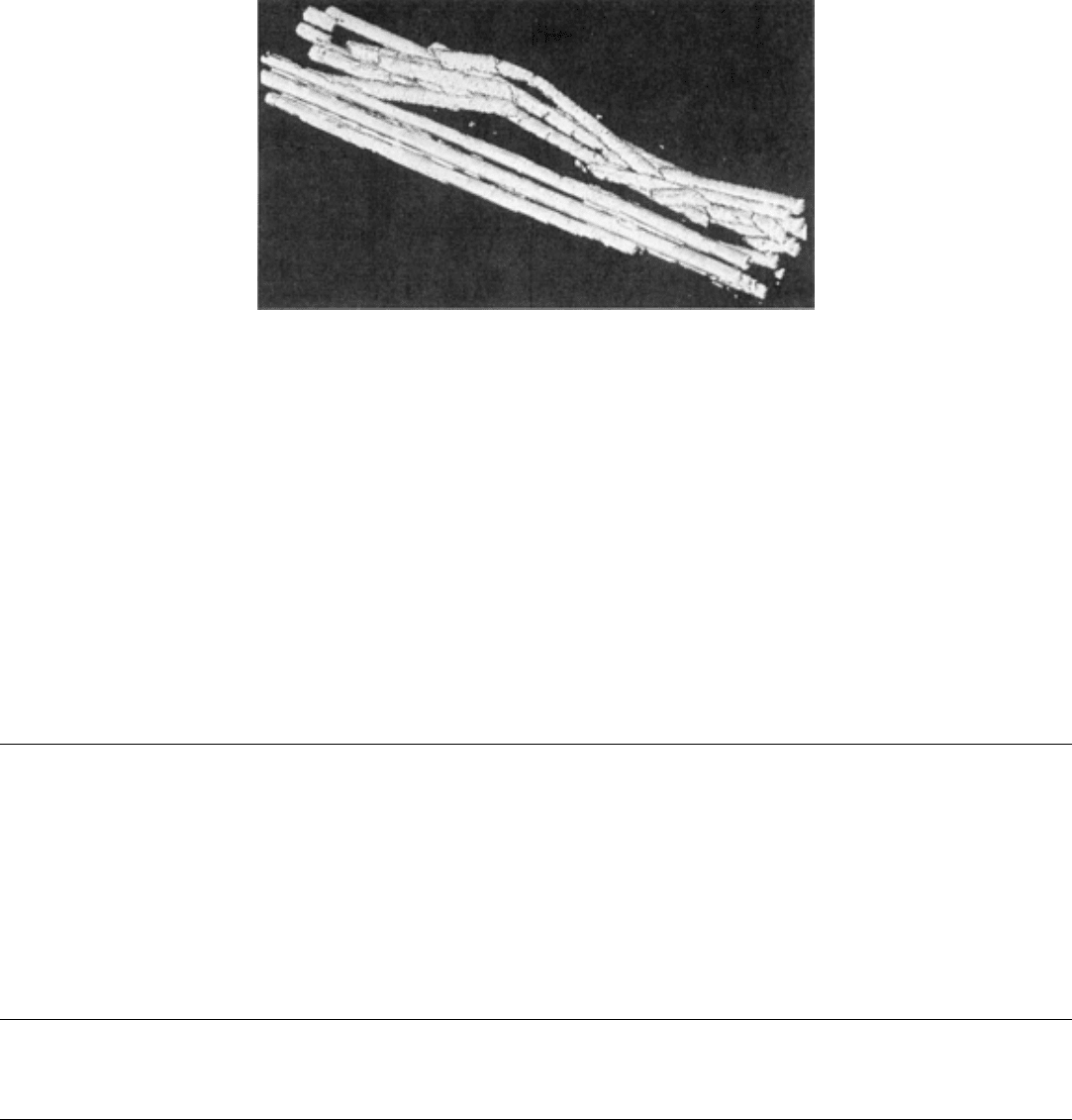
Fig. 11 Three-
dimensional perspective plot representation of a bundle of failed, bent fuel rods. The
representation was reconstructed from three-dimension
al data sets obtained from a series of 250 individual 128
× 128 pixel tomographic slices of the rod bundle.
Figure 8 in the article "Use of Color for NDE" shows a tomogram of a turbine blade that was displayed in terms of voxel
imaging (also known as volume rendering), rather than modeling the surfaces by polygons or other shapes. Volume
elements are created from the raw data, and these voxels are then projected to a view surface to produce an image. The
raw data are not actually displayed. Figure 8 also demonstrates how the surfaces can be made transparent and the lighting
model adjusted for a realistic effect. Graphics accelerators are available so that the images shown in Fig. 8 can be rotated,
lighting sources adjusted, and textures added in almost real time (see Fig. 7 of the article "Use of Color for NDE" in this
Volume). Three-dimensional image processing is in its infancy, but many medical and NDE applications are envisioned
(Ref 21).
References cited in this section
18.
J. Gieske, private communication, Sandia National Laboratories, 1988
19.
W.D. Brosey, Ultrasonic Analysis of Spherical Composite Test Specimens, Compos. Sci. Technol.,
Vol 24,
1985, p 161-178; private communication, Sandia National Laboratories, 1988
20.
C. Little, private communication, Sandia National Laboratories, 1988
21.
A.R. Smith, Geometry and Imaging: Two Distinct Kinds of Graphics,
to be published 1989; private
communication
Digital Image Enhancement
T.N. Claytor and M.H. Jones, Los Alamos National Laboratory
References
1. W.K. Pratt, Digital Image Processing, John Wiley & Sons, 1978
2. M.H. Jacoby, Image Data Analysis, in Radiography & Radiation Testing, Vol 3, 2nd ed.,
Nondestructive
Testing Handbook, American Society for Nondestructive Testing, 1985
3. R.C. Gonzalez and P. Wintz, Digital Image Processing, Addison-Wesley, 1977
4. G.A. Baxes, Digital Image Processing: A Practical Primer, Prentice-Hall, 1984
5. W. B. Green, Digital Image Processing: A Systems Approach, Van Nostrand Reinhold, 1983
6. H.K. Huang, Element of Digital Radiology: A Professional Handbook and Guide, Prentice-Hall, 1987

7. A. Rosenfeld and A.A. Kak, Digital Picture Processing, Academic Press, 1982
8. K.R. Castleman, Digital Image Processing, Prentice-Hall, 1979
9. B.R. Frieden, Image Enhancement and Restoration, in Picture Processing and Digital Filtering,
Vol 6,
Topics in Applied Physics, T.S. Huang, Ed., Springer-Verlag, 1979, p 177
10.
D.H. Janney and R.P. Kruger, Digital Image Analysis Applied to Industrial Nondestructive Evaluation and
Automated Parts Assembly, Int. Adv. Nondestr. Test., Vol 6, 1979, p 39-93
11. J.J. Dongarra
, "Performance of Various Computers Using Standard Linear Equations Software in a Fortran
Environment," Technical Memorandum 23, Argonne National Laboratory, 1989
12. J.R. Janesick, T. Elliott, S. Collins, M.M. Blouke, and J. Freeman, Scientific Charge Co
upled Devices,
Opt. Eng., Vol 26 (No. 8), 1987, p 692-714
13. I.P. Csorba, Image Tubes, Howard W. Sams & Co., 1985
14.
G.I. Yates, S.A. Jaramillo, V.H. Holmes, and J.P. Black, "Characterization of New FPS Vidicons for
Scientific Imaging Applications," LA-11035-MS, US-37, Los Alamos National Laboratory, 1988
15. L.E. Rovich, Imaging Processes and Materials, Van Nostrand Reinhold, 1989
16. K. Thompson, private communication, Sandia National Laboratories, 1988
17. C.S. Burrus and T.W. Parks, DFT/FFT and Convolution Algorithms: Theory and Implementation,
John
Wiley & Sons, 1985
18. J. Gieske, private communication, Sandia National Laboratories, 1988
19. W.D. Brosey, Ultrasonic Analysis of Spherical Composite Test Specimens, Compos. Sci. Technol.,
Vol 24,
1985, p 161-178; private communication, Sandia National Laboratories, 1988
20. C. Little, private communication, Sandia National Laboratories, 1988
21. A.R. Smith, Geometry and Imaging: Two Distinct Kinds of Graphics,
to be published 1989; private
communication
Acoustic Microscopy
Lawrence W. Kessler, Sonoscan, Inc.
Introduction
ACOUSTIC MICROSCOPY is the general term applied to high-resolution, high-frequency ultrasonic inspection
techniques that produce images of features beneath the surface of a sample. Because ultrasonic energy requires continuity
of materials to propagate, internal defects such as voids, inclusions, delaminations, and cracks interfere with the
transmission and/or reflection of ultrasound signals. Compared to conventional ultrasound imaging techniques, which
operate in the 1 to 10 MHz frequency range, acoustic microscopes operate up to and beyond 1 GHz, where the
wavelength is very short and the resolution correspondingly high. In the early stages of acoustic microscopy development,
it was envisioned that the highest frequencies would dominate the applications. However, because of the high-attenuation
properties of materials, the lower frequency range of 10 to 100 MHz is extensively used. Acoustic microscopy is
recognized as a valuable tool for nondestructive inspection and materials characterization. Acoustic microscopy
comprises three different methods:
• Scanning laser acoustic microscopy (SLAM), which was first discussed in the literature in 1970 (Ref 1)
• C-mode scanning acoustic microscopy (C-SAM), which is the improved version of the C-
scan
instrumentation (Ref 2)
• Scanning acoustic microscopy (SAM), which was first discussed in the literature in 1974 (Ref 3)
Each of these methods has a specific range of utility, and most often the methods are noncompetitive with regard to
applications. That is, only one method will be best suited to a particular inspection problem.
