ASM Metals HandBook Vol. 17 - Nondestructive Evaluation and Quality Control
Подождите немного. Документ загружается.


of photon counting when signal levels are low. Gas ionization detectors have low detection efficiencies but better long-
term stability than scintillator- and phosphor-photodetector arrays.
A typical phosphor-photodetector array for the radiographic inspection of welds consists of 1024 pixel elements with 25
m (1 mil) spacing, covering 25 mm (1 in.) in length perpendicular weld seam. The linear photodiode array is covered
with a fiber-optic faceplate and can be cooled in order to reduce noise. For the conversion of the x-rays to visible light,
fluorescent screens are coupled to the array by means of the fiber optics. A linear collimator parallel to the array is
arranged in front of the screen. The resolution perpendicular to the array is defined by the width of this slit and the speed
of the manipulator. A second, single-element detector is provided to detect instabilities of the x-ray beam. Using 100-kV
radiation, a spatial resolution of 0.1 mm (0.004 in.) can be achieved with a scanning speed of 1 to 10 mm/s (0.04 to 0.4
in./s).
The data from the detector system are digitized and then stored in a fast dual-ported memory. This permits quasi-
simultaneous access to the data during acquisition. Before the image is stored in the frame buffer and displayed on the
monitor, simple preprocessing can be done, such as intensity correction of the x-ray tube by the data of the second
detector and correction of the sensitivity for different array elements. If further image processing or automatic defect
evaluation is required, the system can be equipped with fast image-processing hardware. All standard devices for digital
storage can be utilized.
Image Processing. Because real-time systems generally do not provide the same level of image quality and contrast as
radiographic film, image processing is often used to enhance the images from image intensifiers, fluorescent screens, and
detector arrays. With image processing, the video images from real-time systems can compete with the image quality of
film radiography. Moreover, image processing also increases the dynamic range of real-time systems beyond that of film
(which typically has a dynamic range of about 1000:1).
Images can be processed in two ways: as an analog video signal and as a digitized signal. An example of analog
processing is to shade the image after the signal leaves the camera. Shading compensates for irregularities in brightness
across the video image due to thickness or density variations in the testpiece. This increases the dynamic range (or
latitude) of the system, which allows the inspection of parts having larger variations in thickness and density.
After analog image processing, the images can be digitized for further image enhancement. This digitization of the signal
may involve some detector requirements. In all real-time radiological applications, the images have to be obtained at low
dose rates (around 20 R/s). In digital x-ray imaging, however, the most often used dose is around 1 mR, in order to
reduce the signal fluctuations that would result from a weak x-ray flux. This means that only a short exposure time (of the
order of a few milliseconds) and a frequency of several images can be used if kinetic (motion) blurring is to be avoided.
The resulting requirements for the x-ray detector are therefore:
• The capability of operating properly in a pulsed mode, which calls for a fast temporal response
• An excellent linearity, to allow the use of the simplest and most efficient form of signal processing
• A wide operating dynamic range in terms of dose output (around 2000:1)
Once the image from the video camera has been digitized in the image processor, a variety of processing techniques can
be implemented. The image-processing techniques may range from the relatively simple operation of frame integration to
more complex operations such as automatic defect evaluation. The following discussions briefly describe three image-
processing operations. More detailed information on image processing is available in the article "Digital Image
Enhancement" in this Volume.
Frame integration (or summing) is a simple technique that can improve the signal-to-noise ratio and decrease (or
eliminate) the screen mottle associated with low levels of radiation intensity. This technique adds the digital images from
a number of frames and displays the result on a video monitor. Frame integration can be done in real time, that is, at
standard television frequency of 30 frames per second. The number of television frames that can be integrated depends on
the number of frame buffers available for this procedure. Typical summation times range from a few seconds for
applications using x-rays with high intensities up to 1 min or more for applications using radiation from low-activity
radionuclide sources. However, these times are small compared to the time required for exposing and developing x-ray
film.
Image subtraction can be used to improve sensitivity through the subtraction of one integrated image from a second
integrated image. The advantage of the subtraction technique is that all areas of the scene having the same brightness
cancel each other to produce an extremely flat or uniformly gray image. With such an image available, the contrast and
brightness can then be electronically enhanced to reveal details not visible in either a real-time or a simple integrated
image. Example 1 in this article describes one method called pairs subtraction.
Automated defect evaluation systems have been developed for the radiographic inspection of welds (Ref 5). The
automatic detection of defects with such systems is based on a mathematical algorithm that evaluates a set of parameters
for each indication and compares this set to a catalog of known defects. For any particular problem, the inspection
parameters must be selected to provide optimum and reproducible results. To compare different radiographs and to extract
features such as the depth of a defect, it is necessary to calibrate the gray levels against material thickness and to calibrate
the linear dimensions. Once established, the optimized conditions must be consistently used for all inspections.
Figure 31 shows a flow chart of an automatic image evaluation procedure, and Fig. 32 shows the images from different
steps in the procedure. The first step is to remove irrelevant details from the digitized image. This can be done by
subtracting a background image. This background image is generated by creating an unsharp image with a low-pass filter.
Although radiographs usually have a center brightness that differs from that at the edges, this gradient is eliminated by
subtracting the background image from the original image. At this state (Fig. 32b), it is possible to set a threshold and to
identify as possible defects all pixels whose gray levels exceed this threshold.
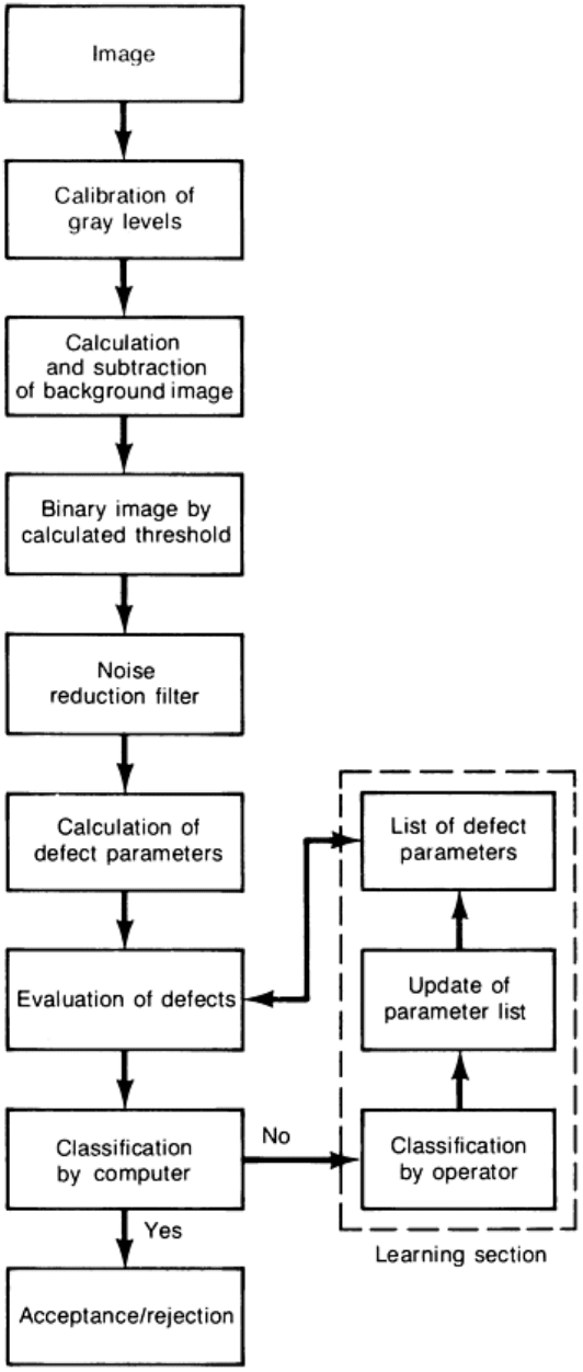
Fig. 31 Flow chart of an automatic image evaluation procedure
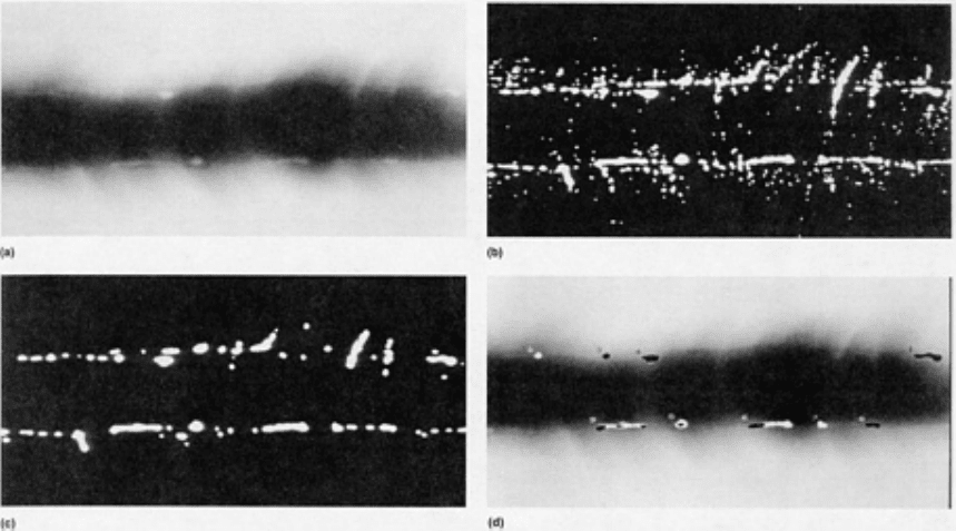
Fig. 32 Images developed by an image-processing procedure that identifies and evalu
ates the images of weld
defects in real-
time radiographs. (a) Original image. (b) Binary image. (c) Binary image after noise reduction.
(d) Original image with defects identified. Courtesy of RTS Technology, Inc
In the next step, these potential defects are examined. First, all very small indications are examined, such as single pixels
or groups of only a few pixels, which result from image noise. Larger details, which are obviously separated by one or
two missing pixels, are connected (Fig. 32c). This algorithm prevents the computer from spending time analyzing noise
that exceeds the threshold.
For each potential defect, a number of parameters are evaluated. These parameters include position, orientation, and a set
of descriptors for size and shape. The values of these parameters are compared with the mean value and standard
deviation for each parameter in the catalog data already stored. Where some consistency is found between measured and
stored data, the program identifies the defects (Fig. 32d) and calculates the probability that the indication is a particular
type of defect. Where there is insufficient consistency between the measured and stored data, the program calls for the
assistance of an interpreter and enters a learn mode. Once the interpreter has evaluated the defect, the program stores the
values for each parameter of the defect, along with the evaluation in the catalog. This information is now considered in
future automated evaluations. Relevant defects cause the rejection of the workpiece.
Applications. Real-time radiography is often applied to objects on assembly lines for rapid inspection. Digital
radiography with discrete detectors, in particular, is being more widely used because it provides an unlimited field of
view and the largest dynamic range of all the radiographic methods. Because a larger dynamic range allows the inspection
of parts having a wider range of thicknesses, digital radiography has found application in the inspection of castings (see
the article "Castings" in this Volume).
Radiography is also an accepted method of detecting internal discontinuities in welds. Image intensifiers are the most
widely used system for the real-time radiographic inspection of welds, although digital radiography with discrete
detectors is also used. Projective magnification with microfocus x-ray sources is also useful in weld and diffusion bond
inspection.
Advantages and Limitations. Real-time radiography provides immediate information without the delay and expense
of film development. Real-time systems also allow the manipulation of the testpiece during inspection and the
improvement of resolution and dynamic range by digital image enhancement. Real-time radiography with discrete
detectors (that is, digital radiography) also provides good scatter rejection.
Nevertheless, film radiography is a simpler technique that represents less of a capital investment. The sensitivity and
resolution of real-time systems are usually not as good as those of film, although real-time radiography can approach the
sensitivity and resolution of film radiography when good inspection techniques are used. Good technique is often more
important than the details of the imaging system itself.
Example 1: Design of a Rapid, High-Sensitivity Real-Time Radiographic System.
A system was needed for the real-time radiographic inspection of jet-engine turbine blades. These were critical nickel
alloy castings that required inspection to at least a 2-1T (1.4%) penetrameter sensitivity level through thicknesses that
ranged from about 0.5 to more than 13 mm (0.02 to 0.5 in.). The volume of parts produced necessitated inspection at a
rate of 60 parts per hour or more. For consistency, all aspects of the inspection were to be computer controlled. In
addition, it was necessary to visually compare the actual parts with their radiographic images to separate castings with
excess metal due to mold flaws that might be acceptable from castings with high-density inclusions that might be
rejectable.
Image Processing. A preliminary evaluation of the parts indicated that the 2-1T penetrameter sensitivity requirement
could be met with suitable image-processing techniques. Both analog and digital processing were used for these complex
parts.
The analog processing technique used was an electronic adjustment called shading. Shading is performed in the camera
control unit after the signal leaves the camera and before it is sent to the image processor for digitizing and further
processing. Shading compensates for variations in brightness across the video image due to thickness variations in the
part. In effect, proper shading adjustment increases the latitude (dynamic range) of the image. In the case of the airfoils to
be inspected with the real-time radiographic system, shading permitted blade thicknesses ranging from 0.5 to 4 mm (0.02
to 0.16 in.) to be adequately imaged in a single view.
Once the image from the video camera had been digitized in the image processor, a great many processing techniques
were implemented. The simplest was to integrate the image. This technique added the digital images from a number of
frames and displayed the result on the video monitor. Integration improved the signal-to-noise ratio over that of a single
frame from the video camera. For these castings, 128 frames of integration produced good results.
To achieve the required sensitivity, an image subtraction technique was also used. This was an extremely useful
processing technique in which one integrated image was subtracted from a second integrated image. There are a number
of variations of this technique. It was experimentally determined that the best technique for this application was the one
termed "pairs subtraction." To produce a "pairs subtraction," the part was displaced slightly between the two integrated
images that were subsequently subtracted from each other.
X-Ray Source. Because of the range of thicknesses to be inspected, two x-ray sources were selected for the system--one
rated at 160 kV for the thinner sections and the other at 320 kV for the thicker sections. The use of 0.4 mm (0.015 in.)
focal spots for the 160-kV unit and 0.8 mm (0.03 in.) focal spots for the 320-kV unit provided sufficient resolution to
detect 0.25 mm (0.01 in.) defects even at the relatively short source-to-screen distances available in the final real-time
radiographic system design. Because it was required that the blade be exposed to only one source of radiation at a time
and that the x-ray tubes be run continuously, two computer-controlled, high-speed lead shutters were designed to block
the unwanted radiation.
Imaging System. The imaging camera, which was housed in its own shielded enclosure, included a 100 × 100 mm (4
× 4 in.) rare-earth fluorescent screen for converting x-rays to visible light. Light from the screen was directed to the
objective lens of an image isocon camera by a 45° front surface mirror (Fig. 33). An isocon camera was selected over an
image intensifier for two reasons. First, an isocon camera is much less susceptible to blooming due to bypass radiation.
This permitted the use of relatively loose masking around the part. Second, computer-selectable shading channels were
available with the isocon camera; therefore, shading could be preadjusted for different views. The computer program
could then set the required shading channel during an inspection. The camera system was designed to include remote
controls to facilitate focusing of the camera and adjustment of the field of view from the full-screen size down to a 25-mm
(1-in.) square to increase the optical magnification if necessary.
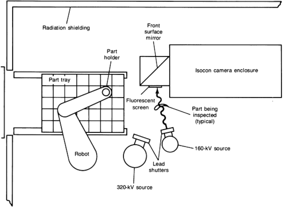
Fig. 33 Schematic of the equipment described in Example 1
Part Handling. To minimize part-handling time, a very accurate, high-speed, commercially available robot with four
axes of movement (X, Y, Z, and rotation) was selected. The robot was equipped with a computer and sophisticated
operating programs. After the robot was taught a few fixed points, it determined its own path from one position to
another. This was especially convenient because a large number of parts were to be placed into the real-time radiographic
system in a tray. The robot only needed to be told the number of rows and columns in the tray and taught three corner
pickup locations. From this information, it could calculate the exact pickup points for all other locations in the tray.
The part containers required considerable development before consistent robot pickup was achieved. In the final design,
parts were dropped into molded plastic holders with no special care, and they were positioned well enough for the robot to
pick them up with a high degree of reliability.
System Layout and Operating Console. To permit installation in a factory, the x-ray sources, imaging camera, and
robot were located in an exempt, radiation-shielded enclosure. Trays of parts to be inspected were inserted through a
sliding access door from an adjacent environmental enclosure.
An environmental enclosure was included for the operator's station to provide a relatively clean environment in the
manufacturing area. The inspection operator's station was designed to include a desktop console with a video monitor on
which either real-time or processed images could be viewed. The console also included a computer terminal and
keyboard; a joystick control was used for performing various image processor functions and for manual control of the
robot. The x-ray controls, the robot controller, the isocon camera controls, and the computer and image processor were
located near the console.
A console for reviewing images while examining parts visually was also provided in the environmental enclosure. In
addition to the video monitor, image processor, and computer terminal, a voice recognition system was added so that
commands and information could be input to the computer without using the keyboard.
System Software. Three separate programs were developed for the real-time radiographic system: Inspect, Review,
and Supervisor. The computer selected included a multiuser operating system to allow the Inspect and Review programs
to be run concurrently at different terminals. All the programs were menu driven; the operator simply entered information
or exercised options when prompted to do so by the menu on the computer terminal.
The Inspect program was designed for day to day inspection operation. In this program, a preestablished scan plan
controlled all the parameters required for an inspection. As long as the operator continued accepting views, the program
moved through the plan for each part in turn until all parts in the tray had been completed.
In running the scan plan, the computer interfaced with the x-ray machines to control the kilovolt and milliamp settings,
the shutters, and x-ray on and off. It also interfaced with the isocon camera control unit to turn the camera beam on and
off and to call the required shading channel, and it interfaced with the robot controller to direct the part movements.
The software was designed so that the scan plan could be interrupted at any time. One option in the interrupt mode gave
the operator complete manual control of all system functions through the terminal keyboard. The ability to call an image
quality indicator (IQI) to check for proper radiographic sensitivity was also designed into the interrupt mode. When the
IQI was called for, the part was returned to its tray position, and the IQI was picked up and moved into the x-ray beam.
An image of the IQI was generated using the same x-ray parameters and image processing as employed for the part view
being inspected at the time.
The Review program was designed to allow stored images to be recalled when the parts were available for visual review.
In addition to accessing images, this program allowed various enhancement techniques to be performed on the image. All
program functions could be exercised using the voice recognition capability so that the operator's hands were free to
handle parts. Once a final disposition for a part was entered, the views for that part were automatically deleted from
memory.
The Supervisor program was written to include a number of program functions that were to be protected from normal
operator access. These included changing the lists of authorized users and their passwords, access to an operations log,
and creation or modification of scan plans.
System Performance. The sensitivity of the system was demonstrated with plaque penetrameters, a wire
penetrameter, and an actual part with small holes. The plaque penetrameter sensitivity of the system is shown in Fig. 34.
Figure 34(a) shows a pairs-subtracted image of a 0.025 mm (0.001 in.) thick penetrameter through 1.25 mm (0.05 in.) of
material, and Fig. 34(b) shows a 0.125 mm (0.005 in.) penetrameter through 6.4 mm (0.25 in.) of material. All three holes
can be seen near the centers of the circles as adjacent white and black areas that result from the pairs subtraction
technique.
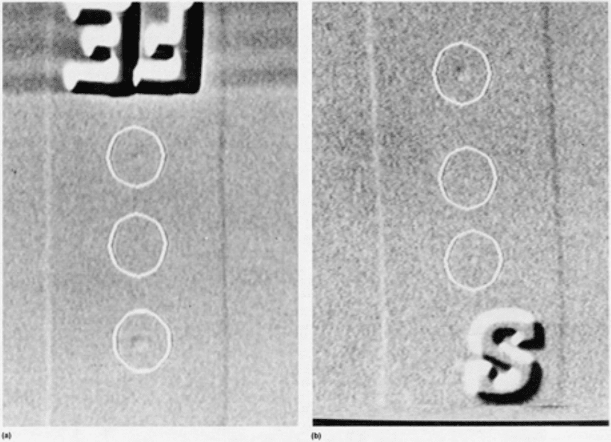
Fig. 34
Examples of plaque penetrameter indications. (a) 0.025 mm (0.001 in.) thick penetrameter on a
testpiece with a thickness of 1.3 mm (0.050 in.
). (b) 0.125 mm (0.005 in.) penetrameter on a testpiece with a
thickness of 6.4 mm (0.25 in.)
Figure 35 shows a pairs-subtracted image of an actual part in which a series of holes was drilled using the electrical
discharge machining process. The holes have a nominal diameter of 0.25 mm (0.01 in.) and a depth that is 2% of the
material thickness at the location where they were drilled. The material thickness in this airfoil view ranged from 1 to
approximately 4 mm (0.04 to 0.16 in.).
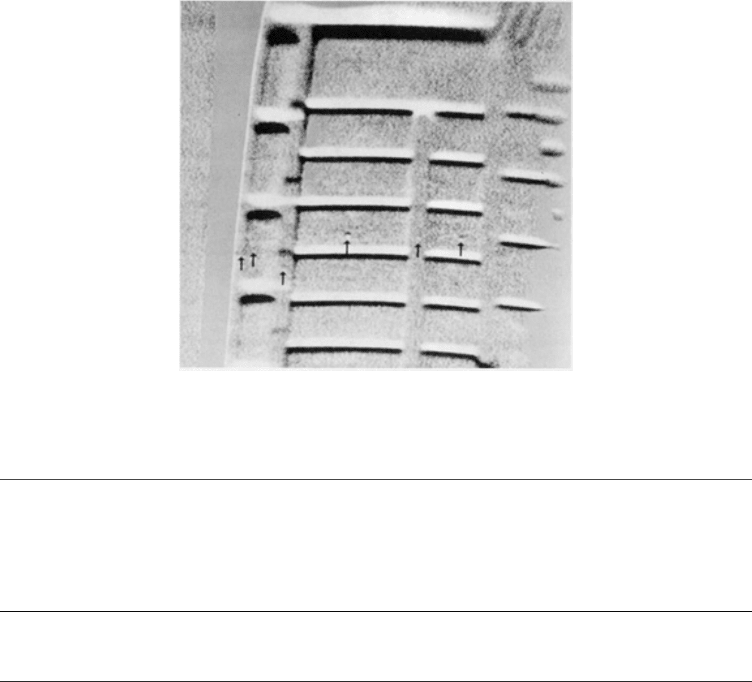
Fig. 35 Radi
ographic image of holes with a nominal diameter of 0.25 mm (0.01 in.) and a depth of 2% of wall
thickness. Holes are indicated with arrows. Courtesy of Lockheed Missiles & Space Company, Inc.
References cited in this section
3.
R. Link, W. Nuding, and K. Sauerwein, Br. J. Non-Destr. Test., Vol 26, 1984, p 291-295
4.
L.H. Brixner, Mater. Chem. Phys., Vol 16, 1987, p 253-281
5.
W. Daum, P. Rose, H. Heidt, and J.H. Builtjes, Br. J. Non-Destr. Test., Vol 29, 1987, p 79-82
Radiographic Inspection
Revised by the ASM Committee on Radiographic Inspection
*
Film Radiography
Film selection and exposure time are two major factors in film radiography. The selection of radiographic film for a
particular application is generally a compromise between the desired quality of the radiograph and the cost of exposure
time. The quality of the radiograph depends mainly on film density, gradient, graininess, and fog, which are functions of
film type and development procedure. Exposure time depends mainly on film speed and on radiation intensity at the film
surface. The factors affecting film selection and exposure time are discussed in more detail in this section.
Characteristics of X-Ray Film
Three general characteristics of film--speed, gradient, and graininess--are primarily responsible for the performance of the
film during exposure and processing and for the quality of the resulting image. Film speed, gradient, and graininess are
interrelated; that is, the faster the film, the larger the graininess and the lower the gradient--and vice versa. Film speed and
gradient are derived from the characteristic curve for a film emulsion, which is a plot of film density versus the exposure
required for producing that density in the processed film. Graininess is an inherent property of the emulsion, but can be
influenced somewhat by the conditions of exposure and development.

There are other characteristics of x-ray film that can be used to produce special effects. However, the following
discussion is mainly confined to film speed, gradient, graininess, and the factors that affect them.
Density. The quantitative measure of the blackening of a photographic emulsion is called density. Density is usually
measured directly with an instrument called a densitometer.
There are two kinds of density:
• Transmission density, which is associated with transparent-base radiographic film
• Reflection density, which is associated with opaque-base imaging material such as radiographic paper
In the following discussion, the unqualified term density will refer to transmission density.
Transmission density is defined by:
(Eq 14)
where D is the density, I
0
is the intensity of light incident on the film, and I
t
is the intensity of light transmitted through
the film. Reflection density is defined by:
(Eq 15)
where D
r
is the reflection density, I
0
is the light intensity incident on the radiographic image, and I
r
is the light intensity
reflected from the image. Although Eq 14 and 15 are similar, the densities are quite different.
The ratio of incident intensity, I
0
, to transmitted intensity, I
t
, in Eq 14 is called opacity. The inverse of this ratio, I
t
/I
0
, is
called transmittance and indicates the fraction of incident light transmitted through the film. The relationship of density to
opacity and transmittance is as tabulated below:
Density, log
(I
0
/I
t
)
Opacity,
I
0
/I
t
Transmittance,
I
t
/I
0
0 1
1.00
0.3 2
0.50
0.6 4
0.25
1.0 10
0.10
2.0 100
0.01
