Voet D., Voet Ju.G. Biochemistry
Подождите немного. Документ загружается.

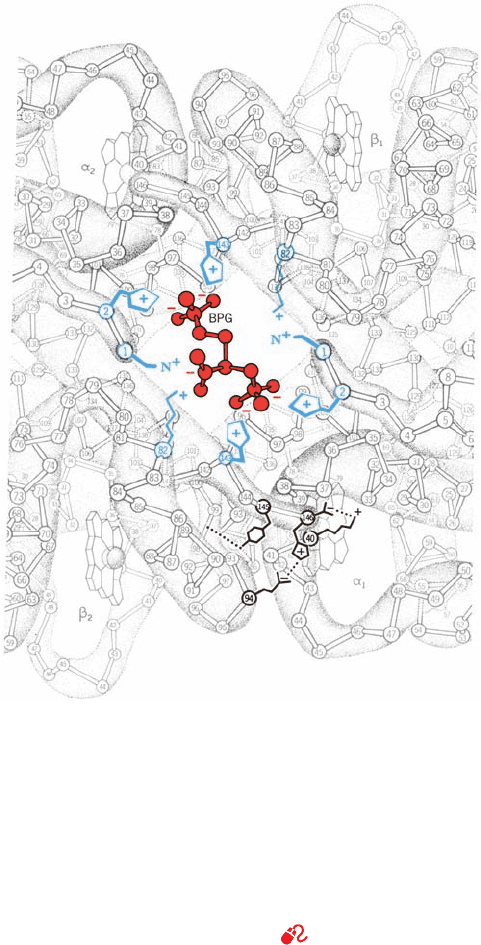
F. Structural Basis of BPG Binding
BPG decreases the oxygen-binding affinity of Hb by prefer-
entially binding to its deoxy state (Section 10-1D). The
binding of the physiologically quadruply charged BPG to
deoxyHb is weakened by high salt concentrations, which
suggests that this association is ionic in character. This ex-
planation is corroborated by the X-ray structure of a BPG–
deoxyHb complex, which indicates that BPG binds in the
central cavity of deoxyHb on its 2-fold axis (Fig. 10-21).The
anionic groups of BPG are within hydrogen bonding and
salt bridging distances of the cationic Lys EF6(82), His
H21(143), His NA2(2), and N-terminal amino groups of
both  subunits (Fig. 10-21). The T S R transformation
brings the two  H helices together, which narrows the cen-
tral cavity (compare Figs. 10-13a and 10-13b) and expels the
BPG. It also widens the distance between the  N-terminal
amino groups from 16 to 20 Å, which prevents their simul-
taneous hydrogen bonding with BPG’s phosphate groups.
BPG therefore stabilizes the T conformation of Hb by cross-
linking its  subunits. This shifts the T 34 R equilibrium to-
ward the T state, which lowers hemoglobin’s O
2
affinity.
The structure of the BPG–deoxyHb complex also indi-
cates why fetal hemoglobin (HbF) has a reduced affinity
for BPG relative to HbA (Section 10-1D).The cationic His
H21(143) of HbA is changed to an uncharged Ser residue
in HbF’s -like ␥ subunit, thereby eliminating a pair of
ionic interactions stabilizing the BPG–deoxyHb complex
(Fig. 10-21).
The excess positive charge lining Hb’s central cavity is
also partially responsible for the allosteric effect of Cl
⫺
ions in stabilizing the T state relative to the R state (the re-
mainder being due to the participation of Cl
⫺
in the T-state
salt bridge networks; Fig. 10-18a). The central cavity is
larger in the T state than in the R state (Fig. 10-13), so that
more Cl
⫺
ions occupy this channel in the T state than in the
R state. The additional Cl
⫺
ions, through electrostatic
shielding, reduce the mutual repulsions of the positive
charges, thereby stabilizing the T state.
G. Role of the Distal Histidine Residue
O
2
binding paradoxically protects the heme iron from au-
tooxidation: The rate of Mb oxidation decreases as the
partial pressure of O
2
increases. This is because heme iron
oxidation is catalyzed by protons that are reduced by the
heme iron and that in turn reduce O
2
in the solvent to su-
peroxide ion (O
2
). Bound O
2
evidently shields the Fe
from the attacking protons.
The mutagenic replacement of the distal His residue in
Mb by any other residue reduces Mb’s oxygen affinity and
increases its rate of autooxidation.Asp, a proton source, at
this position increases the rate of Mb autooxidation by 350-
fold, the largest increase of all residue replacements,
whereas Phe, Met, and Arg provide only 50-fold accelera-
tions, the smallest observed increases.The imidazole ring of
the distal His, which has a pK of 5.5 and is therefore neutral
at neutral pH and whose unprotonated N
ε
atom faces the
heme pocket (Fig. 10-12), acts as a proton trap, thereby
ⴢ
⫺
protecting the Fe from protons. Thus, to quote Perutz,
“Evolution is a brilliant chemist.”
3 ABNORMAL HEMOGLOBINS
Mutant hemoglobins provided the original opportunity to
study structure–function relationships in proteins because
Hb is a readily isolated protein of known structure that has
a large number of well-characterized naturally occurring
variants. The examination of individuals with physiological
disabilities, together with the routine electrophoretic
screening of human blood samples, has led to the discovery
of over 1000 variant hemoglobins, ⬎90% of which result
from single amino acid substitutions in a globin polypeptide
Section 10-3. Abnormal Hemoglobins 341
Figure 10-21 Binding of BPG to deoxyHb. The view is down
the molecule’s exact twofold axis (the same view as in Fig. 10-13a).
BPG (red), with its five anionic groups, binds in the central cavity
of deoxyHb, where it is surrounded by a ring of eight cationic
side chains (blue) extending from the two  subunits. In the R
state, the central cavity is too narrow to admit BPG (Fig. 10-13b).
The arrangement of salt bridges and hydrogen bonds between
the ␣
1
and 
2
subunits that partially stabilizes the T state (Fig.
10-18b) is indicated at the lower right. [Illustration, Irving Geis.
Image from the Irving Geis Collection, Howard Hughes Medical
Institute. Reprinted with permission.]
See Kinemage
Exercise 6-3
JWCL281_c10_323-358.qxd 8/10/10 9:41 AM Page 341
chain (a compendium of variant human hemoglobins is lo-
cated at http://globin.cse.psu.edu/). In this section, we con-
sider the nature of these hemoglobinopathies. Hemoglobin
diseases characterized by defective globin synthesis, the
thalassemias, are the subject of Section 34-2G. It should be
noted that ⬃300,000 individuals with serious hemoglobin
disorders are born every year and that ⬃5% of the world’s
population are carriers of an inherited variant hemoglobin.
A. Molecular Pathology of Hemoglobin
The physiological effect of an amino acid substitution on
Hb can, in most cases, be understood in terms of its molec-
ular location:
1. Changes in surface residues
Changes of surface residues are usually innocuous because
most of these residues have no specific functional role
[although sickle-cell Hb (HbS) is a glaring exception to this
generalization; Section 10-3Ba]. For example, HbE [Glu
B8(26) S Lys], the most common human Hb mutant af-
ter HbS (possessed by up to 10% of the population in parts
of Southeast Asia), has no clinical manifestations in either
heterozygotes or homozygotes. About half of the known
Hb mutations are of this type and have been discovered
only accidentally or through surveys of large populations.
2. Changes in internally located residues
Changing an internal residue often destabilizes the Hb
molecule. The degradation products of these hemoglobins,
particularly those of heme, form granular precipitates
(known as Heinz bodies) that hydrophobically adhere to
the erythrocyte cell membrane. The membrane’s perme-
ability is thereby increased, causing premature cell lysis.
Carriers of unstable hemoglobins therefore suffer from
hemolytic anemia of varying degrees of severity.
The structure of Hb is so delicately balanced that small
structural changes may render it nonfunctional.This can oc-
cur through the weakening of the heme–globin association
or as a consequence of other conformational changes. For in-
stance, the heme group is easily dislodged from its closely fit-
ting hydrophobic binding pocket. This occurs in Hb Ham-
mersmith (Hb variants are often named after the locality of
their discovery), in which Phe CD1(42), an invariant
residue that wedges the heme into its pocket (see Fig.10-12),
is replaced by Ser. The resulting gap permits water to enter
the heme pocket, which causes the hydrophobic heme to
drop out easily (Phe CD1 and the proximal His F8 are the
only invariant residues among all known hemoglobins).Sim-
ilarly, in Hb Bristol, the substitution of Asp for Val E11(67),
which partially occludes the O
2
pocket, places a polar group
in contact with the heme. This weakens the binding of the
heme to the protein, probably by facilitating the access of
water to the subunit’s otherwise hydrophobic interior.
Hb may also be destabilized by the disruption of ele-
ments of its 2°, 3°, and/or 4° structures.The instability of Hb
Bibba results from the substitution of a helix-breaking Pro
for Leu H19(136)␣. Likewise, the instability of Hb Savan-
nah is caused by the substitution of Val for the highly con-
served Gly B6(24), which is located on the B helix where
it crosses the E helix with insufficient clearance for side
chains larger than an H atom (Fig. 10-13). The ␣
1
–
1
con-
tact, which does not significantly dissociate under physio-
logical conditions, may do so on structural alteration. This
occurs in Hb Philly, in which Tyr C1(35), which partici-
pates in the hydrogen bonded network that helps knit to-
gether the ␣
1
–
1
interface, is replaced by Phe.
3. Changes stabilizing methemoglobin
Changes at the O
2
-binding site that stabilize the heme in the
Fe(III) oxidation state eliminate the binding of O
2
to the de-
fective subunits. Such methemoglobins are designated
HbM and individuals carrying them are said to have
methemoglobinemia. These individuals usually have bluish
skin, a condition known as cyanosis, which results from the
presence of deoxyHb in their arterial blood.
All known methemoglobins arise from substitutions that
provide the Fe atom with an anionic oxygen atom ligand. In
Hb Boston, the substitution of Tyr for His E7(58)␣ (the dis-
tal His, which protects the heme from oxidation; Section 10-
2G) results in the formation of a 5-coordinate Fe(III) com-
plex, with the phenolate ion of the mutant Tyr E7 displacing
the imidazole ring of His F8(87) as the apical ligand (Fig.10-
22a). In Hb Milwaukee, the ␥-carboxyl group of the Glu
that replaces Val E11(67) forms an ion pair with a 5-coor-
dinate Fe(III) complex (Fig. 10-22b). Both the phenolate
and glutamate ions in these methemoglobins so stabilize the
Fe(III) oxidation state that methemoglobin reductase is in-
effective in converting them to the Fe(II) form.
Individuals with HbM are alarmingly cyanotic and have
blood that is chocolate brown, even when their normal sub-
units are oxygenated. In northern Japan, this condition is
named “black mouth” and has been known for centuries;it is
caused by the presence of HbM Iwate [His F8(87)␣ S Tyr].
Methemoglobins have Hill constants of ⬃1.2.This indicates
a reduced cooperativity in comparison with HbA even
though HbM, which can bind only two oxygen molecules,
can have a maximum Hill constant of 2 (the unmutated ␣ or
 chains remain functional). Surprisingly, heterozygotes with
HbM, which have an average of one nonfunctional ␣ or 
subunit per Hb molecule, have no apparent physical disabil-
ities. Evidently, the amount of O
2
released in their capillaries
is within normal limits. Homozygotes of HbM, however, are
unknown; this condition is, no doubt, lethal.
4. Changes at the ␣
1
–
2
contact
Changes at the ␣
1
–
2
contact often interfere with hemoglo-
bin’s quaternary structural changes. Most such hemoglobins
have an increased O
2
affinity so that they release less than
normal amounts of O
2
in the tissues. Individuals with such
defects compensate for it by increasing their hematocrit
(concentration of erythrocytes in their blood). This condi-
tion, which is named polycythemia, often gives them a
ruddy complexion. Some amino acid substitutions at the
␣
1
–
2
interface instead result in a reduced O
2
affinity. Indi-
viduals carrying such hemoglobins are cyanotic.
Amino acid substitutions at the ␣
1
–
2
contact may change
the relative stabilities of hemoglobin’s R and T forms,
342 Chapter 10. Hemoglobin: Protein Function in Microcosm
JWCL281_c10_323-358.qxd 2/24/10 1:58 PM Page 342
thereby altering its O
2
affinity. For example, the replacement
of Asp G1(99) by His in Hb Yakima eliminates the hydro-
gen bond at the ␣
1
–
2
contact that stabilizes the T form of Hb
(Fig. 10-17a). The interloping imidazole ring also acts as a
wedge that pushes the subunits apart and displaces them to-
ward the R state.This change shifts the T S R equilibrium al-
most entirely to the R state, which results in Hb Yakima hav-
ing an increased O
2
affinity (p
50
⫽ 12 torr under physiological
conditions vs 26 torr for HbA) and a total lack of cooperativ-
ity (Hill constant ⫽ 1.0). In contrast, the replacement of Asn
G4(102) by Thr in Hb Kansas eliminates the hydrogen bond
in the ␣
1
–
2
contact that stabilizes the R state (Fig.10-17b),so
that this Hb variant remains in the T state on binding O
2
.Hb
Kansas therefore has a low O
2
affinity (p
50
⫽ 70 torr) and a
low cooperativity (Hill constant ⫽ 1.3).
B. Molecular Basis of Sickle-Cell Anemia
Most harmful Hb variants occur in only a few individuals,
in many of whom the mutation apparently originated.
However, ⬃10% of American blacks and as many as 25%
of African blacks are heterozygotes for sickle-cell hemo-
globin (HbS). HbS arises, as we have seen (Section 7-3Aa),
from the substitution of a hydrophobic Val residue for the
hydrophilic surface residue Glu A3(6) (Fig. 10-13). The
prevalence of HbS results from the protection it affords
heterozygotes against malaria. However, homozygotes for
HbS, of which there are ⬃ 50,000 in the United States, are
severely afflicted by hemolytic anemia together with painful,
debilitating, and sometimes fatal blood flow blockages
caused by the irregularly shaped and inflexible erythro-
cytes characteristic of the disease (Fig. 7-19b).
a. HbS Fibers Are Stabilized by Intermolecular
Contacts Involving Val 6 and Other Residues
The sickling of HbS-containing erythrocytes results from
the aggregation (polymerization) of deoxyHbS into rigid
fibers that extend throughout the length of the cell (Fig. 10-23).
Section 10-3. Abnormal Hemoglobins 343
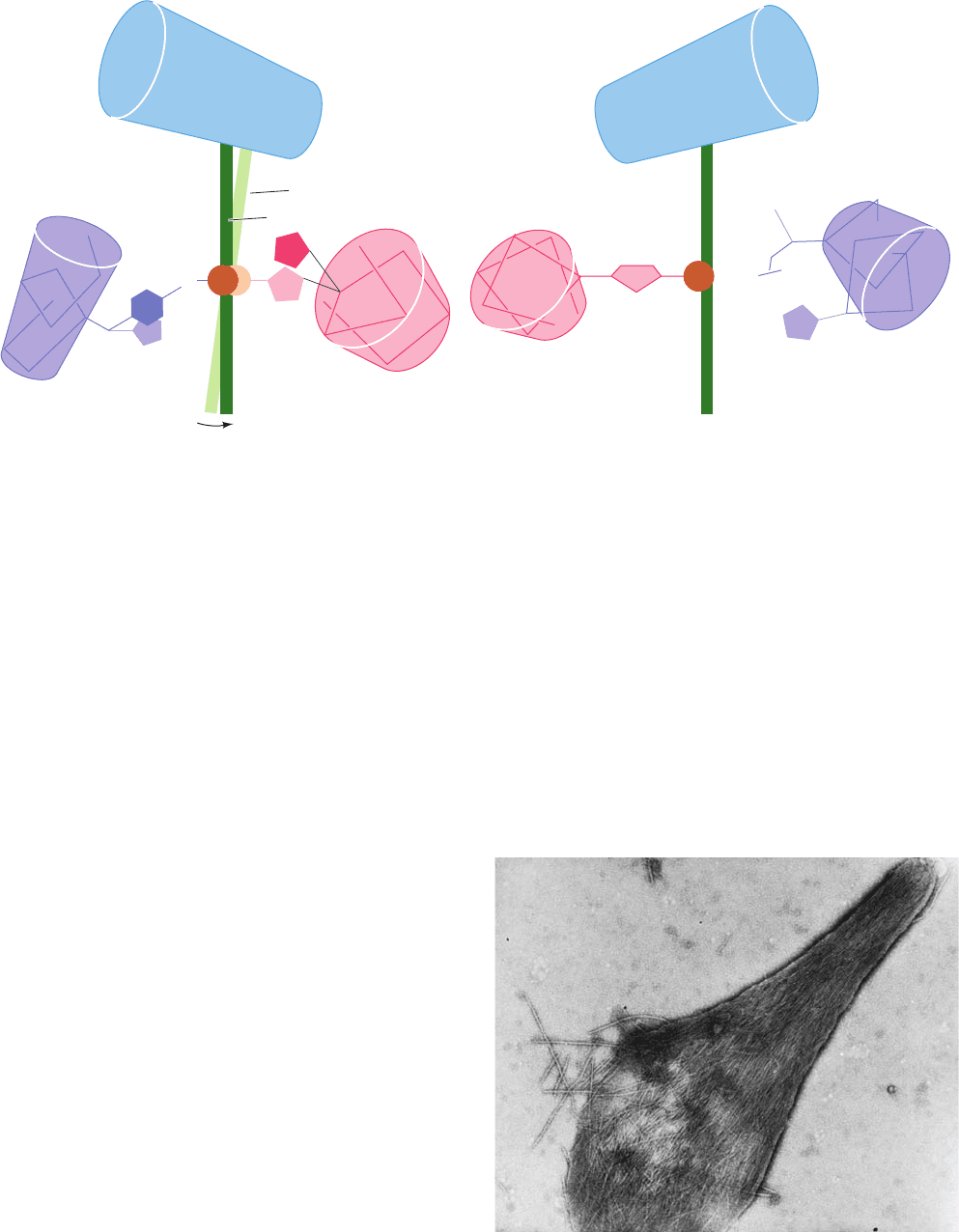
Figure 10-22 Mutations stabilizing the Fe(III) oxidation state
of heme. (a) Alterations in the heme pocket of the ␣ subunit on
changing from deoxyHbA to Hb Boston [His E7(58)␣ S Tyr].
The phenolate ion of the mutant Tyr becomes the fifth ligand of
the Fe atom, thereby displacing the proximal His [F8(87)a].
[After Pulsinelli, P.D., Perutz, M.F., and Nagel, R.L., Proc. Natl.
HbA
Fe
3+
(a) (b)
Hb Boston
Heme
E Helix
His E7
Tyr E7
α Subunit
F Helix
F Helix
β Subunit
His F8
Fe
3+
His F8
G Helix G Helix
Glu E11
His E7
E Helix
Heme
CH
3
Val E11
–
O
–
O
O
Hb
Milwaukee
Hb
Boston
Acad. Sci. 70, 3870 (1973).] (b) The structure of the heme pocket
of the  subunit in Hb Milwaukee [Val E11(67) S Glu]. Here
the mutant Glu residue’s carboxyl group forms an ion pair with
the heme iron atom so as to stabilize its Fe(III) state. [From
Perutz, M.F., Pulsinelli, P.D., and Ranney, H.M., Nature New Biol.
237, 259 (1972).]
Figure 10-23 Electron micrograph of deoxyHbS fibers spilling
out of a ruptured erythrocyte. [Courtesy of Robert Josephs,
University of Chicago.]
JWCL281_c10_323-358.qxd 2/24/10 1:58 PM Page 343
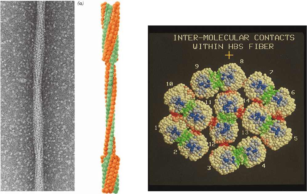
Electron microscopy indicates that these fibers are
⬃220-Å-diameter elliptical rods consisting of 14 hexago-
nally packed and helically twisting strands of deoxyHbS
molecules that associate in parallel pairs (Figs. 10-24 and
10-25a).
The structural relationship among the HbS molecules in
the pairs of parallel HbS strands has been established by
the X-ray structure analysis of deoxyHbS crystals. When
this crystal structure was first determined, it was unclear
whether the intermolecular contacts in the crystal resem-
bled those in the fiber. However, the subsequent observa-
tion that HbS fibers slowly convert to these crystals with
little change in their overall X-ray diffraction pattern indi-
cates that the fibers structurally resemble the crystals. The
crystal structure of deoxyHbS consists of double filaments
of HbS molecules whose several different intermolecular
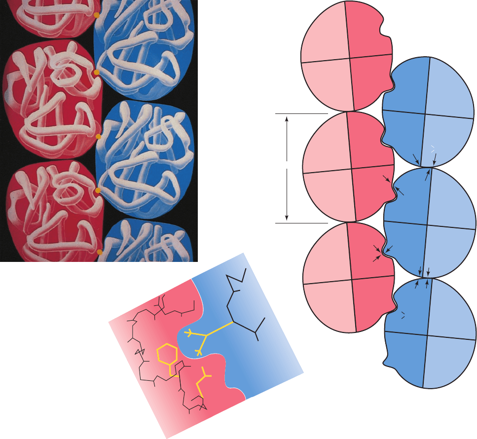
contacts are diagrammed in Fig. 10-25b. Only one of the
two Val 6’s per Hb molecule contacts a neighboring mol-
ecule. In this contact, the mutant Val side chain occupies a
hydrophobic surface pocket on the  subunit of an adja-
cent molecule whose Val 6 does not make an intermolec-
ular contact (Fig. 10-25c). This pocket is absent in oxyHb.
Other contacts involve residues that also occur in HbA, in-
cluding Asp 73 and Glu 23␣ (Fig. 10-25b). The observa-
tion that deoxyHbA does not aggregate into fibers, how-
ever, even at very high concentrations, indicates that the
contact involving Val 6b is essential for fiber formation.This
conclusion is corroborated by the observation that a genet-
ically engineed human Hb in which Glu 6 is replaced by
Ile (which differs from Val by an additional CH
2
group and
is therefore even more hydrophobic) has half the solubility
of HbS in 1.8M phosphate.
The importance of the other intermolecular contacts to
the structural integrity of HbS fibers has been demon-
strated by studying the effects of other mutant hemoglo-
bins on HbS gelation (polymerization). For example, the
doubly mutated Hb Harlem (Glu 6 S Val ⫹ Asp
73 S Asn) requires a higher concentration to gel than does
HbS (Glu 6 S Val); similarly, mixtures of HbS and Hb
Korle-Bu (Asp 73 S Asn) gel less readily than equivalent
344 Chapter 10. Hemoglobin: Protein Function in Microcosm
Figure 10-24 The 220-Å-diameter fibers of deoxyHbS. (a) An
electron micrograph of a negatively stained fiber.The
accompanying cutaway interpretive drawing indicates the
relationship between the inner and outer strands; each sphere
represents an individual HbS molecule. The fiber has a layer
repeat distance of 64 Å and a moderate twist such that it repeats
every 350 Å along the fiber axis. [Courtesy of Stuart Edelstein,
University of Geneva.] (b) A model, viewed in cross section, of
the HbS fiber based on the crystal structure of HbS and three-
dimensional reconstructions of electron micrographs of HbS
fibers.The residues in the 14 HbS molecules are represented by
spheres centered on their C
␣
positions.The residues making
inter-double strand, intra-double strand lateral, and intra-double
strand axial contacts are colored red, green, and blue, respectively,
with lighter and darker toned residues making intermolecular
contacts of ⬍8 Å and ⬍5 Å, respectively. The ␣ and  chain
residues outside the contact regions are colored white. [Courtesy
of Stanley Watowich, Leon Gross, and Robert Josephs,
University of Chicago.]
(b)
JWCL281_c10_323-358.qxd 2/24/10 1:58 PM Page 344
mixtures of HbS and HbA.These observations suggest that
Asp 73 occupies an important intermolecular contact site
in HbS fibers (Fig. 10-25b). Likewise, the observation that
hybrid tetramers consisting of ␣ subunits from Hb Memphis
(Glu 23␣ S Gln) and  subunits from HbS gel less readily
than does HbS indicates that Glu 23␣ also participates in
the polymerization of HbS fibers (Fig. 10-25b). The other
white-lettered residues in Fig. 10-25b have been similarly
implicated in sickling interactions.
b. The Initiation of HbS Gelation Is
a Complex Process
The gelation of HbS, both in solution and within the red
cell, follows an unusual time course.A solution of HbS can
be brought to conditions under which it will gel by lower-
ing the pO
2
, raising the HbS concentration, and/or raising
the temperature. On achieving gelation conditions, there is a
reproducible delay that varies according to conditions from
milliseconds to days: During this time, no HbS fibers can be
Section 10-3. Abnormal Hemoglobins 345
Figure 10-25 Structure of the deoxyHbS fiber. (a) The
arrangement of the deoxyHbS molecules in the fiber.The yellow
dots represent the side chains of Glu 6
2
. [Illustration, Irving
Geis/Geis Archives Trust. Copyright Howard Hughes Medical
Institute. Reproduced with permission.] (b) A schematic diagram
indicating the intermolecular contacts in the crystal structure of
deoxyHbS. The white-lettered residues are implicated in forming
these contacts. Note that the only intermolecular association in
which the mutant residue Val 6 participates involves subunit 
2
;
63 Å
α
1
β
1
β
2
α
2
α
1
β
1
β
2
α
2
β
1
α
1
α
2
β
2
β
1
α
1
α
2
β
2
β
1
α
1
α
2
β
2
α
1
β
1
β
2
α
2
His
116
Pro 114
( C=0)
( C=0)
Glu
121
Glu
23
Glu
121
Asp 73
Val 6
Thr 4
His
20
Gly
16
Glu
22
Phe
85
Leu
88
Val 6
Leu
88
Phe
85
(c)
(a)
(b)
Val 6 of subunit 
1
is free. [After Wishner, B.C.,Ward, K.B.,
Lattman, E.E., and Love,W.E., J. Mol. Biol. 98, 192 (1975).]
(c) The mutant Val 6
2
fits neatly into a hydrophobic pocket
formed mainly by Phe 85 and Leu 88 of an adjacent 
1
subunit.
This pocket, which is located between helices E and F at the
periphery of the heme pocket, is absent in oxyHb and is too
hydrophobic to contain the normally occurring Glu 6 side chain.
[Illustration, Irving Geis. Image from the Irving Geis Collection,
Howard Hughes Medical Institute. Reprinted with permission.]
JWCL281_c10_323-358.qxd 8/10/10 9:41 AM Page 345
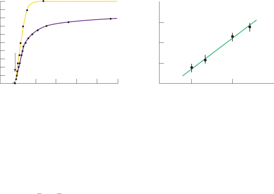
detected. Only after the delay do fibers first appear, and
gelation is then completed in about half the delay time
(Fig. 10-26a).
William Eaton and James Hofrichter discovered that the
delay time, t
d
, has a concentration dependence described by
[10.13]
where c
t
is the total deoxyHbS concentration prior to gela-
tion, c
s
is the solubility of deoxyHbS measured after gela-
tion is complete, and k and n are constants. Graphical
analysis of the data indicates that k ⬇ 10
⫺7
s
⫺1
and that n is
between 30 and 50 (Fig. 10-26b). This is a remarkable re-
sult: No other known solution process even approaches a
30th power concentration dependence.
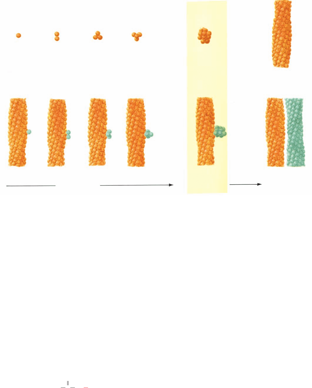
A two-stage process accounts for Eq. [10.13]:
1. At first, HbS molecules sequentially aggregate to
form a nucleus consisting of m HbS molecules (Fig. 10-27a):
Prenuclear aggregates are unstable and easily decompose,
but once a nucleus has formed it assumes a stable structure
that rapidly elongates to form an HbS fiber.
2. Once a fiber has formed, it can nucleate the growth
of other fibers (Fig. 10-27b). These newly formed fibers, in
turn, nucleate the growth of yet other fibers, etc., so that
this latter process is autocatalytic.
The initial homogeneous nucleation process (taking place
in solution) accounts for the very high concentration de-
pendence in Eq. [10.13], whereas the secondary heteroge-
neous nucleation process (taking place on a surface—that
of a fiber in this case) is responsible for the rapid onset of
gelation (Fig. 10-26a).
Δ (HbS)
m
¡
Growth
HbS Δ (HbS)
2
Δ (HbS)
3
Δ
p
1
t
d
⫽ k a
c
t
c
s
b
n
The foregoing kinetic hypothesis suggests why sickle-
cell anemia is characterized by episodic “crises” caused by
blood flow blockages. HbS fibers dissolve essentially in-
stantaneously on oxygenation, so that none are present in
arterial blood. Erythrocytes take from 0.5 to 2 s to pass
through the capillaries, where deoxygenation renders HbS
insoluble. If the delay time, t
d
, for sickling is greater than
this transit time, no blood flow blockage occurs (although
sickling that occurs in the veins damages the erythrocyte
membrane). However, Eq. [10.13] indicates that small in-
creases in HbS concentration, c
t
, and/or small decreases in
HbS solubility, c
s
, caused by conditions known to trigger
sickle-cell crises, such as dehydration, O
2
deprivation, and
fever, result in significant decreases of t
d
. Once a blockage
occurs, the resulting lack of O
2
and slowdown of blood flow
in the area compound the situation.
The kinetic hypothesis of sickling has profound clinical
implications for the treatment of sickle-cell anemia. Het-
erozygotes of HbS, whose blood usually contains ⬃60%
HbA and 40% HbS, rarely show any symptoms of sickling.
The t
d
for the gelation of their Hb is ⬃10
6
-fold greater than
that of homozygotes.Accordingly, a treatment of sickle-cell
anemia that increases t
d
by this amount, which corresponds
to decreasing the ratio c
t
/c
s
by a factor of ⬃1.6, would re-
lieve the symptoms of this disease.This has suggested three
different therapeutic strategies (besides gene therapy; Sec-
tion 5-5Hb) to increase t
d
, and thus inhibit HbS gelation:
1. The disruption of intermolecular interactions, thus in-
creasing c
s
. Of particular interest are compounds that have
been designed with the aid of the X-ray structure of HbS to
bind stereospecifically to its intermolecular contact regions.
However, a large amount of any such compound would be
necessary to bind to the ⬃400 g of hemoglobin in the human
body. Consequently, no antisickling drug yet tested has had a
sufficiently high ratio of efficacy to toxicity to merit clinical use.
346 Chapter 10. Hemoglobin: Protein Function in Microcosm
Figure 10-26 Time course of deoxyHbS gelation. (a) The
extent of gelation as monitored calorimetrically (yellow) and
optically (purple). Gelation of the 0.233 g ⴢ mL
⫺1
deoxyHbS
solution was initiated by rapidly increasing the temperature from
0ºC, where HbS is soluble, to 20ºC; t
d
is the delay time. (b) A
1.0
0.5
0 100 200 300
Time (min)
Fractional change
Calorimetric
Optical
(a)
t
d
1.30 1.35 1.40
0.001
0.01
0.1
1
10
1/t
d
(min
-1
)
log c
t
Slope≈30
(b)
log–log plot showing the concentration dependence of 1/t
d
for
the gelation of deoxyHbS at 30ºC.The slope of this line is ⬃30.
[After Hofrichter, J., Ross, P.D., and Eaton, W.A., Proc. Natl.
Acad. Sci. 71, 4865, 4867 (1974).]
JWCL281_c10_323-358.qxd 2/24/10 1:58 PM Page 346
2. The use of agents that increase hemoglobin’s O
2
affin-
ity, thus decreasing c
t
. For example, the administration of
cyanate carbamoylates the N-terminal amino groups of Hb
(Fig. 10-20). This treatment eliminates some of the salt
bridges that stabilize the T state (Section 10-2E) and thereby
increases the O
2
affinity of Hb.Although cyanate is an effec-
tive in vitro antisickling agent, its clinical use has been dis-
continued because of toxic side effects, cataract formation
and peripheral nervous system damage, that probably result
from the carbamoylation of proteins other than Hb.
3. Lowering the HbS concentration (c
t
) in erythrocytes.
Agents that alter erythrocyte membrane permeability so as
to permit the influx of water have promise in this regard.
The first, and as yet the only, effective treatment for
sickle-cell anemia is a variation of the latter strategy
through the administration of hydroxyurea.
Adults with sickle-cell anemia have two types of red blood
cells: S cells, which contain only HbS; and F cells, which
Hydroxyurea
H
2
N
O
CNHOH
contain ⬃20% HbF and the remainder HbS. In most
adults, the fraction of F cells is ⬃30%. However, in those
treated with hydroxyurea, this fraction increases to ⬃50%.
Although the mechanism by which hydroxyurea stimulates
the production of F cells is unknown, the mechanism by
which increased levels of F cells prevent sickling seems
clear. F cells contain three species of hemoglobin: HbS
(␣
2

S
2
), HbF (␣
2
␥
2
), and their hybrid (␣
2

S
␥), where 
S
sub-
units are the sickle-cell variants of the normal  subunits.
Since neither HbF nor the ␣
2

S
␥ hybrid Hb can form
sickle-cell fibers, they act to dilute the HbS in a cell.This, in
turn, increases the time it takes the F cells to sickle by a fac-
tor of ⬃1000, so that F cells do not significantly sickle in the
period (10–20 s) it takes them pass from the tissues to the
lungs, where they are oxygenated.Thus, the greater the pro-
portion of F cells in the blood,the smaller the proportion of
S cells that can sickle.
4 ALLOSTERIC REGULATION
One of the outstanding characteristics of life is the high de-
gree of control exercised in almost all of its processes.
Through a great variety of regulatory mechanisms, the
Section 10-4. Allosteric Regulation 347
(a) Homogeneous nucleation
(b) Heterogeneous nucleation
Growth of
thermodynamically
unstable aggregates
Critical
nucleus
Increasing
stability and
rapid growth
Figure 10-27 Double nucleation mechanism for deoxyHbS
gelation. (a) The initial aggregation of HbS molecules (spheres)
occurs very slowly because this process is thermodynamically
unfavorable and hence the intermediates tend to decompose
rather than grow. However, once an aggregate reaches a certain
size, the critical nucleus, its further growth becomes
thermodynamically favorable, leading to rapid fiber formation.
(b) Each fiber, in turn, can nucleate the growth of other fibers,
leading to the explosive appearance of polymer. [After Ferrone,
F.A., Hofrichter, J., and Eaton,W.A., J. Mol. Biol. 183, 614 (1985).]
JWCL281_c10_323-358.qxd 2/24/10 1:58 PM Page 347

exploration of which constitutes a significant portion of
this textbook, an organism is able to respond to changes in
its environment, maintain intra- and intercellular commu-
nications, and execute an orderly program of growth and
development. Regulation is exerted at every organiza-
tional level in living systems, from the control of rates of
reactions on the molecular level, through the control of ex-
pression of genetic information on the cellular level, to the
control of behavior on the organismal level. It is therefore
not surprising that many, if not most, diseases are caused by
aberrations in biological control processes.
Our exploration of the structure and function of hemo-
globin continues with a theoretical discussion of the regu-
lation of ligand binding to proteins through allosteric inter-
actions (Greek: allos, other ⫹ stereos, solid or space).These
cooperative interactions occur when the binding of one lig-
and at a specific site is influenced by the binding of another
ligand, known as an effector or modulator, at a different
(allosteric) site on the protein. If the ligands are identical,
this is known as a homotropic effect, whereas if they are
different, it is described as a heterotropic effect. These
effects are termed positive or negative depending on
whether the effector increases or decreases the protein’s
ligand-binding affinity.
Hemoglobin, as we have seen, exhibits both homotropic
and heterotropic effects. The binding of O
2
to Hb results in
a positive homotropic effect since it increases hemoglo-
bin’s O
2
affinity. In contrast, BPG, CO
2
,H
⫹
, and Cl
⫺
are
negative heterotropic effectors of O
2
binding to Hb be-
cause they decrease its affinity for O
2
(negative) and are
chemically different from O
2
(heterotropic). The O
2
affin-
ity of Hb, as we have seen, depends on its quaternary struc-
ture. In general, allosteric effects result from interactions
among subunits of oligomeric proteins.
Even though hemoglobin catalyzes no chemical reac-
tion, it binds ligands in the same manner as do enzymes.
Since an enzyme cannot catalyze a reaction until after it
has bound its substrate(s) [the molecule(s) undergoing re-
action], the enzyme’s catalytic rate varies with its substrate-
binding affinity. Consequently, the cooperative binding of
O
2
to Hb is taken as a model for the allosteric regulation of
enzyme activity. Indeed, in this section, we shall consider
several models of allosteric regulation that, for the most
part, were formulated to explain the O
2
-binding properties
of Hb. Following this, we shall compare these models with
the realities of Hb behavior.
A. The Adair Equation
The derivation of the Hill equation (Section 10-1B) is pred-
icated on the assumption of all-or-none O
2
binding.The ob-
servation of partially oxygenated Hb molecules, however,
led Gilbert Adair, in 1924, to propose that the binding of
ligands to proteins occurs sequentially with dissociation
constants that are not necessarily equal.The expression for
the saturation function under this model is straightfor-
wardly derived.
For a protein such as Hb with four ligand-binding sites,
the reaction sequence is
where the K
i
are the macroscopic or apparent dissociation
constants for binding the ith ligand to the protein,
[10.14]
and the k
i
are the microscopic or intrinsic dissociation con-
stants, that is, the individual dissociation constants for the
ligand-binding sites.The intrinsic dissociation constants are
equal to the apparent dissociation constants multiplied by
statistical factors, 4, and that account for the number
of ligand-binding sites on the protein molecule. The statis-
tical factor 4 derives from the fact that a tetrameric protein
E bears four sites that can bind ligand to form ES (that is,
the concentration of ligand-binding sites is 4[E]) but only
one site from which ES can dissociate ligand to form E
(that is, the concentration of bound ligand is 1[E]); the sta-
tistical factor is a result of there being three remaining
sites on ES that can bind ligand to form ES
2
and two sites
from which ES
2
can dissociate ligand to form ES; etc. In
general, for a protein with n equivalent binding sites:
[10.15]
since (n ⫺ i ⫹ 1)[ES
i⫺1
] is the concentration of free ligand-
binding sites in ES
i⫺1
and i[ES
i
] is the concentration of
bound ligand on ES
i
.Therefore,solving sequentially for the
concentration of each protein–ligand species in a tetrameric
protein, we obtain:
The fractional saturation of ligand binding, the fraction of
occupied ligand-binding sites divided by the total concen-
tration of ligand-binding sites, is expressed
[10.16]
so that, substituting in the above relationships and cancel-
ing terms, we obtain
[10.17]
This is the Adair equation for four ligand-binding sites.
Equations describing ligand binding to proteins with dif-
ferent numbers of binding sites are similarly derived.
If the microscopic dissociation constants of the Adair
equation are not equal, the fractional saturation curve will
Y
S
⫽
[S]
k
1
⫹
3[S]
2
k
1
k
2
⫹
3[S]
3
k
1
k
2
k
3
⫹
[S]
4
k
1
k
2
k
3
k
4
1 ⫹
4[S]
k
1
⫹
6[S]
2
k
1
k
2
⫹
4[S]
3
k
1
k
2
k
3
⫹
[S]
4
k
1
k
2
k
3
k
4
Y
S
⫽
[ES] ⫹ 2[ES
2
] ⫹ 3[ES
3
] ⫹ 4[ES
4
]
4([E] ⫹ [ES] ⫹ [ES
2
] ⫹ [ES
3
] ⫹ [ES
4
])
[ES
4
] ⫽ [ES
3
][S]>K
4
⫽
1
4
[ES
3
][S]>k
4
⫽ [E] [S]
4
>k
1
k
2
k
3
k
4
[ES
3
] ⫽ [ES
2
][S]>K
3
⫽
2
3
[ES
2
][S]>k
3
⫽ 4[E][S]
3
>k
1
k
2
k
3
[ES
2
] ⫽ [ES][S]>K
2
⫽
3
2
[ES][S]>k
2
⫽ 6[E][S]
2
>k
1
k
2
[ES] ⫽ [E][S]>K ⫽ 4[E][S]>k
1
k
i
⫽
(n ⫺ i ⫹ 1)[ES
i⫺1
][S]
i[ES
i
]
⫽ a
n ⫺ i ⫹ 1
i
bK
i
3
2
1
4
,
3
2
,
2
3
,
K
i
⫽
[ES
i⫺1
][S]
[ES
i
]
ES
3
⫹ S Δ ES
4
k
4
⫽
1
4
K
4
ES
2
⫹ S Δ ES
3
k
3
⫽
2
3
K
3
ES ⫹ S Δ ES
2
k
2
⫽
3
2
K
2
E⫹ S Δ ES
k
1
⫽ 4K
1
348 Chapter 10. Hemoglobin: Protein Function in Microcosm
JWCL281_c10_323-358.qxd 2/24/10 1:58 PM Page 348
describe cooperative ligand binding. Decreasing and in-
creasing values of these constants lead to positive and neg-
ative cooperativity, respectively. Of course, the values of
the microscopic dissociation constants may also alternate
so that, for example, k
1
⬍ k
2
⬎ k
3
⬍ k
4
.
In our discussion of the O
2
-dissociation curve of Hb
(Section 10-1B), we have seen how its values of k
1
and k
4
may be obtained by extrapolating the lower and upper
asymptotes of the Hill plot to the log pO
2
axis. The remain-
ing microscopic dissociation constants can be evaluated by
fitting Eq. [10.17] to the Hill plot.The values of these Adair
constants for Hb are given in Table 10-2. Note that k
4
is rel-
atively insensitive to the presence of BPG. Hb therefore
binds and releases its last O
2
almost independently of the
BPG concentration.
Although the Adair equation is the most general rela-
tionship describing ligand binding to a protein and is
widely used to do so, it provides no physical insight as to
why the various microscopic dissociation constants differ
from each other.Yet, if the protein consists, as so many do,
of identical subunits that are symmetrically related, it is de-
sirable to understand how ligand binding at one site influ-
ences the ligand-binding affinity at a seemingly identical
site. This need led to the development of models for ligand
binding that rationalize how the binding sites of oligomeric
proteins can exhibit different affinities. Two of these mod-
els are described in the following sections.
B. The Symmetry Model
Perhaps the most elegant model for describing cooperative
ligand binding to a protein is the symmetry model of
allosterism, which was formulated in 1965 by Jacques
Monod, Jeffries Wyman, and Jean-Pierre Changeux. This
model, alternatively termed the MWC model, is defined by
the following rules:
1. An allosteric protein is an oligomer of protomers
that are symmetrically related (for hemoglobin, we shall
assume, for the sake of algebraic simplicity, that all four
subunits are functionally identical).
2. Each protomer can exist in (at least) two conforma-
tional states, designated T and R; these states are in equilib-
rium whether or not ligand is bound to the oligomer.
3. The ligand can bind to a protomer in either confor-
mation. Only the conformational change alters the affinity
of a protomer for the ligand.
4. The molecular symmetry of the protein is conserved
during conformational change. Protomers must therefore
change conformation in a concerted manner,which implies
that the conformation of each protomer is constrained by
its association with the other protomers; in other words,
there are no oligomers that simultaneously contain R-
and T-state protomers.
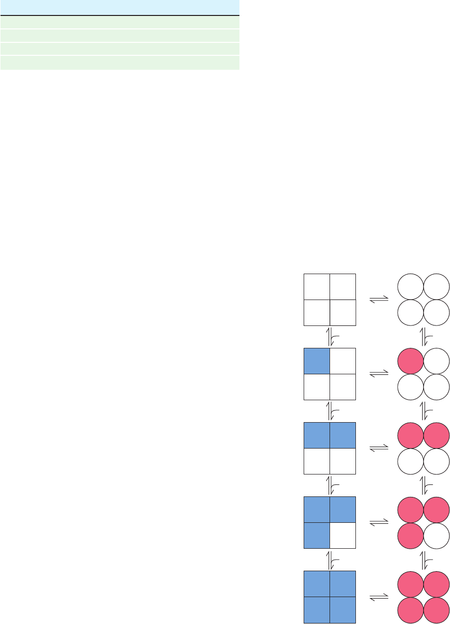
For a ligand S and an allosteric protein consisting of n
protomers, these rules imply the following equilibria for
conformational conversion and ligand-binding reactions
(for the sake of brevity, and ).
[10.18]
This is illustrated in Fig. 10-28 for a tetramer.
T
n⫺1
⫹ S Δ T
n
R
n⫺1
⫹ S Δ R
n
oo
T
1
⫹ S Δ T
2
R
1
⫹ S Δ R
2
T
0
⫹ S Δ T
1
R
0
⫹ S Δ R
1
T
0
Δ R
0
R
i
⬅ RS
i
T
i
⬅ TS
i
Section 10-4. Allosteric Regulation 349
Figure 10-28 The species and reactions permitted under the
symmetry model of allosterism. Squares and circles represent
T- and R-state protomers, respectively.
Table 10-2 Adair Constants for Hemoglobin A at pH 7.40
Solution k
1
(torr) k
2
(torr) k
3
(torr) k
4
(torr)
Stripped 8.8 6.1 0.85 0.25
0.1M NaCl 41. 13. 12. 0.14
2 mM BPG 74. 112. 23. 0.24
0.1M NaCl ⫹ 2 mM BPG 97. 43. 119. 0.09
Source: Tyuma, I., Imai, K., and Shimizu, K., Biochemistry 12, 1493,
1495 (1973).
T-state
subunits
R-state
subunits
S
SS
S
SS
SS
SS
S
S
SS
S
SS
SSS
S S
S S
S S
S S
JWCL281_c10_323-358.qxd 2/24/10 1:58 PM Page 349

The equilibrium constant L for the conformational in-
terconversion of the oligomeric protein in the absence of
ligand is expressed
[10.19]
The microscopic dissociation constant for the R state, k
R
,
which according to Rule 3 is independent of the number of
ligands bound to R, is expressed according to Eq. [10.15]:
[10.20]
The microscopic dissociation constant for ligand binding to
the T state, k
T
, is similarly expressed.The fractional satura-
tion, Y
s
, for ligand binding is
[10.21]
We shall make two definitions:
␣ may be considered a normalized ligand concentration. c is
the ratio of the ligand-binding dissociation constants; c in-
creases with the ligand-binding affinity of the T state rela-
tive to that of the R state. Then, combining the foregoing
relationships as is shown in Section A of the Appendix to
this chapter, we obtain the equation describing the symme-
try model of allosterism for homotropic interactions:
[10.22]
Note that this equation depends on three parameters, ␣, c,
and L, which are, respectively, the normalized ligand con-
centration, the relative affinities of the T and R states for
Y
S
⫽
␣(1 ⫹␣)
n⫺1
⫹ Lc␣(1 ⫹ c␣)
n⫺1
(1 ⫹␣)
n
⫹ L(1 ⫹ c␣)
n
␣⫽[S]>k
R
c ⫽ k
R
>k
T
([R
1
] ⫹ 2[R
2
] ⫹
p
⫹ n[R
n
]) ⫹ ([T
1
] ⫹ 2[T
2
] ⫹
p
⫹ n[T
n
])
n5([R
0
] ⫹ [R
1
] ⫹
p
⫹ [R
n
]) ⫹ ([T
0
] ⫹ [T
1
] ⫹
p
[T
n
])6
Y
S
⫽
k
R
⫽ a
n ⫺ i ⫹ 1
i
b
[R
i⫺1
] [S]
[R
i
]
(i ⫽ 1, 2, 3, p , n)
L ⫽
[T
0
]
[R
0
]
ligand, and the relative stabilities of the T and R states. In
contrast, the Hill equation (Section 10-1B) has but two pa-
rameters, K and n, whereas the number of parameters in
the Adair equation is equal to the number of ligand-
binding sites on the protein.
a. Homotropic Interactions
Let us examine the nature of the symmetry model by plot-
ting Eq. [10.22] for a tetramer (n ⫽ 4) as a function of ␣ for
different values of the parameters L and c (Fig. 10-29).Three
major points are evident from an inspection of these plots:
1. The degree of upward curvature exhibited by the ini-
tial sections of these sigmoid curves is indicative of their
level of cooperativity.
2. When only the R state binds ligand (c ⫽ 0), the ligand-
binding cooperativity increases as the oligomer’s conforma-
tional preference for the non–ligand-binding T state in-
creases (L increases; Fig. 10-29a). For high L values, if a
single ligand is to bind, it must “force” the protein into its less
preferred R state.The requirement that all protomers change
their conformational states in a concerted manner causes the
remaining three ligand-binding sites to become available.
The binding of the first ligand therefore promotes the bind-
ing of subsequent ligands, which is the essence of a positive
homotropic effect. Note that cooperativity and ligand-bind-
ing affinity are different quantities; in fact, for c ⫽ 0, curves
indicative of high ligand-binding affinity (those with low L)
exhibit low cooperativity and vice versa.
3. When the T state is highly preferred (L is large), lig-
and-binding cooperativity increases with the R state’s lig-
and-binding affinity relative to that of the T state (decreas-
ing c; Fig. 10-29b). At low ligand concentrations (low ␣)
the amount of ligand bound (Y
S
) increases with the lig-
and-binding affinity of the T state (increasing c) since the
protein is largely in the T state.As ␣ increases, however,the
amount of ligand bound to the intrinsically less stable R
state eventually surpasses that of the T state, thereby
resulting in a cooperative effect. This is because the free
350 Chapter 10. Hemoglobin: Protein Function in Microcosm
Figure 10-29 Symmetry model
saturation function curves for
tetramers according to Eq.
[10.22]. Here L ⫽ [T
0
]/[R
0
],
c ⫽ k
R
/k
T
, and ␣⫽[S]/k
R
.
(a) Their variation with L when
c ⫽ 0. (b) Their variation with c
when L ⫽ 1000. [After Monod,
J., Wyman, J., and Changeux, J.P.,
J. Mol. Biol. 12, 92 (1965).]
1
0.5
0
0 2 5 0 2 5 10 2010 15
Y
S
αα
(a) (b)
L
= 1
L
= 10
L
= 100
L
= 1000
L
= 10,000
= 0.00
= 0.10
= 0
= 4
= 1000
= 4
= 0.04
c
c
c
L
n
c
n
JWCL281_c10_323-358.qxd 2/24/10 1:58 PM Page 350
