Voet D., Voet Ju.G. Biochemistry
Подождите немного. Документ загружается.

Section 11-2. Polysaccharides 371
embedded in a gel-like matrix known as ground substance.
Ground substance is composed largely of glycosaminogly-
cans (GAGs; alternatively, mucopolysaccharides), un-
branched polysaccharides of alternating uronic acid and
hexosamine residues. Solutions of GAGs have a slimy, mu-
cuslike consistency that results from their high viscosity
and elasticity. In the following paragraphs, we discuss the
structural origin of these important mechanical properties.
a. Hyaluronic Acid
Hyaluronic acid (also called hyaluronan) is an important
GAG component of ground substance, synovial fluid (the
fluid that lubricates the joints), and the vitreous humor of
the eye. It also occurs in the capsules surrounding certain,
usually pathogenic, bacteria. Hyaluronic acid molecules are
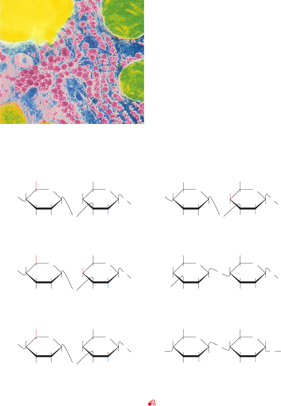
composed of 250 to 25,000 (1 S 4)-linked disaccharide
units that consist of
D-glucuronic acid and N-acetyl-D-
glucosamine linked by a (1 S 3) bond (Fig. 11-21). The
anionic character of its glucuronic acid residues causes
hyaluronic acid to bind cations such as K
⫹
,Na
⫹
, and Ca
2⫹
tightly. X-ray fiber analysis indicates that Ca
2⫹
hyaluronate
Figure 11-20 Photomicrograph showing the glycogen granules
(pink) in the cytoplasm of a liver cell. The greenish objects are
mitochondria and the yellow object is a fat globule. Note that the
glycogen granules tend to aggregate. The glycogen content of
liver may reach as high as 10% of its net weight. [CNRI/Science
Photo Library/Photo Researchers, Inc.]
Figure 11-21 The disaccharide repeating units of the common glycosaminoglycans. The anionic groups
are drawn in red and the N-acetylamido groups are drawn in blue.
See Kinemage Exercise 7-3
OH H
H
H
HOH
H
O
41
COO
–
H
H
H
HO
H
H
O
3
CH
2
OH CH
2
OH
1
O
β
NHCOCH
3
NHCOCH
3
O
β
D-Glucuronate N-Acetyl-D-glucosamine N-Acetyl-D-galactosamine-
4-sulfate
N-Acetyl-D-galactosamine-
4-sulfate
N-Acetyl-D-glucosamine-
6-sulfate
N-Acetyl-D-galactosamine-
6-sulfate
N-Sulfo-D-glucosamine-
6-sulfate
L-Iduronate-2-sulfate
Hyaluronate
OH H
H
H
HOH
H
O
41
COO
–
H
H
H
H
O
3
1
O
α
O
β
L-Iduronate
Dermatan sulfate
–
O
3
SO
–
O
3
SO
H
OH H
H
H
HOH
H
O
41
COO
–
COO
–
COO
–
H
H
H
H
O
3
1
O
β
O
β
D-Glucuronate
D-Glucuronate
Chondroitin-4-sulfate
H
HO
HOH
H
O
3
3
H
H
H
H
O
1O
β
NHCOCH
3
NHCOCH
3
NHCOCH
3
NHOSO
3
–
O
β
D-Galactose
Keratan sulfate
OH H
H
H
HOH
H
O
41
H
HO
H
H
H
O
1
O
β
O
β
Chondroitin-6-sulfate
OH H
H
H
H
O
41
H
H
H
H
O
2
1
O
α
O
α
Heparin
H
H
4
4
4
4
4
H
CH
2
OHCH
2
OH CH
2
OSO
3
–
CH
2
OSO
3
–
CH
2
OSO
3
–
OSO
3
–
OH
H
H
6
H
H
OH
2
JWCL281_c11_359-385.qxd 6/3/10 10:35 AM Page 371
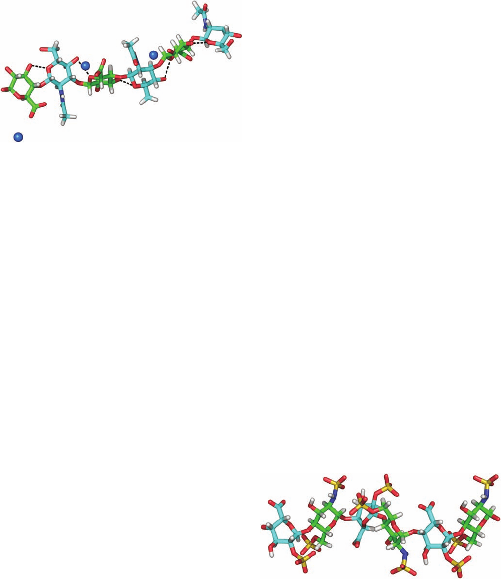
forms an extended, left-handed, single-stranded helix with
⬃3 disaccharide units per turn (Fig. 11-22).
Hyaluronate’s structural features suit it to its biological
function. Its high molecular mass and numerous mutually
repelling anionic groups make hyaluronate an extended,
rigid, and highly hydrated molecule which, in solution,
occupies a volume ⬃1000 times that in its dry state.
Hyaluronate solutions therefore have a viscosity that is
shear dependent (an object under shear stress has equal
and opposite forces applied across its opposite faces). At
low shear rates, the hyaluronate molecules form tangled
masses that greatly impede flow; that is, the solution is
quite viscous. As the shear rate increases, the stiff rodlike
hyaluronate molecules tend to line up with the flow and
thus offer less resistance to it. This viscoelastic behavior
makes hyaluronate solutions excellent biological shock ab-
sorbers and lubricants.
Hyaluronic acid and other GAGs (see below) are de-
graded by hyaluronidase, which hydrolyzes their (1 S 4)
linkages. Hyaluronidase occurs in a variety of animal tis-
sues, in bacteria (where it presumably expedites their inva-
sion of animal tissue), and in snake and insect toxins.
b. Other Glycosaminoglycans
Other GAG components of ground substance consist of
50 to 1000 sulfated disaccharide units which occur in pro-
portions that are both tissue and species dependent. The
most prevalent structures of these generally heterogeneous
substances are (Fig. 11-21)
1. Chondroitin-4-sulfate (Greek: chondros, cartilage), a
major component of cartilage and other connective tissue,
has N-acetyl-
D-galactosamine-4-sulfate residues in place of
hyaluronate’s N-acetyl-
D-glucosamine residues.
2. Chondroitin-6-sulfate is instead sulfated at the C6
position of its N-acetyl-
D-galactosamine residues. The two
chondroitin sulfates occur separately or in mixtures de-
pending on the tissue.
3. Dermatan sulfate (Greek: derma, skin), which is so
named because of its prevalence in skin, differs from chon-
droitin-4-sulfate only by an inversion of configuration
about C5 of the -
D-glucuronate residues to form ␣-L-
iduronate.This results from the enzymatic epimerization of
these residues after the formation of chondroitin. The
epimerization is usually incomplete, so dermatan sulfate
also contains glucuronate residues.
4. Keratan sulfate (Greek: keras, horn; not to be con-
fused with the protein keratin) consists mainly of alternating
(1 S 4)-linked
D-galactose and N-acetyl-D-glucosamine-6-
sulfate residues (and hence lacks uronic acid residues). It is
a component of cartilage, bone, cornea, as well as hair,
nails, and horn. Keratan sulfate is the most heteroge-
neous of the major GAGs in that its sulfate content is
variable and it contains small amounts of fucose, man-
nose, N-acetylglucosamine, and sialic acid.
5. Heparin is a variably sulfated GAG that consists pre-
dominantly of alternating ␣(1 S 4)-linked residues of
L-
iduronate-2-sulfate and N-sulfo-
D-glucosamine-6-sulfate.
It has an average of 2.5 sulfate residues per disaccharide
unit, which makes it the most negatively charged polyelec-
trolyte in mammalian tissues (Fig. 11-23). Heparin, in con-
trast to the above GAGs, is not a constituent of connective
tissue, but occurs almost exclusively in the intracellular
granules of the mast cells that line arterial walls, especially
in the liver, lungs, and skin. It inhibits the clotting of blood,
and its release, through injury, is thought to prevent run-
away clot formation (Section 35-1Ea). Heparin is therefore
in wide clinical use to inhibit blood clotting, for example, in
372 Chapter 11. Sugars and Polysaccharides
Figure 11-22 X-ray fiber structure of Ca
2⫹
hyaluronate. Three
consecutive disaccharide units of the hyaluronate fiber are drawn
in stick form with atoms colored according to type with
glucuronate C green, N-acetyl-
D-glucosamine C cyan, H white,
N blue, and O red. Ca
2⫹
ions are represented by blue spheres.
The hyaluronate polyanion forms an extended, left-handed,
single-stranded helix with a pitch of 28.3 Å and ⬃3 disaccharide
units per turn that is stabilized by intramolecular hydrogen
bonds (dashed lines).The positions of the H atoms are inferred
and hence the H atoms of the OH groups are not shown. [Based
on a fiber X-ray structure by Struther Arnott, Purdue University.
PDBid 4HYA.]
Figure 11-23 NMR structure of heparin. Three consecutive
disaccharide units of this helical polymer are shown in stick
form.Atoms are colored according to type with glucosamine
C green, iduronate C cyan, H white, N blue, O red, and S yellow.
The helical repeat unit is two disaccharides with a pitch of 17.5
Å. Note the high density of anionic sulfate groups. [Based on an
NMR structure by Barbara Mulloy and Mark Forster, National
Institute for Biological Standards and Control, Herts, U.K.
PDBid 1HPN.]
JWCL281_c11_359-385.qxd 6/3/10 10:35 AM Page 372

postsurgical patients. Heparan sulfate, a ubiquitous cell-
surface component as well as an extracellular substance in
blood vessel walls and brain, resembles heparin but has a
far more variable composition with fewer N- and O-sulfate
groups and more N-acetyl groups.
3 GLYCOPROTEINS
Until about 1960, carbohydrates were thought to be
rather dull compounds that were probably some sort of
inert filler. Protein chemists therefore considered them to
be a nuisance that complicated protein “purification.” In
fact, most eukaryotic proteins are glycoproteins, that is,
they are covalently associated with carbohydrates. Glyco-
proteins vary in carbohydrate content from ⬍1% to
⬎90% by weight. They occur in all forms of life and have
functions that span the entire spectrum of protein activi-
ties, including those of enzymes, transport proteins, recep-
tors, hormones, and structural proteins. Their carbohy-
drate moieties, as we shall see, have several important
biological roles, but in many cases their functions remain
enigmatic.
The polypeptide chains of glycoproteins, like those of
all proteins, are synthesized under genetic control. Their
carbohydrate chains, in contrast, are enzymatically gener-
ated and covalently linked to the polypeptide without the
rigid guidance of nucleic acid templates. The processing
enzymes are generally not available in sufficient quanti-
ties to ensure the synthesis of uniform products. Glyco-
proteins therefore have variable carbohydrate composi-
tions, a phenomenon known as microheterogeneity, that
compounds the difficulties in their purification and char-
acterization.
In this section we consider the structures and properties
of glycoproteins. In particular, we shall study the glycopro-
teins of connective tissues, those of bacterial cell walls, and
several soluble glycoproteins. We end by discussing the
general principles of glycoprotein structure and function.
A. Proteoglycans
Proteins and glycosaminoglycans in ground substance, in
basal laminae [basement membranes; the thin matlike ex-
tracellular matrix separating epithelial cells (the cells lining
body cavities and free surfaces) from underlying cells], and
in cell-surface membranes aggregate covalently and non-
covalently to form a diverse group of macromolecules
known as proteoglycans. Proteoglycans consist of a core
protein to which at least one glycosaminoglycan chain, most
often keratan sulfate and/or chondroitin sulfate, is cova-
lently linked. Numerous types of core proteins have been
characterized (Table 11-1). Proteoglycans appear to have
multiple roles, most notably as organizers of tissue mor-
phology via their interactions with molecules such as colla-
gen; as selective filters that regulate the traffic of molecules
according to their size and/or charge; and as regulators of
the activities of other proteins, particularly those involved
in signaling (see below).
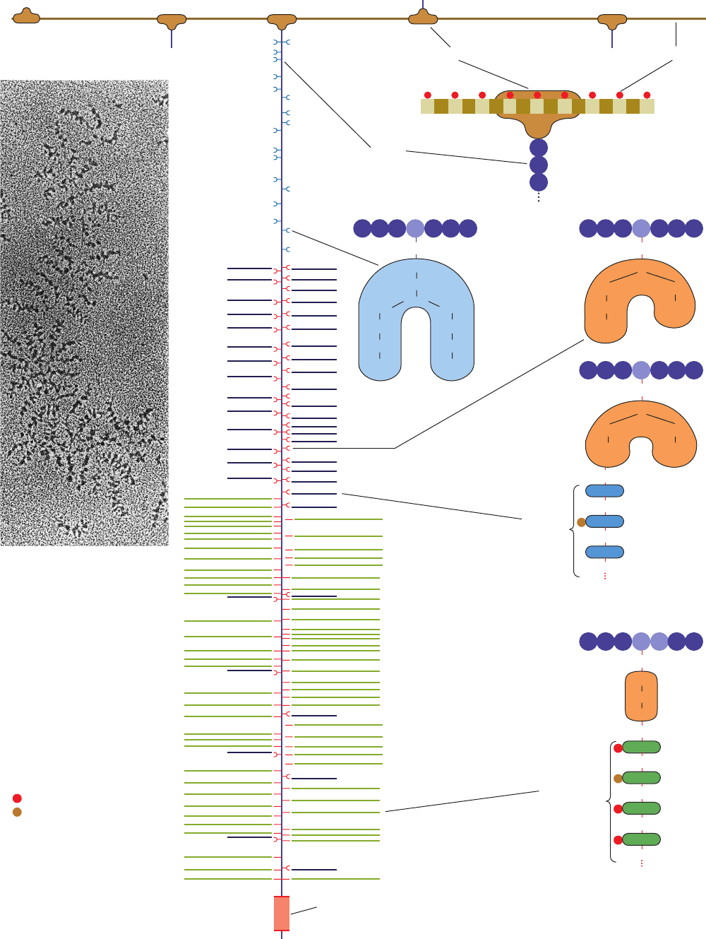
Electron micrographs such as Fig. 11-24a together with
reconstitution experiments indicate that proteoglycans can
form huge complexes. For example, aggrecan, the main pro-
teoglycan component of cartilage, has a bottlebrush-like mo-
lecular architecture (Fig. 11-24b), whose proteoglycan sub-
unit “bristles” are noncovalently attached to a filamentous
hyaluronic acid “backbone” at intervals of 200 to 300 Å.
Aggrecan has three domains. Its N-terminal domain forms
a globular region of 60 to 70 kD that binds noncovalently
Section 11-3. Glycoproteins 373
Approximate Core Protein Glycosaminoglycan
Proteoglycan Molecular Mass (kD) Type (Number)
a
Proteoglycans interacting with
hyaluronic acid
Aggrecan 220 CS (⬃100), KS (⬃30)
Versican 265–370 CS/DS (10–30)
Neurocan 136 CS (3–7)
Proteoglycans of the basal laminae
Perlecan 400–467 HS/CS (3)
Agrin 250 HS (3)
Bamacan 138 CS (3)
Small leucine-rich proteoglycans
Decorin 40 DS/CS (1)
Fibromodulin 42 KS (2–3)
Osteoglycin 35 KS (2–3)
Table 11-1 Properties of Some Proteoglycans
a
Abbreviations: CS, chondroitin sulfate; DS, dermatan sulfate; HS, heparan sulfate; KS, keratan sulfate.
Source: Iozzo, R.V., Annu. Rev. Biochem. 67, 611, 626, and 624 (1998).
JWCL281_c11_359-385.qxd 6/3/10 10:35 AM Page 373
374 Chapter 11. Sugars and Polysaccharides
(b)
Hyaluronic acidLink protein
Core
Protein
N
GlcNAc
GlcNAc
Man
Man
GlcNAc
NeuNAc
Gal
Man
GlcNAc
NeuNAc
Gal
N-linked oligosaccharides
O
GalNAc
GlcNAc
NeuNAc
Gal
NeuNAc
Gal
Ser
O
O
GalNAc
NeuNAc
Gal
NeuNAc
Keratan
sulfate
Gal
Ser
Gly
O
Ser
Asn
O-linked oligosaccharides
Asn
Gal
GalNAc
GlcNAc
GluA
Man
N
NeuNAc
O
Ser
Xyl
Asparagine
Galactose
N-Acetyl-
galactosamine
N-Acetyl-
glucosamine
Glucuronate
Mannose
Nitrogen atom
Sialic acid
Oxygen atom
Serine
Xylose
Carboxyl group
Sulfate group
Gal
O
GlcNAc
O
O
Gal
O
Chondroitin
sulfate
Lectinlike
module
COO
–
GluA
O
GlcNAc
O
GluA
O
O
GlcNAc
Xyl
Gal
Gal
(a)
JWCL281_c11_359-385.qxd 6/3/10 10:35 AM Page 374
to hyaluronic acid. This attachment is stabilized by the
40- to 60-kD link protein, which is similar in sequence to
aggrecan’s N-terminal domain. Aggrecan’s highly ex-
tended central domain is covalently linked to a series of
polysaccharides, which comprise nearly 90% of this glyco-
protein’s mass. They divide the central domain into three
regions:
1. An N-terminal region, which overlaps the globular
hyaluronic acid–binding domain, binds a relatively few car-
bohydrate chains. These tend to be oligosaccharides that
are covalently bonded to the protein via the amide N atoms
of specific Asn residues (Section 11-3Ca).
2. A region rich in oligosaccharides, many of which
serve as anchor points for keratan sulfate chains. These
oligosaccharides are covalently bonded to side chain O
atoms of Ser and Thr residues.
3. A C-terminal region rich in chondroitin sulfate
chains, which are covalently linked to the side chain O
atoms of Ser residues in Ser-Gly dipeptides via galactose–
galactose–xylose trisaccharides.
Aggrecan’s C-terminal domain contains a lectinlike mod-
ule, which binds certain monosaccharide units.Thus, aggre-
can probably functions to bind together various con-
stituents of the cell surface and the extracellular matrix
(see below).
Altogether, a central strand of hyaluronic acid, which
varies in length from 4000 to 40,000 Å, noncovalently binds
up to 100 associated aggrecan chains, each of which cova-
lently binds ⬃30 keratan sulfate chains of up to 250 disac-
charide units each and ⬃100 chondroitin sulfate chains of
up to 1000 disaccharide units each. This accounts for the
enormous molecular masses of the aggrecans, which range
up to 220,000 kD, and for their high degree of polydisper-
sity (range of molecular masses).Note,however, that many
proteoglycans do not bind to hyaluronic acid (Table 11-1)
and hence function as monomers.
a. Cartilage’s Mechanical Properties Are Explained
by Its Molecular Structure
Cartilage consists largely of a meshwork of collagen fib-
rils that is filled in by proteoglycans whose chondroitin sul-
fate and core protein components specifically interact with
the collagen. The tensile strength of cartilage and other
connective tissues is, as we have seen (Section 8-2Ba), a
consequence of their collagen content. Cartilage’s charac-
teristic resilience, however, results from its high proteogly-
can content. The extended brushlike structure of proteo-
glycans, together with the polyanionic character of keratan
sulfate and chondroitin sulfate, cause this complex to be
highly hydrated. The application of pressure on cartilage
squeezes water away from these charged regions until
charge–charge repulsions prevent further compression.
When the pressure is released, the water returns. Indeed,
the cartilage in the joints, which lack blood vessels, is nour-
ished by this flow of liquid brought about by body move-
ments. This explains why long periods of inactivity cause
joint cartilage to become thin and fragile.
b. Proteoglycans Modulate the Effects of Protein
Growth Factors
Proteoglycans have been implicated in a great variety
of cellular processes. For example, fibroblast growth factor
(FGF; growth factors are proteins that function to induce
their specific target cells to grow and/or differentiate; Sec-
tion 19-3Aa) binds to heparin or to the heparan sulfate
chains of proteoglycans and is only bound to its cell-surface
receptor in complex with these glycosaminoglycans. Since
the binding of FGF to heparin or heparan sulfate protects
FGF from degradation, the release of this growth factor
from the extracellular matrix by the proteolysis of proteo-
glycan core proteins or by the partial degradation of he-
paran sulfate probably provides an important source of
active FGF–glycosaminoglycan complexes. Several other
growth factors interact similarly with proteoglycans.
Apparently, the abundant and ubiquitous distribution of
proteoglycans limits the action of these growth factors
on their target cells to short distances from the cells se-
creting the growth factors, a phenomenon that probably
greatly influences the formation and maintenance of tis-
sue architecture.
B. Bacterial Cell Walls
Bacteria are surrounded by rigid cell walls (Fig. 1-13) that
give them their characteristic shapes (Fig. 1-1) and permit
them to live in hypotonic (less than intracellular salt con-
centration) environments that would otherwise cause them
to swell osmotically until their plasma (cell) membranes
lysed (burst). Bacterial cell walls are of considerable med-
ical significance because they are responsible for bacterial
virulence (disease-evoking power). In fact, the symptoms
of many bacterial diseases can be elicited in animals merely
by the injection of bacterial cell walls. Furthermore, the
Section 11-3. Glycoproteins 375
Figure 11-24 (Opposite) Proteoglycans. (a) An electron
micrograph showing a central strand of hyaluronic acid, which
runs down the field of view, supporting numerous projections,
each of which consists of a core protein to which many bushy
polysaccharide protrusions are linked. [From Caplan,A.I., Sci.
Am. 251(4); 87 (1984). Copyright © 1984 Scientific American,
Inc. Used by permission.] (b) The bottlebrush model of the
proteoglycan aggrecan.The core proteins, one of which is shown
extending down through the middle of the diagram, project from
the central hyaluronic acid strand.The core is noncovalently
anchored to the hyaluronic acid via its globular N-terminal end
in an association that is stabilized by link protein.The core has
three saccharide-binding regions: (1) the inner region
predominantly binds oligosaccharides via the side chain N atoms
of Asn residues; (2) the central region binds oligosaccharides,
many of which bear keratan sulfate chains, via the side chain
O atoms of Ser and Thr residues; and (3) the outer region mainly
binds chondroitin sulfate chains that are linked to the core
protein via a galactose–galactose–xylose trisaccharide that is
bonded to side chain O atoms of Ser residues in the sequence
Ser-Gly.The C-terminal end of the aggrecan core protein consists
of a lectinlike sequence.
JWCL281_c11_359-385.qxd 6/3/10 10:35 AM Page 375
characteristic antigens (immunological markers; Section
35-2) of bacteria are components of their cell walls and
capsules, so that injection of preparations of these sub-
stances into an animal often invokes its immunity against
these bacteria. Consequently, several vaccines that are
based on purified bacterial polysaccharides have recently
become available, including those against Streptococcus
pneumoniae, a major cause of pneumonia, and Neisseria
meningitidis, a major cause of meningitis.
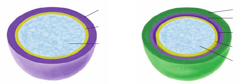
Bacteria are classified as gram-positive or gram-negative
depending on whether or not they take up gram stain
(Section 1-1B). Gram-positive bacteria (Fig. 11-25a) have
a thick (⬃250 Å) cell wall surrounding their plasma
membrane, whereas gram-negative bacteria (Fig. 11-25b)
have a thin (⬃30 Å) cell wall covered by a complex outer
membrane.
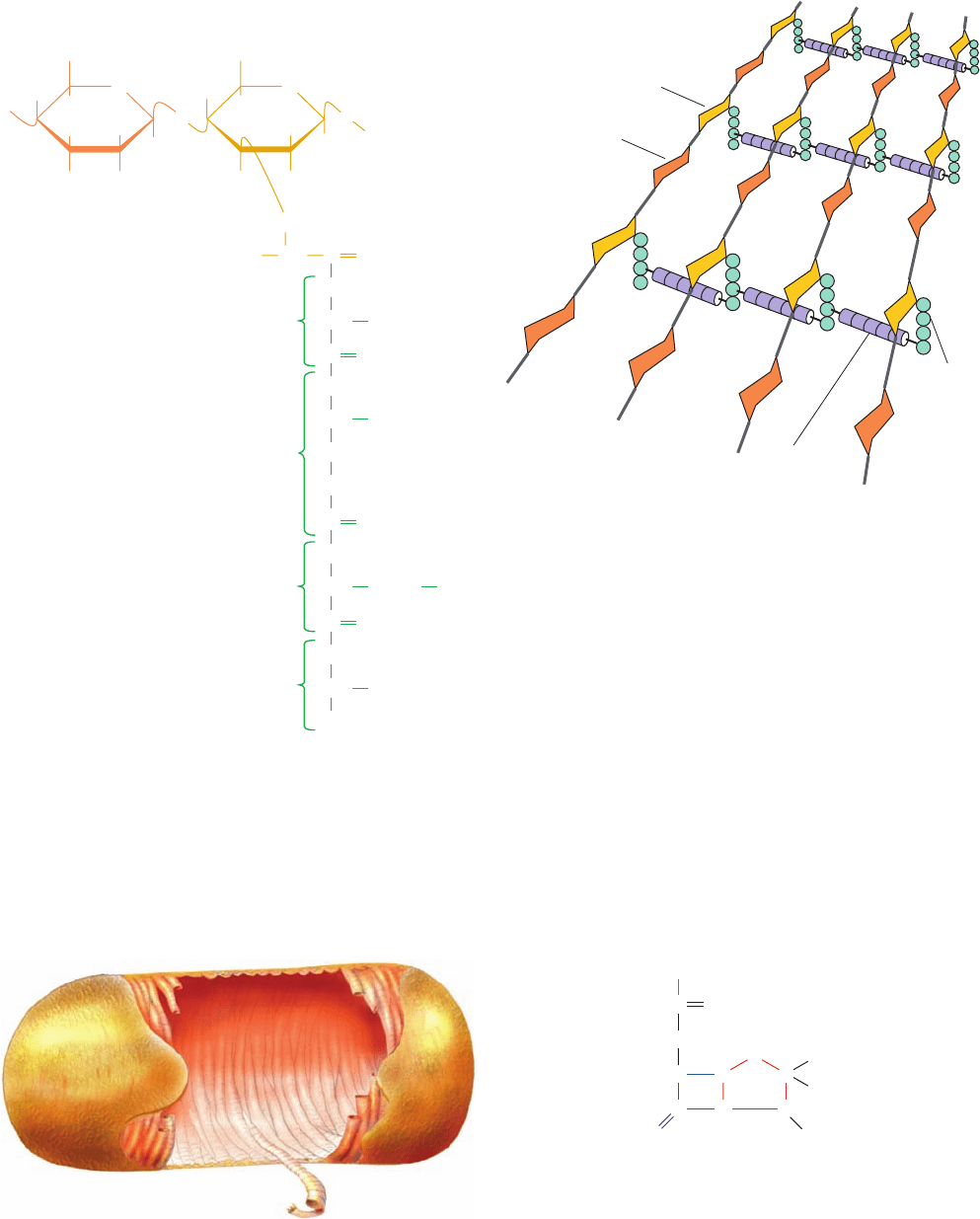
a. Bacterial Cell Walls Have a
Peptidoglycan Framework
The cell walls of both gram-positive and gram-negative
bacteria consist of covalently linked polysaccharide and
polypeptide chains that form a framework that completely
encases the cell. This substance, whose molecular structure
was elucidated in large part by Jack Strominger, is known
as a peptidoglycan or murein (Latin: murus, wall). Its
polysaccharide component consists of linear chains of al-
ternating (1 S 4)-linked N-acetylglucosamine (NAG)
and N-acetylmuramic acid (NAM). The NAM’s lactic acid
residue forms an amide bond with a
D-amino acid–contain-
ing tetrapeptide to form the peptidoglycan repeating unit
(Fig. 11-26). Neighboring parallel peptidoglycan chains
are covalently cross-linked through their tetrapeptide
side chains. In the gram-positive bacterium Staphylococ-
cus aureus, whose tetrapeptide has the sequence
L-Ala-D-
isoglutamyl-
L-Lys-D-Ala, this cross-link consists of a pen-
taglycine chain that extends from the terminal carboxyl
group of one tetrapeptide to the ε-amino group of the Lys
in a neighboring tetrapeptide.
Atomic force microscopy (AFM; an imaging technique
that reports the variation in the force between a probe that
is several nanometers in diameter and a surface of interest
as the probe is scanned over the surface; its resolution is as
little as several Ångstroms) was used by Simon Foster to
image the cell wall of the gram-negative bacterium Bacillus
subtilis leading to the following model (Fig. 11-27). Several
glycan chains are cross-linked much as described above to
form a peptidoglycan “rope,” which due to its natural twist,
forms an ⬃50-nm-diameter helical cable of up to 50 m in
length that coils around the long axis of the bacterium to
form its cell wall. This structure is presumably stabilized by
the formation of covalent cross-links between neighboring
segments of the coil.The cell walls of gram-negative bacteria
appear to be only one layer thick, whereas as those of
gram-positive bacteria are postulated to consist of several
such layers. How the peptidoglycan imposes cell shape is
unknown.
The
D-amino acids of peptidoglycans render them resist-
ant to proteases. However, lysozyme, an enzyme which is
present in tears, mucus, and other vertebrate body secre-
tions, as well as in egg whites, catalyzes the hydrolysis of the
(1 S 4) glycosidic linkage between NAM and NAG.
Consequently, treatment of gram-positive bacteria with
lysozyme degrades their cell walls, which results in their ly-
sis (gram-negative bacteria are resistant to lysozyme
degradation). Lysozyme was discovered in 1922 by the
British bacteriologist Alexander Fleming after he noticed
that a bacterial culture had dissolved where mucus from a
sneeze had landed. It was Fleming’s hope that lysozyme
would be a universal antibiotic but, unfortunately, it is clin-
ically ineffective against pathogenic bacteria.The structure
and mechanism of lysozyme are examined in detail in Sec-
tion 15-2.
b. Penicillin Kills Bacteria by Inhibiting
Cell Wall Biosynthesis
In 1928, Fleming noticed that the chance contamination
of a bacterial culture plate with the mold Penicillium
notatum lysed nearby bacteria (a clear demonstration of
Pasteur’s maxim that chance favors a prepared mind).This
was caused by the presence of penicillin (Fig. 11-28), an
antibiotic secreted by the mold. Yet the difficulties of
isolating and characterizing penicillin, owing to its instabil-
ity, led to the passage of over 15 years before penicillin was
available for routine clinical use. Penicillin specifically
binds to and inactivates enzymes that function to cross-link
the peptidoglycan strands of bacterial cell walls. Since cell
376 Chapter 11. Sugars and Polysaccharides
Figure 11-25 Schematic diagram comparing the cell envelopes of (a) gram-positive bacteria
and (b) gram-negative bacteria.
Peptidoglycan
(cell wall)
Plasma
membrane
Cytoplasm
Peptidoglycan
(cell wall)
Plasma
membrane
Cytoplasm
Periplasmic
space
Outer membrane
(a) Gram-positive bacteria (b) Gram-negative bacteria
JWCL281_c11_359-385.qxd 6/3/10 10:35 AM Page 376
wall expansion also requires the action of enzymes that de-
grade cell walls, exposure of growing bacteria to penicillin
results in their lysis; that is, penicillin disrupts the normal
balance between cell wall biosynthesis and degradation.
However, since no human enzyme binds penicillin, it is of
low human toxicity, a therapeutic necessity.
Penicillin-treated bacteria that are kept in a hypertonic
medium remain intact, even though they have no cell wall.
Section 11-3. Glycoproteins 377
Figure 11-26 Chemical structure of peptidoglycan. (a) The
repeating unit of peptidoglycan is an NAG–NAM disaccharide
whose lactyl side chain forms an amide bond with a tetrapeptide.
The tetrapeptide of S. aureus is shown.The isoglutamate is so
designated because it forms an amide link via its ␥-carboxyl
group. In some species, its ␣-carboxylate group is replaced by an
amide group to form
D-isoglutamine and/or the L-Lys residue
may have a carboxyl group appended to its C
ε
to form
diaminopimelic acid. (b) The S. aureus bacterial cell wall
peptidoglycan. In other gram-positive bacteria, the Gly
5
connecting bridges shown here may contain different amino acid
residues such as Ala or Ser. In gram-negative bacteria, the
peptide chains are directly linked via peptide bonds.
Figure 11-27 Model of the B. subtilis cell wall. The cell wall
consists of a right-handed helical cable composed of several
peptidoglycan strands that wraps about the bacterium’s plasma
membrane. The cell is ⬃3 m long. [Courtesy of Simon Foster,
University of Sheffield, U.K.]
Figure 11-28 Structure of penicillin. Penicillin contains a
thiazolidine ring (red) fused to a -lactam ring (blue).A variable
R group is bonded to the -lactam ring via a peptide linkage. In
benzyl penicillin (penicillin G), one of several naturally occurring
derivatives that are clinically effective, R is the benzyl group
(¬CH
2
). In ampicillin, a semisynthetic derivative, R is the
aminobenzyl group [¬CH(NH
2
)].
Peptide
chain
Pentaglycine
bridge
N
-Acetylmuramic acid
N
-Acetylglucosamine
(b)
OH H
H
H
H
H
O
CH
2
OH
CH
C
O
H
H
H
H
H
O
CH
2
OH
O
NHCOCH
3
COO
–
NHCOCH
3
O
H
3
C
CH C O
O
O
C
NH
CH
3
NH
CH
CH
3
CH
2
CH
2
NH
CH
(CH
2
)
4
NH
3
+
O
C
NH
CH
COO
–
L-Ala
Isoglutamate
L-Lys
N-Acetylglucosamine N-Acetylmuramic acid
D-Ala
N
-A
(a) (b)
C
HN
HC CC
H
S
N
CHC
O
O
R
Penicillin
CH
3
CH
3
COO
–
JWCL281_c11_359-385.qxd 6/3/10 10:35 AM Page 377
Such bacteria, which are called protoplasts or spheroplasts,
are spherical and extremely fragile because they are encased
by only their plasma membranes. Protoplasts immediately
lyse on transfer to a normal medium.
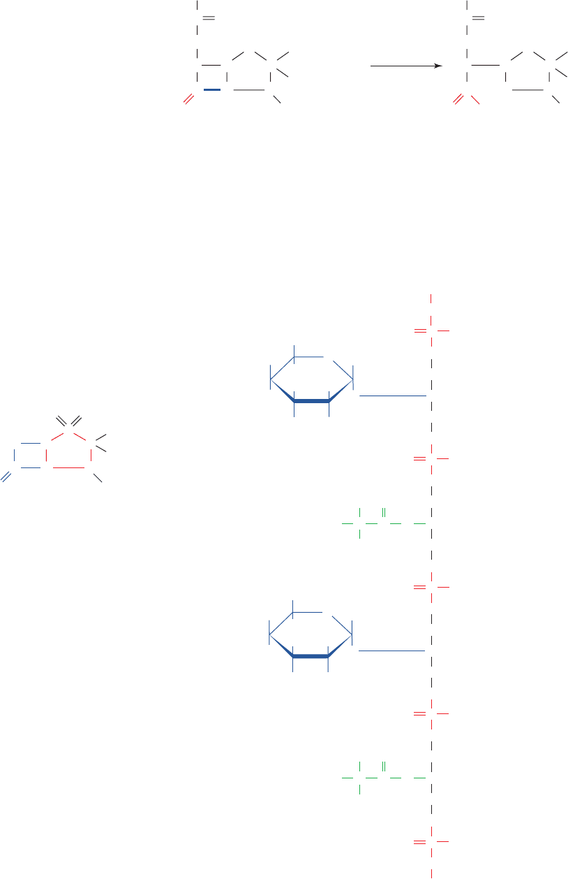
Most bacteria that are resistant to penicillin secrete a
-lactamase (also known as penicillinase), which inactivates
penicillin by hydrolytically cleaving the amide bond of its
-lactam ring (Fig. 11-29). However, the observation that
penicillinase activity varies with the nature of penicillin’s R
group has prompted the semisynthesis of penicillins, such
as ampicillin (Fig. 11-28), which are clinically effective
against penicillin-resistant strains of bacteria. In addition,
penicillins are often administered in combination with
-lactamase inhibitors such as sulbactam.
c. Bacterial Cell Walls Are Studded with
Antigenic Groups
The surfaces of gram-positive bacteria are covered by
teichoic acids (Greek: teichos, city walls),which account for
up to 50% of the dry weight of their cell walls. Teichoic
acids are polymers of glycerol or ribitol linked by phospho-
diester bridges (Fig. 11-30). The hydroxyl groups of this
sugar–phosphate chain are substituted by
D-Ala residues
and saccharides such as glucose or NAG.Teichoic acids are
anchored to the peptidoglycans via phosphodiester bonds
to the C6-OH groups of their NAG residues. They often
terminate in lipopolysaccharides (lipids that contain poly-
saccharides; Section 12-1).
The outer membranes of gram-negative bacteria (Fig.
11-25b) are composed of complex lipopolysaccharides,
proteins, and phospholipids that are organized in a compli-
cated manner. The periplasmic space, an aqueous com-
partment that lies between the plasma membrane and the
peptidoglycan cell wall, contains proteins that transport
sugars and other nutrients. The outer membrane functions
as a barrier to exclude harmful substances (such as gram
stain).This accounts for the observation that gram-negative
bacteria are less affected by lysozyme and penicillin, as well
as by other antibiotics, than are gram-positive bacteria.
HC CC
H
S
N
CHC
O
OO
Sulbactam
CH
3
CH
3
COO
–
The outer surfaces of gram-negative bacteria are coated
with complex and often unusual polysaccharides known as
O-antigens that uniquely mark each bacterial strain (Fig.
11-31). The observation that mutant strains of pathogenic
bacteria lacking O-antigens are nonpathogenic suggests
378 Chapter 11. Sugars and Polysaccharides
Figure 11-29 Enzymatic inactivation of
penicillin. Penicillinase inactivates penicillin by
catalyzing the hydrolysis of its -lactam ring to
form penicillinoic acid.
Figure 11-30 Structure of teichoic acid. A segment of a
teichoic acid molecule with a glycerol phosphate backbone that
bears alternating residues of
D-Ala and NAG.
R
C
O
C CHN
HN
HC C C
CH
3
CH
3
H
2
O
H
O
S
COO
⫺
R
C
OO
⫺
C CHN
HN
HC C C
CH
3
CH
3
H
H
O
S
COO
⫺
⫹
Penicillinoic acidPenicillin
penicillinase
HO
OH
CH
2
OH
CH
2
CH
2
CH
H
H
H
H
H
HO
NHCOCH
3
O
O
O
O
O
PO
⫺
(NAG)
D-Ala
O
O
O
PO
⫺
CH
2
CH
3
CH
2
CH
O
O
O
PO
⫺
CH
2
CH
2
CH
O
O
O
PO
⫺
CH
2
CH
2
CH
O
O
O
PO
⫺
HO
OH
CH
2
OH
H
H
H
H
H
NHCOCH
3
O
O
(NAG)
CNH
3
⫹
CO
HO
D-Ala
CH
3
CNH
3
⫹
CO
JWCL281_c11_359-385.qxd 6/3/10 10:35 AM Page 378
that O-antigens participate in the recognition of host cells.
O-Antigens, as their name implies, are also the means by
which a host’s immunological defense system recognizes
invading bacteria as foreign (Section 35-2A).As part of the
ongoing biological warfare between pathogen and host,
O-antigens are subject to rapid mutational alteration so as
to generate new bacterial strains that the host does not ini-
tially recognize (the mutations are in the genes specifying
the enzymes that synthesize the O-antigens).
C. Glycoprotein Structure and Function
a. Glycoprotein Carbohydrate Chains Are
Highly Diverse
Almost all the secreted and membrane-associated pro-
teins of eukaryotic cells are glycosylated. Indeed, protein
glycosylation is more abundant than all other types of post-
translational modifications combined. Oligosaccharides
form two types of direct attachments to these proteins:
N-linked and O-linked. Sequence analyses of glycopro-
teins have led to the following generalizations about these
attachments.
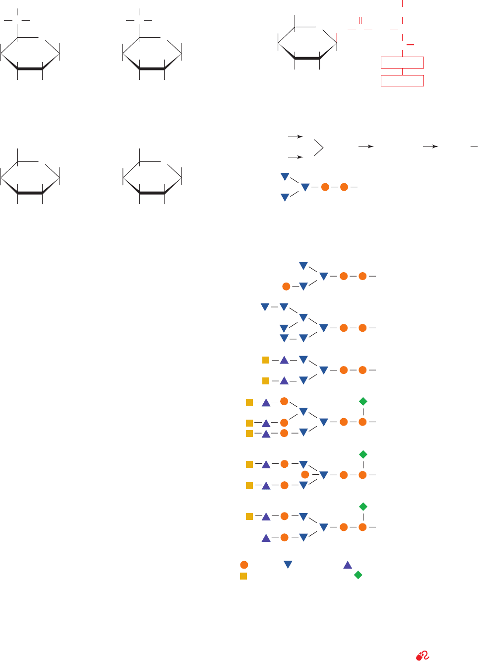
1. In the vast majority of N-glycosidic (N-linked) attach-
ments, an NAG is -linked to the amide nitrogen of an Asn
in the sequence Asn-X-Ser or Asn-X-Thr, where X is any
amino acid residue except Pro and only rarely Asp, Glu,
Leu, or Trp (Fig. 11-32a). The oligosaccharides in these
linkages usually have a distinctive core (innermost se-
quence; Fig. 11-32b) whose peripheral mannose residues
are linked to either mannose or NAG residues.These latter
residues may, in turn, be linked to yet other sugar residues,
Section 11-3. Glycoproteins 379
H
H
CH
3
H
H
HO
OH
H
H
OH
O
Abequose
(Abe)
HO
H
CH
3
H
OH
H
OH
H
H
H
O
Tyvelose
H
OH
CHHO
CH
2
OH
H
H
HO
OH
COO
⫺
H
H
O
2-Keto-3-deoxyoctanoate
(KDO)
HO
OH
COHH
CH
2
OH
H
OH
H
OH
H
H
H
O
L-Glycero-D-mannoheptose
Figure 11-31 Some of the unusual monosaccharides that occur
in the O-antigens of gram-negative bacteria. These sugars rarely
occur in other organisms.
Figure 11-32 N-Linked oligosaccharides. (a) All N-glycosidic
protein attachments occur through a -N-acetylglucosamino–Asn
bond in which the Asn occurs in the sequence Asn-X-Ser/Thr
(red) where X is any amino acid. (b) N-Linked oligosaccharides
usually have the branched (mannose)
3
(NAG)
2
core shown. (c)
Some examples of N-linked oligosaccharides. [After Sharon, N.
and Lis, H., Chem. Eng. News 59(13), 28 (1981).]
See
Kinemage Exercise 7-4
HO
OH
CH
2
OH
H
H
H
H
NH
NH
CCH
2
CH
C
X
Ser or Thr
O
H
NHCOCH
3
O
O
Asn
(NAG)
(a)
(b) Man ␣ (1
(c) Type Occurrence
Human
immunoglobulin M (IgM),
Bovine rhodopsin
Chicken ovalbumin,
Sindbis virus
Human and rabbit
transferrin,
Rat liver plasma
membrane
Vesicular
stomatitis
virus
Human immunoglobulin
G (IgG)
Bovine immunoglobulin
G (IgG)
6)
Man ␣ (13)
Man  (14) NAG  (1 4) NAG
= NAG, = Mannose, = Galactose,
= N-Acetylneuraminic acid, = Fucose
JWCL281_c11_359-385.qxd 6/3/10 10:35 AM Page 379
so that an enormous diversity of N-linked oligosaccharides
is possible (e.g., there are ⬃10
12
possible hexasaccharides,
although only a small fraction of them are actually synthe-
sized). Several N-linked oligosaccharides are shown in
Fig. 11-32c.
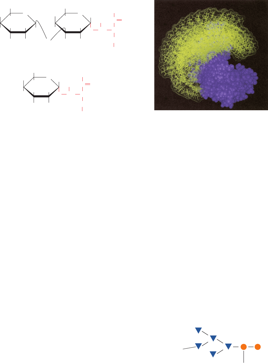
2. The most common O-glycosidic (O-linked) attach-
ment involves the disaccharide core -galactosyl-(1 S 3)-␣-
N-acetylgalactosamine ␣-linked to the OH group of either
Ser or Thr (Fig. 11-33a). Less commonly, glucose, galactose,
mannose, and xylose form ␣-O-glycosides with Ser or Thr
(Fig. 11-33b). All other hydroxyl-bearing amino acid side
chains occasionally form O-glycosidic bonds: those with Tyr
(e.g., in the protein glycogenin; Section 18-2B), 5-hydroxy-
Lys (Hyl; e.g., in collagen; Section 8-2Bb), and 4-hydroxy-
Pro (Hyp). However,there seem to be few, if any, additional
generalizations that can be made about O-glycosidically
linked oligosaccharides. They vary in size from a single
galactose residue in collagen to chains of up to 1000 disac-
charide units in proteoglycans.
N-Linked glycans are around 5-fold more common than O-
linked glycans with only ⬃10% of glycoproteins having
both types of attachments.
Oligosaccharides tend to attach to proteins at sequences
that form  bends. Taken with their hydrophilic character,
this observation suggests that oligosaccharides extend from
the surfaces of proteins rather than participate in their internal
structures. Indeed, the relatively few glycoprotein X-ray
structures that have yet been reported, for example, those of
immunoglobulin G (Section 35-2Ba) and the influenza virus
hemagglutinin (Section 33-4Bb), are consistent with this hy-
pothesis. This accounts for the observation that the protein
structures of most glycoproteins are unaffected by the re-
moval of their associated oligosaccharides. Both experimen-
tal and theoretical studies indicate that oligosaccharides have
mobile and rapidly fluctuating conformations (Fig. 11-34;
which accounts for the difficulty in crystallizing them). Thus,
representations in which oligosaccharides are shown as
having fixed three-dimensional structures do not tell the
whole story.
b. Glycoprotein Carbohydrates Have a
Variety of Functions
Cells tend to synthesize a large repertoire of a given N-
linked glycoprotein, in which each variant species (glyco-
form) differs somewhat in the sequences, locations, and num-
bers of its covalently attached oligosaccharides. For example,
one of the simplest glycoproteins, bovine pancreatic ribonu-
clease B (RNase B), differs from the well-characterized and
carbohydrate-free enzyme RNase A (Section 9-1A) only
by the attachment of a single N-glycosidically linked
oligosaccharide chain.The oligosaccharide has the core se-
quence diagrammed in Fig. 11-35 with considerable micro-
heterogeneity in the position of a sixth mannose residue.
The oligosaccharide does not affect the native enzyme’s
380 Chapter 11. Sugars and Polysaccharides
Figure 11-33 Some common O-glycosidic attachments of
oligosaccharides to glycoproteins (red).
Figure 11-34 Model of oligosaccharide dynamics in
bovine pancreatic ribonuclease B (RNase B). The allowed
conformations of the (mannose)
5
(NAG)
2
oligosaccharide
(yellow) that is linked to a single site on the protein (purple) are
shown in superimposed snapshots. [Courtesy of Raymond Dwek,
Oxford University, U.K.]
Figure 11-35 The microheterogeneous N-linked
oligosaccharide of RNase B has the (mannose)
5
(NAG)
2
core
shown. A sixth mannose residue occurs at various positions on
this core.
OH H
H
H
H
O
CH
2
OH CH
2
OH
HO
CH
NH
CH
C
H
H
O
H
O
NHCOCH
3
β-Galactosyl-(1n3)-α-N-acetylgalactosaminyl-Ser/
Thr
HO
OH
H
R
O
R = H or CH
3
H
H
O
(a)
CH
2
OH
CH
3
H
H
H
O
HO
H
α-Mannosyl-Ser/Thr
(b)
CH
NH
CH
C
O
R
O
R = H or
H
OH HO
NAG
Mannose
JWCL281_c11_359-385.qxd 6/3/10 10:35 AM Page 380
