Voet D., Voet Ju.G. Biochemistry
Подождите немного. Документ загружается.

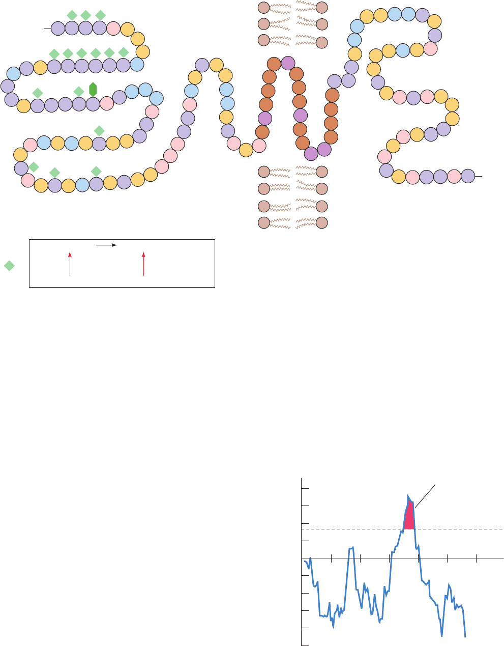
domain that has a high proportion of charged and polar
residues. The transmembrane domain, as is common in
many integral proteins, forms an ␣ helix, thereby satisfying
the hydrogen bonding requirements of its polypeptide
backbone. Indeed, the existence of glycophorin A’s single
transmembrane helix is predicted by computing the free
energy change in transferring ␣ helically folded polypep-
tide segments from the nonpolar interior of a membrane to
water (Fig. 12-22). Similar computations on other integral
proteins have also identified their transmembrane helices.
In many integral proteins, the hydrophobic segment(s)
anchors the active region of the protein to the membrane.
For instance, trypsin cleaves the membrane-bound enzyme
cytochrome b
5
into a polar, enzymatically active ⬃85-
residue N-terminal fragment and an ⬃50-residue C-termi-
nal fragment that remains embedded in the membrane
Section 12-3. Biological Membranes 401
Figure 12-21 The amino acid sequence and membrane
location of human erythrocyte glycophorin A. The protein,
which is ⬃60% carbohydrate by weight, bears 15 O-linked
oligosaccharides (green diamonds) and one that is N-linked (dark
green hexagon). The predominant sequence of the O-linked
oligosaccharides is given below. The protein’s transmembrane
portion (brown and purple) consists of 19 sequential predominantly
hydrophobic residues. Its C-terminal portion, which is located on
Ser
Ser
Ser
Ser
Ser
Ser
Asn
Ser
Thr
Thr Gln
Ser
Thr
Leu
Ser
Thr
Thr
Thr
Thr
Thr Asn
Asp
Asp
Ser
Ser
Tyr
Gly
Ser
Ser
Ser
Ser
Ser
Ser
Ser
Gln
Gln
Ser
Tyr
Tyr
Tyr
Ala
Ala
Phe
Ala
Ala
Pro
Pro
Pro
Pro
Pro
Pro
Pro
Pro
Pro
Asp
Asp
Asp
Asp
Met
His
His
His
Arg
Arg
Arg
Arg
Lys
Glu
Gly
Val
Val
Val
Val
Val
Leu
Val
Val
Val
Val
Ile
Ile
Ile
Ile
Ile
Ile
Ile
Ile
Val
Met
Ala
Val
Phe
Leu
Leu
Leu
Ile
Ile
Ile
Leu
Leu
Leu
Thr
Thr
Thr
Thr
Thr
Thr
Gly
Gly
Gly
Gly
His
Lys
Lys Lys
Lys
Ala Pro
Arg
Arg
His
Glu
Glu
=
Glu
Glu
Glu
Glu
Glu
Glu
Glu
Glu
Glu
10
30
20
40
50
70
60
80
90
100
110
120
130
Outside InsideBilayer
H
3
+
N
COO
–
NeuNAc –α NeuNAc–α
2,3
Gal –β (1 3) – GalNAc – α – Ser/Thr
2,6
Thr
Figure 12-22 A plot, for glycophorin A, of the calculated free
energy change in transferring 20-residue-long ␣ helical segments
from the interior of a membrane to water versus the position of
the segment’s first residue. Peaks higher than ⫹85 kJ ⴢ mol
⫺1
are
indicative of a transmembrane helix. [After Engleman, D.M.,
Steitz,T.A., and Goldman, A., Annu. Rev. Biophys. Biophys.
Chem. 15, 343 (1986).]
the membrane’s cytoplasmic face, is rich in anionic (pink) and
cationic (blue) amino acid residues.There are two common
genetic variants of glycophorin A: glycophorin A
M
has Ser and
Gly at positions 1 and 5, respectively, whereas they are Leu and
Glu in glycophorin A
N
. [Abbreviations: Gal ⫽ galactose, GalNAc
⫽ N-acetylgalactosamine, NeuNAc ⫽ N-acetylneuraminic acid
(sialic acid)]. [After Marchesi, V.T., Semin. Hematol. 16, 8 (1979).]
0 20 40 60 80 100 120
–100
+100
+200
–200
0
Free energy of transfer to water (kJ
.
mol
–1
)
First amino acid in 20-residue segment
85 kJ
.
mol
–1
cutoff
Position of
predicted
transmembrane
helix
JWCL281_c12_386-466.qxd 6/9/10 12:05 PM Page 401
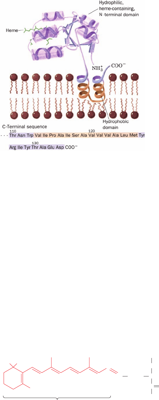
(Fig. 12-23). The asymmetric orientation of integral proteins
in the membrane is maintained by their infinitesimal flip-
flop rates (even slower than those of lipids), which result
from the greater sizes of the membrane protein “head
groups” in comparison to those of lipids. The origin of this
asymmetry is discussed in Section 12-4.
Relatively few integral proteins have yet been crystal-
lized—and then usually in the presence of detergents,
which are but poor substitutes for lipid bilayers. Thus, de-
spite their biological abundance, only ⬃0.8% of the pro-
teins of known structure are integral proteins (⬎80% of
which are bacterial proteins). A database of these proteins
is maintained at http://blanco.biomol.uci.edu/Membrane_
Proteins_xtal.html. In the remainder of this subsection, we
discuss the structures of four integral proteins: bacteri-
orhodopsin, the bacterial photosynthetic reaction center,
porins, and fatty acid amide hydrolase.
b. Bacteriorhodopsin Contains a Bundle of Seven
Hydrophobic Helical Rods
One of the structurally most studied integral proteins is
bacteriorhodopsin (BR) from the halophilic (salt loving)
bacterium Halobacterium salinarium that inhabits such
salty places as the Dead Sea (it grows best in 4.3M NaCl
and is nonviable below 2.0M NaCl;seawater contains 0.6M
NaCl). Under low O
2
conditions, its cell membrane devel-
ops ⬃0.5-m-wide patches of purple membrane whose
only protein component is BR. This 247-residue protein is
a light-driven proton pump; it generates a proton concen-
tration gradient across the membrane that powers the syn-
thesis of ATP (by a mechanism discussed in Section 22-3Bh).
Bacteriorhodopsin’s light-absorbing element, retinal, is
covalently bound to its Lys 216 (Fig. 12-24). This chro-
mophore (light-absorbing group), which is responsible for
the membrane’s purple color, is also the light-sensitive ele-
ment in vision.
The purple membrane, which is 75% protein and 25%
lipid, has an unusual structure compared to most other
membranes (Section 12-3C): Its BR molecules are
arranged in a highly ordered two-dimensional array (a
two-dimensional crystal). This permitted Richard Hender-
son and Nigel Unwin, through electron crystallography (a
technique they devised, resembling X-ray crystallography,
in which the electron beam of an electron microscope is
used to elicit diffraction from two-dimensional crystals), to
determine the structure of BR to near-atomic resolution
(3.0 Å). The more recently determined 1.9-Å-resolution
X-ray structure of BR, based on single crystals of BR dis-
solved in lipidic cubic phases (mixtures of lipids and water
that form a highly convoluted but continuous bilayer that is
interpenetrated by aqueous channels), closely resembles
that determined by electron crystallography.
Bacteriorhodopsin forms a homotrimer. Each of its sub-
units consists mainly of a bundle of seven ⬃25-residue
␣ helical rods that each span the lipid bilayer in directions
almost perpendicular to the bilayer plane (Fig. 12-25). BR
is therefore said to be polytopic (multispanning; Greek:
topos, place). The ⬃20-Å spaces between the protein
molecules in the purple membrane are occupied by this
bilayer (Fig. 12-25b). Adjacent ␣ helices, which are largely
402 Chapter 12. Lipids and Membranes
Figure 12-23 Liver cytochrome b
5
in association with a
membrane. The protein’s enzymatically active N-terminal
domain (purple), whose X-ray structure has been determined, is
anchored in the membrane by a hydrophobic and presumably ␣
helical C-terminal segment (brown) that begins and ends with
hydrophilic segments (purple).The amino acid sequence of the
horse enzyme indicates that this hydrophobic anchor consists of
a 13-residue segment ending 9 residues from the polypeptide’s
C-terminus (below). [Ribbon diagram of the N-terminal domain
after a drawing by Jane Richardson, Duke University. Amino
acid sequence from Ozols, J. and Gerard, C., J. Biol. Chem. 253,
8549 (1977).]
Figure 12-24 Molecular formula of retinal. Retinal, the
prosthetic group of bacteriorhodopsin, forms a Schiff base
with Lys 216 of the protein.A similar linkage occurs in
rhodopsin, the photoreceptor of the eye (Section 19-2B).
NCH
NH
CO
(CH
2
)
4
H
H
C
H
3
CCH
3
CH
3
CH
3
CH
3
Retinal residue
Lys 216
+
JWCL281_c12_386-466.qxd 6/9/10 12:05 PM Page 402
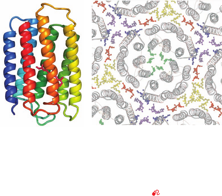
hydrophobic in character, are connected in a head-to-tail
fashion by short polypeptide loops. This arrangement
places the protein’s charged residues near the surfaces of
the membrane in contact with the aqueous solvent. The
internal charged residues line the center of the helix
bundle of each monomer so as to form a hydrophilic
channel that facilitates the passage of protons. Other
membrane pumps and channels (Chapter 20) have similar
structures.
c. The Photosynthetic Reaction Center Contains
Eleven Transmembrane Helices
The primary photochemical process of photosynthesis
in purple photosynthetic bacteria is mediated by the so-
called photosynthetic reaction center (PRC; Section 24-2B),
a transmembrane (TM) protein consisting of at least
three nonidentical ⬃300-residue subunits that collectively
bind four chlorophyll molecules, four other chromophores,
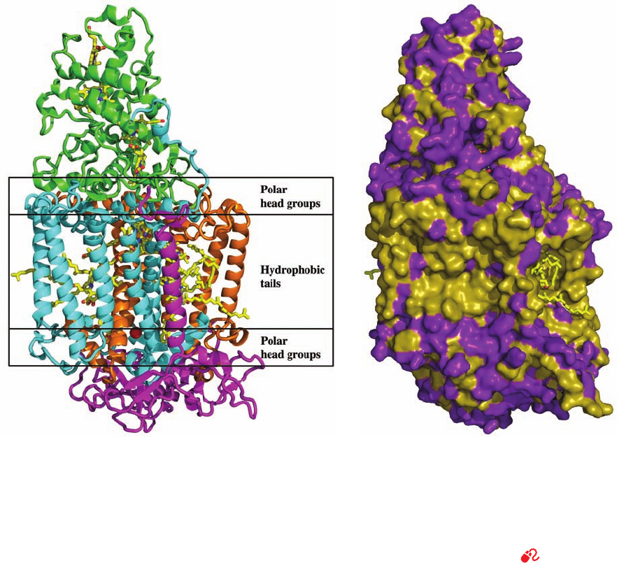
and a nonheme Fe(II) ion. The 1187-residue photosyn-
thetic reaction center of Rhodopseudomonas (Rps.)
viridis, whose X-ray structure was determined in 1984 by
Hartmut Michel, Johann Deisenhofer, and Robert Huber,
was the first TM protein to be described in atomic detail
(Fig. 12-26). The polytopic protein’s TM portion consists
of 11 ␣ helices that form a 45-Å-long flattened cylinder
with the expected hydrophobic surface. In later chapters
we shall see that the transmembrane portions of most TM
proteins consist of bundles of one to ⬎20 helices, most of
which are closely perpendicular to the membrane al-
though some may be obliquely oriented and/or not fully
traverse the membrane.
d. Porins Are Channel-Forming Proteins That
Contain Transmembrane  Barrels
The outer membranes of gram-negative bacteria (Sec-
tion 11-3B) protect them from hostile environments but
must nevertheless be permeable to small polar solutes such
as nutrients and waste products. These outer membranes
consequently contain embedded channel-forming proteins
called porins, which are usually trimers of identical 30- to
50-kD subunits that permit the passage of solutes of less
than ⬃600 D. Porins also occur in eukaryotes in the outer
membranes of mitochondria and chloroplasts (thereby
providing a further indication that these organelles are de-
scended from bacteria; Section 1-2A).
The X-ray structures of several different porins have
been elucidated, among them the Rhodobacter (Rb.) cap-
sulatus porin, determined by Georg Schulz, and the E. coli
OmpF and PhoE porins, determined by Johan Jansonius.
The 340- and 330-residue OmpF and PhoE porins share
63% sequence identity but have little sequence similarity
with the 301-residue Rb. capsulatus porin. Nevertheless, all
Section 12-3. Biological Membranes 403
Figure 12-25 Structure of bacteriorhodopsin. (a) The protein
is shown in ribbon form as viewed from within the membrane
plane and colored in rainbow order from its N-terminus (blue) to
its C-terminus (red). Its covalently bound retinal is drawn in stick
form (magenta). [Based on an X-ray structure by Nikolaus
Grigorieff and Richard Henderson, MRC Laboratory of
Molecular Biology, Cambridge, U.K. PDBid 2BRD.] (b) The
X-ray structure of a bacteriorhodopsin trimer with portions of its
surrounding trimers as viewed from the extracellular side of the
membrane. The protein molecules are shown in ribbon form
(gray) and their associated lipid tails are shown in ball-and-stick
form in different colors with symmetry-related lipid tails the
same color (the lipid head groups are disordered and hence not
seen). Only the lipids in the extracellular leaflet are shown; those
in the cytoplasmic leaflet have a similar distribution. Note how
the seven antiparallel ␣ helices in each BR monomer are
cyclically arranged in two layers of four and three helices with
helices adjacent in sequence also adjacent in space (the N to C
direction circulates clockwise in this view). [Courtesy of Eva
Pebay-Peyroula, Université Joseph Fourier, Grenoble, France.
PDBid 1AP9.]
See Kinemage Exercise 8-1
(a)
(b)
JWCL281_c12_386-466.qxd 6/9/10 12:05 PM Page 403
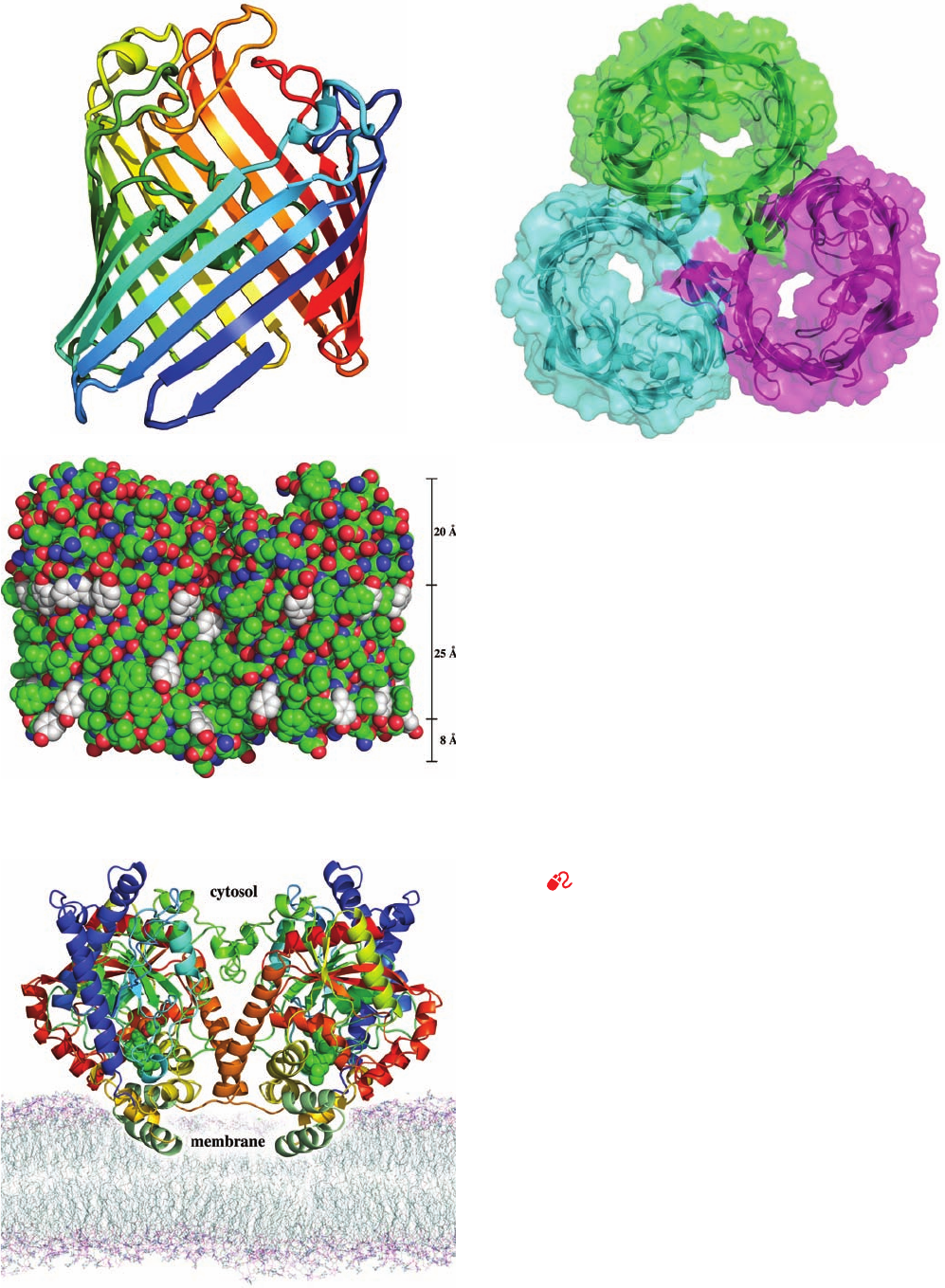
three porins have closely similar structures. Each monomer
of these homotrimeric proteins predominantly consists of a
16-stranded antiparallel  barrel which forms a solvent-
accessible pore along the barrel axis that has a length of
⬃55 Å and a minimum diameter of ⬃7 Å (Fig. 12-27; al-
though note that  barrel membrane proteins with 8, 10, 12,
14, 18, 19, 22, and 24 strands are also known). In the OmpF
and PhoE porins, the N- and C-termini associate via a salt
bridge in the 16th  strand, thereby forming a pseudocyclic
structure (Fig. 12-27a). Note that a  barrel fully satisfies
the polypeptide backbone’s hydrogen bonding potential,
as does an ␣ helix. As expected, the side chains at the pro-
tein’s membrane-exposed surface are nonpolar, thereby
forming an ⬃25-Å-high hydrophobic band encircling the
trimer (Fig. 12-27c). In contrast, the side chains at the sol-
vent-exposed surface of the protein, including those lining
the walls of the aqueous channel, are polar. Possible mech-
anisms for solute selectivity by these porins are discussed
in Section 20-2D.
e. Fatty Acid Amide Hydrolase Binds to Only
One Bilayer Leaflet
Not all integral proteins are TM proteins. For example,
the enzyme fatty acid amide hydrolase (FAAH) is an inte-
gral protein that binds to the cytoplasmic leaflet of the
plasma membrane. It is therefore said to be monotopic as is
cytochrome b
5
(Fig. 12-23). FAAH’s X-ray structure, deter-
mined by Raymond Stevens and Benjamin Cravatt, reveals
that each 537-residue subunit of this homodimer consists
of an 11-stranded mixed  sheet surrounded by 28 ␣ helices
of various lengths (Fig. 12-28). Its membrane-binding seg-
ment consists of a helix–turn–helix motif, whose surface
faces outward from the body of the protein to form a hy-
drophobic plateau. Its component nonpolar residues, many
of which are aromatic, are interspersed with several basic
residues, which presumably interact electrostatically with
the membrane’s phospholipid head groups. The structure
of this hydrophobic motif resembles that observed in the
X-ray structures of two other monotopic integral proteins,
404 Chapter 12. Lipids and Membranes
Figure 12-26 X-ray structure of the photosynthetic reaction
center of Rps. viridis. (a) The H, M, and L subunits, which are
respectively shown as pink, blue, and orange ribbons, collectively
have 11 transmembrane helices.The four-heme c-type
cytochrome (green), which does not occur in all species of
photosynthetic bacteria, is bound to the external face of the
complex.The prosthetic groups are drawn in stick form with C
yellow, N blue, and O red with a bound Fe(II) ion represented by
a red sphere. The position that the transmembrane protein is
thought to occupy in the lipid bilayer is indicated schematically.
(b) A surface diagram, viewed as in Part a, in which hydrophobic
residues are tan and polar residues are purple. Note how few
polar groups are externally exposed in the portion of the protein
that is immersed in the nonpolar region of the lipid bilayer.
[Based on an X-ray structure by Johann Deisenhofer, Robert
Huber, and Hartmut Michel, Max-Planck-Institut für Biochemie,
Martinsreid, Germany. PDBid 1PRC.]
See Kinemage
Exercise 8-2
(a)
(b)
JWCL281_c12_386-466.qxd 6/9/10 12:05 PM Page 404
Section 12-3. Biological Membranes 405
Figure 12-27 X-ray crystal structure of the E. coli OmpF
porin. (a) A ribbon diagram of the monomer colored in rainbow
order from its N-terminus (blue) to its C-terminus (red). Each
strand of this 16-stranded antiparallel  barrel is inclined by
⬃45º to the barrel axis. Its C-terminal strand is continued by the
N-terminal segment (bottom right), thereby forming a pseudo-
continuous strand.All porins of known structure have similar
structural properties. (b) Ribbon diagram of the trimer embedded
in its semitransparent surface and viewed along its threefold axis
of symmetry from the cell’s exterior showing the pore through
each subunit. Each subunit is differently colored. Adjacent
 strands in adjoining subunits extend essentially perpendicularly
to each other. (c) A space-filling model of the trimer viewed
perpendicular to its 3-fold axis. N atoms are blue, O atoms are
red, and C atoms are green, except those of Trp and Tyr side
chains, which are white. These latter groups delimit an ⬃25-Å-high
hydrophobic band (scale at right) that is immersed in the nonpolar
portion of the bacterial outer membrane (with the cell’s exterior
at the tops of Parts a and c). Compare the hydrophobic band in
this figure with that in Fig. 12-26b. [Based on an X-ray structure
by Johan Jansonius, University of Basel, Switzerland. PDBid
1OPF.]
See Kinemage Exercise 8-3
Figure 12-28 X-ray structure of rat liver fatty acid amide
hydrolase indicating its proposed disposition in the cytoplasmic
leaflet of the plasma membrane. This homodimeric enzyme is
viewed along the plane of the membrane with its 2-fold axis of
symmetry vertical. It is drawn in ribbon form with each subunit
colored in rainbow order from its N-terminus (blue) to its
C-terminus (red) except for its putative membrane-binding
motif, which is dark green. The enzyme’s bound inhibitor, methyl
arachidonyl fluorophosphonate, is drawn in space-filling form
with C green, O red, and P orange.The membrane model was
generated by a molecular dynamics simulation of a
palmitoyloleoylphosphatidylethanolamine bilayer. [Based on an
X-ray structure by Raymond Stevens and Benjamn Cravatt,
Scripps Research Institute, La Jolla, California. PDBid 1MT5.]
(a)
(c)
(b)
JWCL281_c12_386-466.qxd 9/9/10 5:57 PM Page 405
prostaglandin H synthase (Section 25-7B) and squalene-
hopene cyclase (Section 25-6Ad). Nevertheless, these en-
zymes have no apparent sequence or structural homology,
which suggests that they have independently evolved simi-
lar modes of membrane integration.
f. Integral Proteins Have Common
Structural Features
Hydrophobic forces, as we have seen in Section 8-4, are
the dominant interactions stabilizing the three-dimensional
structures of water-soluble globular proteins. However,
since the membrane-exposed regions of integral proteins
are immersed in nonpolar environments, what stabilizes
their structures? Analysis of a variety of integral proteins
indicates that their membrane-exposed regions have a
hydrophobic organization opposite to that of water-soluble
proteins: Their membrane-exposed residues are more
hydrophobic, on average, than their interior residues, even
though these interior residues have average hydrophobici-
ties and packing densities comparable to those of water-
soluble proteins. Evidently, the structures of integral and
water-soluble proteins are both stabilized by the exclusion
of their interior residues from the surrounding solvent,
although in the case of integral proteins, the solvent is the
lipid bilayer. In addition, the low polarity and anhydrous
environments of transmembrane proteins are likely to
strengthen their hydrogen bonds relative to those of solu-
ble proteins.
In the foregoing TM proteins, those portions of the
transmembrane secondary structural elements (helices in
BR and the PRC, and  strands in the porins) that contact
the bilayer’s hydrocarbon core consist mainly of the hy-
drophobic residues Ala, Ile, Leu,Val, and Phe.The flanking
residues, which penetrate the bilayer’s interface region, are
enriched with Trp, and Tyr. Hence, TM proteins’ hydropho-
bic transmembrane bands are bordered by rings of Trp and
Tyr side chains (e.g., Fig. 12-27c) that delineate the water–
bilayer interface. Note that these side chains are oriented
such that their polar portions (N and O atoms) extend into
the polar regions of the membrane, a phenomenon named
snorkeling. Lys and Arg side chains near the interface tend
to be similarly oriented. In contrast, Phe, Leu, and Ile side
chains tend to point toward the membrane core, a phenom-
enon dubbed antisnorkeling.
In each of the foregoing TM proteins, the secondary
structural elements that are adjacent in sequence are also
adjacent in structure and hence tend to be antiparallel.This
relatively simple up–down topology results from the con-
straints associated with the insertion of a folding polypep-
tide chain into the lipid bilayer (Section 12-4Be).
B. Lipid-Linked Proteins
Lipids and proteins associate covalently to form lipid-
linked proteins, whose lipid portions anchor their attached
proteins to membranes and mediate protein–protein interac-
tions. Proteins form covalent attachments with three
classes of lipids: (1) isoprenoid groups such as farnesyl and
geranylgeranyl residues, (2) fatty acyl groups such as myris-
toyl and palmitoyl residues, and (3) glycoinositol phospho-
lipids (GPIs). In this subsection, we discuss the properties
of these lipid-linked proteins.
a. Prenylated Proteins
A variety of proteins have covalently attached iso-
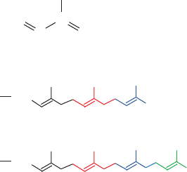
prenoid groups, mainly the C
15
farnesyl and C
20
geranylger-
anyl residues (isoprene, a C
5
hydrocarbon, is the chemical
unit from which many lipids, including cholesterol and
other steroids, are constructed; Section 25-6A).
The most common isoprenylation (or just prenylation) site
in proteins is the C-terminal tetrapeptide CaaX, where C is
Cys, “a” is often an aliphatic amino acid residue, and X is
any amino acid. However, the identity of X is a major
prenylation determinant: Proteins are farnesylated when X
is Gln, Met, or Ser and geranylgeranylated when X is Leu.
In both cases, the prenyl group is enzymatically linked to
the Cys sulfur atom via a thioether linkage.The aaX tripep-
tide is then proteolytically excised and the newly exposed
terminal carboxyl group is esterified with a methyl group
(Fig. 12-29).
Two other types of prenylation sites have also been
characterized: (1) the C-terminal sequence CXC, in which
both Cys residues are geranylgeranylated and the terminal
carboxyl group is methyl esterified; and (2) the C-terminal
sequence CC in which one or both Cys residues are ger-
anylgeranylated but the carboxyl group is not methylated.
Proteins that are so prenylated are almost exclusively
members of the Rab family of small GTP-binding proteins
that participate in intracellular membrane trafficking (Sec-
tion 12-4Db).
What functions are served by protein prenylation?
Many prenylated proteins are associated with intracellular
membranes, and mutating their Cys prenylation sites
blocks their membrane localization. Evidently, the hy-
drophobic prenyl group can act to anchor its attached pro-
tein to a membrane. However, this can only be part of the
story since proteins with the same prenyl groups may be lo-
calized to different intracellular membranes. Moreover,
fusing the CaaX motif from a normally prenylated protein
to the C-terminus of a normally unprenylated protein
yields a hybrid protein that is correctly prenylated and
carboxyl methylated but which remains cytosolic. These
H
2
C
HC
Isoprene
Farnesyl residue
CH
C
CH
2
CH
3
CH
3
CH
3
CH
3
CH
3
HC
Geranylgeranyl residue
CH
3
CH
3
CH
3
CH
3
CH
3
406 Chapter 12. Lipids and Membranes
JWCL281_c12_386-466.qxd 6/9/10 12:06 PM Page 406
observations suggest that prenylated proteins may interact
with specific membrane-bound receptor proteins and
hence that prenylation also facilitates protein–protein inter-
actions. This idea is corroborated by the observation that,
in certain proteins involved in intracellular signaling [for
example, Ras (Section 19-3Cf) and the so-called G proteins
(Section 19-2)], prenylation and carboxyl methylation en-
hance the intersubunit associations that mediate signal
transmission.
b. Fatty Acylated Proteins
Two fatty acids are known to be covalently linked to eu-
karyotic proteins:
1. Myristic acid, a biologically rare saturated C
14
fatty
acid (Table 12-1), which is appended to a protein in amide
linkage to the ␣-amino group of an N-terminal Gly residue.
Myristoylation almost always occurs cotranslationally (as
the protein is being synthesized), and this attachment is
stable, that is, the myristoyl group has a half-life similar to
that of the protein to which it is appended.
2. Palmitic acid, a biologically common saturated C
16
fatty acid, which is joined to a protein in thioester linkage
to a specific Cys residue. In some cases, the palmitoylated
protein is also prenylated. For example, Ras must be farne-
sylated and carboxyl methylated as described above before
it is palmitoylated at a Cys residue that precedes the pro-
tein’s C-terminus by several residues. Palmitoylation oc-
curs post-translationally in the cytosol and is reversible.
Fatty acyl groups are thought to function as membrane an-
chors for proteins, much as do prenyl groups. However, the
requirement of many proteins for specific fatty acyl residues
suggests that these groups also participate in targeting their
attached proteins to specific cellular locations. Indeed,
palmitoylated proteins occur almost exclusively on the cyto-
plasmic face of the plasma membrane, whereas myristoy-
lated proteins are found in a number of subcellular compart-
ments including the cytosol, endoplasmic reticulum, Golgi
apparatus, plasma membrane, and nucleus. Many fatty acy-
lated proteins participate in intracellular signaling processes
through protein–protein interactions in a manner similar to
prenylated proteins. Since the membrane affinities and bio-
logical activities of many proteins are enhanced by palmi-
toylation, the reversibility of palmitoylation appears to be
involved in controlling intracellular signaling processes.
c. GPI-Linked Proteins
Glycosylphosphatidylinositol (GPI) groups function to
anchor a wide variety of proteins to the exterior surface of
the eukaryotic plasma membrane.There is no obvious rela-
tionship among the numerous proteins that have GPI an-
chors, which include enzymes, receptors, immune system
proteins, and recognition antigens. Evidently, GPI groups
simply provide an alternative to transmembrane polypep-
tide domains in binding proteins to the plasma membrane.
The core structure of GPI anchors consists of phos-
phatidylinositol (Table 12-2) glycosidically linked to a lin-
ear tetrasaccharide composed of three mannose residues
Section 12-3. Biological Membranes 407
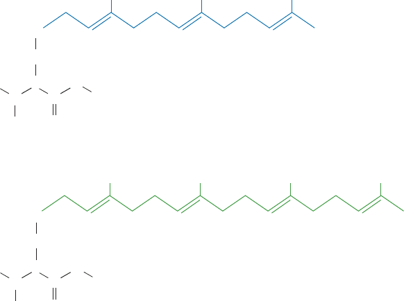
Figure 12-29 Prenylated proteins. (a) A farnesylated protein
and (b) a geranylgeranylated protein. In both cases, the protein is
synthesized with the C-terminal sequence CaaX, where C is Cys,
“a” is often an aliphatic amino acid, and X is any amino acid.
After the prenyl group is appended to the protein in thioether
(a)
(b)
S
CH
2
CHProtein
O
C
N
H
CH
3
O
S–Farnesyl cysteine methyl ester
S
CH
2
CHProtein
O
C
N
H
CH
3
O
S–Geranylgeranyl cysteine methyl ester
linkage with the Cys residue, the aaX tripeptide is hydrolytically
cleaved away and the new carboxyl terminus is methyl esterified.
When X is Ala, Met, or Ser, the protein is farnesylated and when
X is Leu, it is geranylgeranylated.
JWCL281_c12_386-466.qxd 6/9/10 12:06 PM Page 407
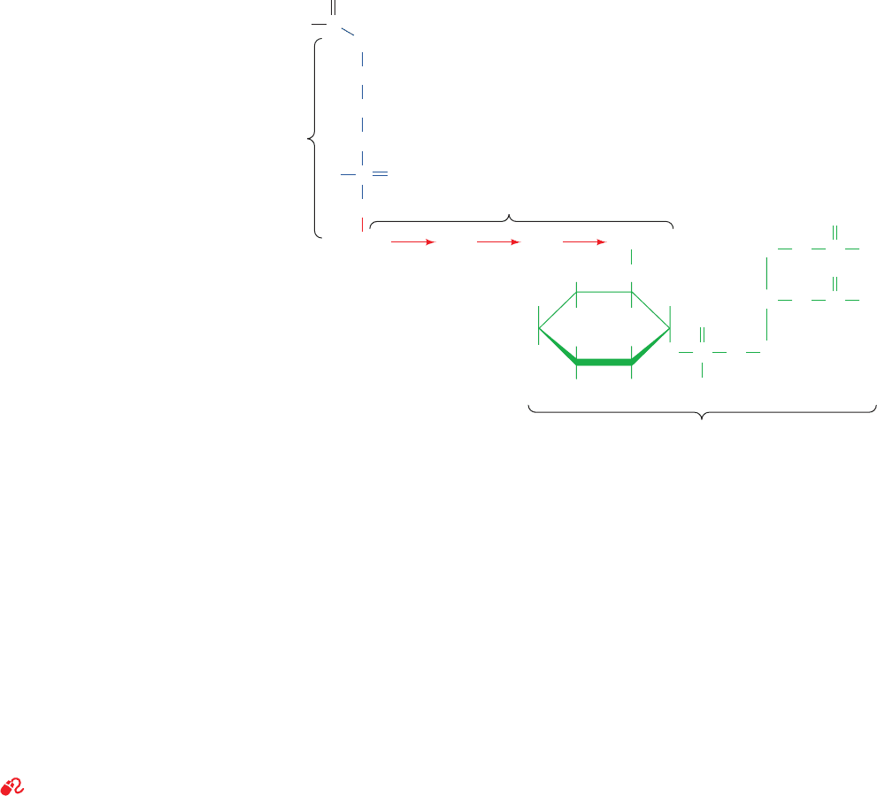
and one glucosaminyl residue (Fig. 12-30).The mannose at
the nonreducing end of this assembly forms a phospho-
ester bond with a phosphoethanolamine residue, which in
turn, is amide-linked to the protein’s C-terminal carboxyl
group. The core tetrasaccharide is generally substituted
with a variety of sugar residues that vary with the identity
of the protein. There is likewise considerable diversity in
the fatty acid residues. The synthesis of GPI anchors is dis-
cussed in Section 23-3Bk.
GPI-anchored proteins occur on the exterior surface
of the plasma membrane for the same reason as do the
carbohydrate residues of glycoproteins (which we discuss
in Section 12-4Ca). Proteins destined to be GPI-anchored
are synthesized with membrane-spanning C-terminal se-
quences of 20 to 30 hydrophobic residues (as described in
Section 12-4Ba) that are removed during GPI addition.
This is corroborated by the observation that GPI-anchored
proteins are released from the plasma membrane by treat-
ment with phosphatidylinositol-specific phospholipases
(Section 19-4B), thereby demonstrating that the mature
polypeptides are not embedded in the lipid bilayer.
C. Fluid Mosaic Model of Membrane Structure
See Guided Exploration 11: Membrane structure and the fluid mosaic
model
The demonstrated fluidity of artificial lipid bilayers
suggests that biological membranes have similar properties.
This seminal idea was proposed in 1972 by S. Jonathan
Singer and Garth Nicolson in their unifying theory of
membrane structure known as the fluid mosaic model. The
theory postulates that integral proteins resemble “ice-
bergs” floating in a two-dimensional lipid “sea” (Fig. 12-20)
and that these proteins freely diffuse laterally in the lipid
matrix unless their movements are restricted by associa-
tions with other cell components.
a. The Fluid Mosaic Model Has Been
Verified Experimentally
The validity of the fluid mosaic model has been estab-
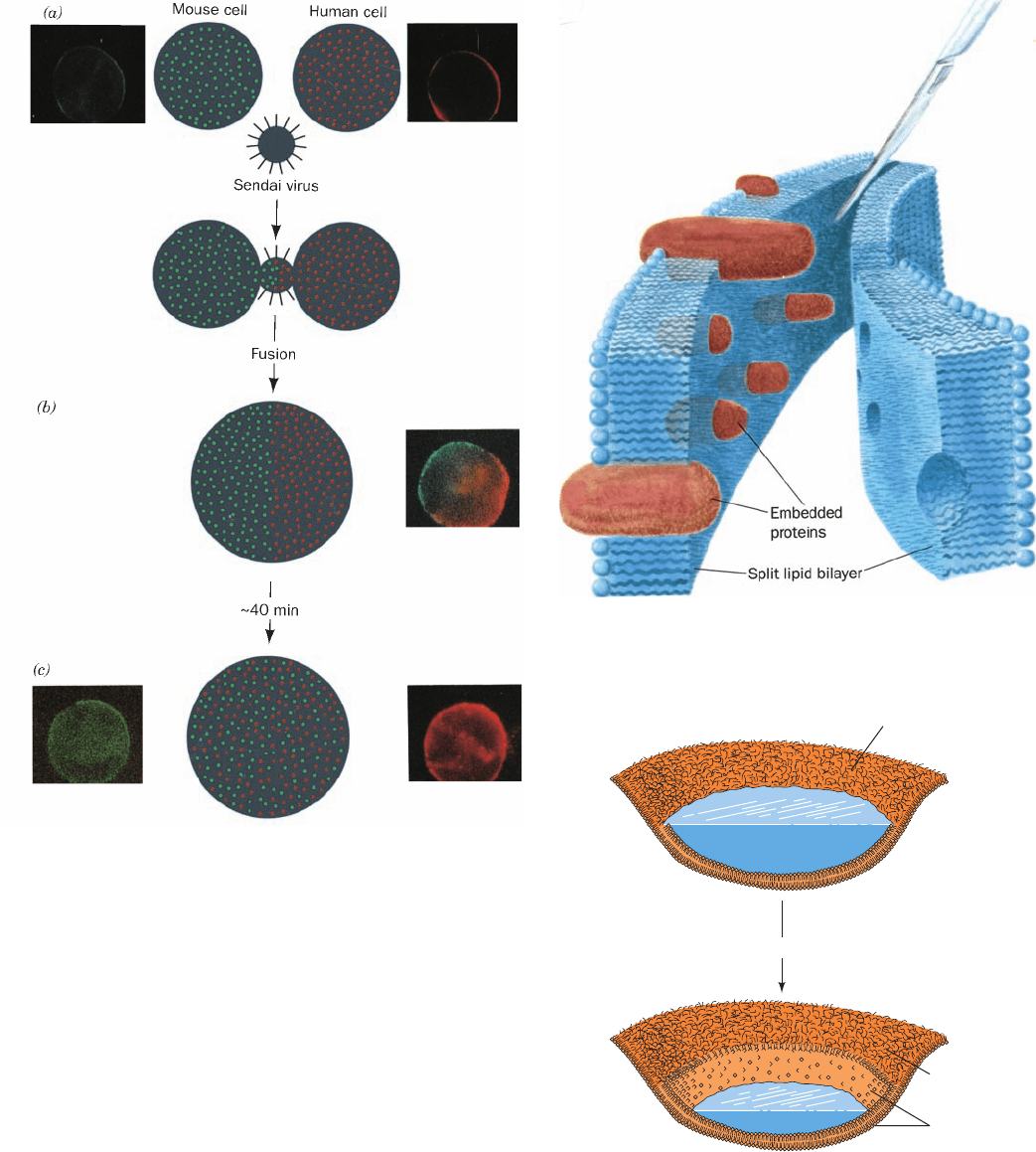
lished in several ways. Perhaps the most vivid is an experi-
ment by Michael Edidin (Fig. 12-31). Cultured mouse cells
were fused with human cells by treatment with Sendai
virus to yield a hybrid cell known as a heterokaryon. The
mouse cells were labeled with mouse protein–specific
antibodies to which a green-fluorescing dye had been cova-
lently linked (immunofluorescence). The proteins on the
human cells were similarly labeled with a red-fluorescing
marker. On cell fusion, the mouse and human proteins, as
seen under the fluorescence microscope, were segregated
on the two halves of the heterokaryon. After 40 min at
37°C, however, these proteins had thoroughly intermin-
gled.The addition of substances that inhibit metabolism or
protein synthesis did not slow this process, but lowering the
temperature below 15°C did. These observations indicate
that the mixing process is independent of both metabolic
energy and the insertion into the membrane of newly syn-
thesized proteins. Rather, it is a result of the diffusion of
existing proteins throughout the fluid membrane, a process
that slows as the temperature is lowered.
Fluorescence photobleaching recovery measurements
(Fig. 12-15) indicate that membrane proteins vary in their
lateral diffusion rates. Some 30 to 90% of these proteins
are freely mobile; they diffuse at rates only an order of
magnitude or so slower than those of the much smaller
lipids, so that they typically take from 10 to 60 min to dif-
fuse the 20-m length of a eukaryotic cell. Other proteins
diffuse more slowly, and some, because of submembrane
attachments, are essentially immobile.
The distribution of proteins in membranes may be visual-
ized through electron microscopy using the freeze-fracture
and freeze-etch techniques. In the freeze-fracture proce-
dure, which was devised by Daniel Branton, a membrane
specimen is rapidly frozen to near liquid nitrogen tempera-
tures (⫺196°C). This immobilizes the sample and thereby
minimizes its disturbance by subsequent manipulations.
The specimen is then fractured with a cold microtome
knife, which often splits the bilayer into monolayers (Fig.
12-32). Since the exposed membrane itself would be
408 Chapter 12. Lipids and Membranes
Man
α1,2 α1,6
α1,6
Man
NH
CH
2
H
HH
H
OH
OH OH
Phosphatidylinositol
Core tetrasaccharide
HO
HO
O
P
O
O
–
H
O
CProtein
Phospho-
ethanolamine
α1,4
Man GlcNH
2
O
CH
2
O
PO
–
O
O
6
O C R
2
O CH
2
HC
O
O C R
1
H
2
C
Figure 12-30 Core structure of the GPI anchors of proteins.
R
1
and R
2
represent fatty acid residues whose identities vary
with the protein.The tetrasaccharide may have a variety of
attached sugar residues whose identities also vary with the
protein.
JWCL281_c12_386-466.qxd 6/9/10 12:06 PM Page 408
destroyed by an electron beam, its metallic replica is made
by coating the membrane with a thin layer of carbon, shad-
owing it (covering it by evaporative deposition under high
vacuum) with platinum, and removing the organic matter
by treatment with acid. Such a metallic replica can be ex-
amined by electron microscopy. In the freeze-etch proce-
dure, the external surface of the membrane adjacent to the
cleaved area revealed by freeze fracture may also be visu-
alized by first subliming (etching) away, at ⫺100°C, some of
the ice in which it is encased (Fig. 12-33).
Section 12-3. Biological Membranes 409
Figure 12-31 Sendai virus–induced fusion of a mouse cell with
a human cell and the subsequent intermingling of their cell-surface
components as visualized by immunofluorescence. Human and
mouse antigens are labeled with red and green fluorescent
markers, respectively. (a) The membrane-encapsulated Sendai
virus specifically binds to cell-surface receptors on both types of
cells and subsequently fuses to their cell membranes. (b) This
results in the formation of a cytoplasmic bridge between the cells
that expands so as to form a heterokaryon. (c) After 40 min, the red
and green markers are fully intermingled.The photomicrographs
were taken through filters that allowed only red or green light to
reach the camera; that in Part b is a double exposure and those in
Part c are of the same cell. [Immunofluorescence photomicrographs
courtesy of Michael Edidin, Johns Hopkins University.]
Figure 12-32 The freeze-fracture technique. A membrane
that has been split by freeze-fracture, as is schematically
diagrammed, exposes the interior of the lipid bilayer and its
embedded proteins.
Figure 12-33 The freeze-etch procedure. The ice that encases
a freeze-fractured membrane (top) is partially sublimed away so
as to expose the outer membrane surface (bottom) for electron
microscopy.
Etching
Ice
Fracture face
Inner
surface
Outer
surface
JWCL281_c12_386-466.qxd 6/9/10 12:06 PM Page 409
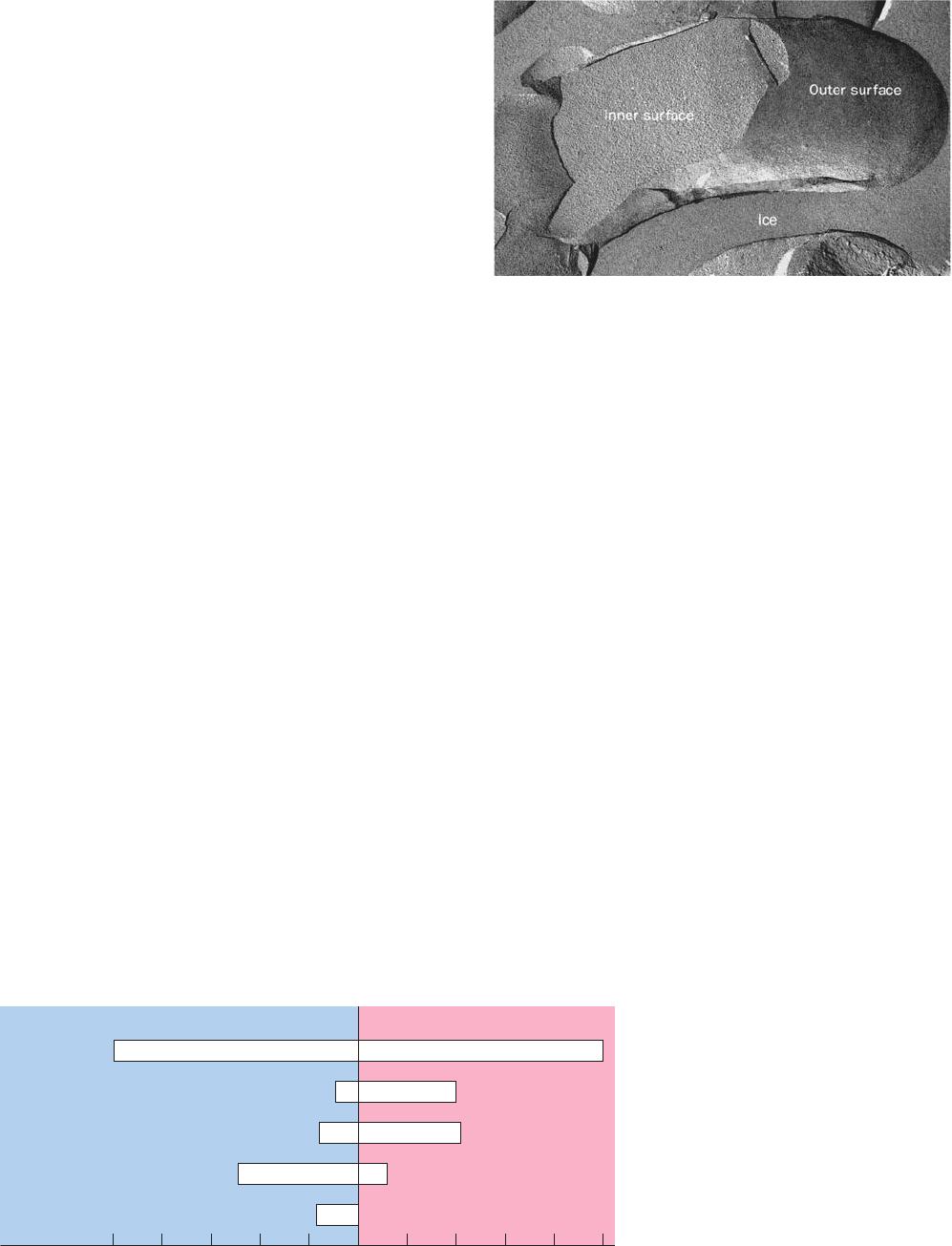
Freeze-etch electron micrographs of most biological mem-
branes show an inner fracture face that is studded with em-
bedded 50- to 85-Å-diameter globular particles (Fig. 12-34)
that appear to be distributed randomly. These particles
correspond to membrane proteins, as is demonstrated by
their disappearance when the membrane is treated with
proteases before its freeze fracture.This is further corrobo-
rated by the observation that the myelin membrane, which
has a low protein content, as well as liposomes composed
of only lipids, have smooth inner fracture faces. Outer
membrane surfaces also have a relatively smooth appear-
ance (Fig. 12-34) because integral proteins tend not to pro-
trude very far beyond them.The distributions of individual
external proteins may be visualized by staining procedures,
such as the use of ferritin-labeled antibodies, to yield elec-
tron micrographs similar in appearance to Fig. 11-36.
b. Membrane Lipids and Proteins Are
Unevenly Distributed
The distribution of lipids between the different sides of bi-
ological membranes has been established through the use
of phospholipid-hydrolyzing enzymes known as phospholi-
pases. Phospholipases cannot pass through membranes, so
that only phospholipids on the external surfaces of intact
cells are susceptible to their action. Such studies indicate
that the lipids in biological membranes, like the proteins, are
asymmetrically distributed between the leaflets of a bilayer
(e.g., Fig. 12-35). Carbohydrates, as we have seen (Section
11-3Cd), are located almost exclusively on the external sur-
faces of plasma membranes.
Lipids and proteins in plasma membranes may also be
laterally organized. Thus, the plasma membranes of most
cells have two or more distinct domains that have different
functions. For example, the plasma membranes of epithe-
lial cells (the cells lining body cavities and free surfaces)
have an apical domain, which faces the lumen of the cavity
and often has a specialized function (e.g., the absorption of
nutrients in intestinal brush border cells; Section 20-4A),
and a basolateral domain, which covers the remainder of
the cell. These two domains, which do not intermix, have
different compositions of both lipids and proteins.
A variety of measurements indicate that the hundreds of
different lipids and proteins within a given plasma mem-
brane domain are not uniformly mixed but instead often seg-
regate to form microdomains that contain only certain types
of lipids and proteins. This may occur for several reasons:
1. Certain integral proteins associate to form aggre-
gates or patches in the membrane (e.g., BR), which in turn
may preferentially associate with specific lipids. Alterna-
tively, some integral proteins are localized by attachments
to elements of the cytoskeleton (which underlies the
plasma membrane; Section 1-2A) or are trapped within the
spaces enclosed by the resulting “fences.”
2. Integral proteins may specifically interact with par-
ticular lipids. For example, mismatches between the length
of an integral protein’s hydrophobic TM band and the av-
erage thickness of a lipid bilayer may result in the selective
accumulation of certain phospholipids around the protein
in an annulus of 10 to 20 layers.
3. Divalent metal ions, notably Ca
2⫹
, selectively ligate
negatively charged head groups such as those of phos-
phatidylserine, thereby causing these phospholipids to ag-
gregate in the membrane. Such metal ion–induced phase
separations are known to regulate the activities of certain
membrane-bound enzymes.
410 Chapter 12. Lipids and Membranes
Figure 12-34 Freeze-etch electron micrograph of a human
erythrocyte plasma membrane. The exposed interior face of the
membrane is studded with numerous globular particles that are
integral proteins (see Fig. 12-32). The outer surface of the mem-
brane appears smoother than the inner surface because proteins
do not project very far beyond the outer membrane surface.
[Courtesy of Vincent Marchesi, Yale University.]
Figure 12-35 Asymmetric distribution of
phospholipids in the human erythrocyte
membrane. The phospholipid content is
expressed as mole percent. [After Rothman,
J.E. and Lenard, J., Science 194, 1744
(1977).]
50 40 30 20 10 0 10 20 30 40 50
Percentage of total
Total
phospholipid
Phosphatidylcholine
Phosphatidylserine
Phosphatidylethanolamine
Sphingomyelin
Inner leaflet Outer leaflet
JWCL281_c12_386-466.qxd 6/9/10 12:06 PM Page 410
