Voet D., Voet Ju.G. Biochemistry
Подождите немного. Документ загружается.

and the heterodimer Sec13/31, which forms polyhedral
cages (see below).
All of the above coated vesicles also carry receptors, which
bind the proteins being transported, as well as fusion pro-
teins, which mediate the fusion of these vesicles with their
target membranes.We discuss these processes below and in
Section 12-4D.
b. Clathrin Cages Are Formed by Overlapping
Heavy Chains
Clathrin-coated vesicles (CCVs) are structurally and
functionally better characterized than those coated with
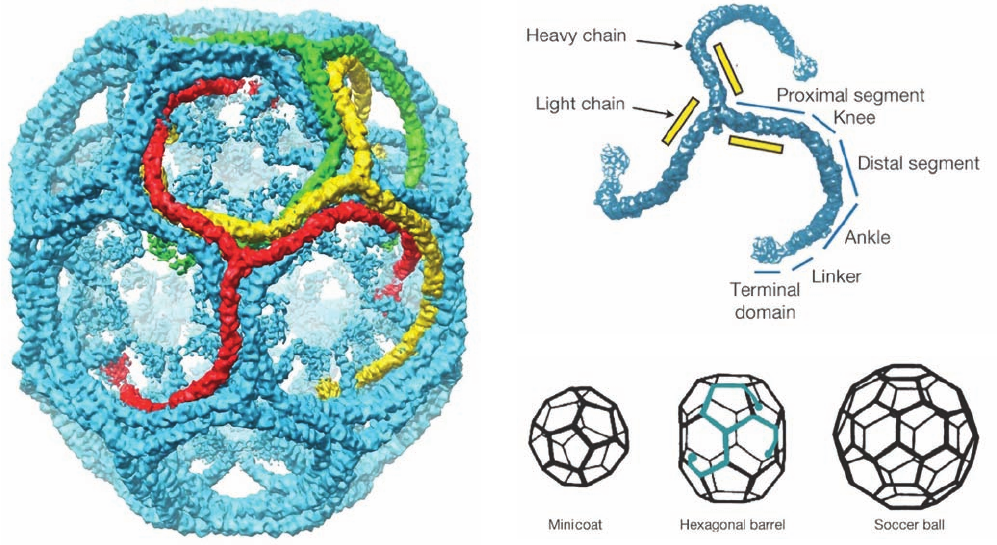
COPI or COPII. Clathrin forms polyhedral cages in which,
as a cryo-EM study by Harrison, Tomas Kirchhausen, and
Thomas Walz has shown most clearly (Fig. 12-62a), each
vertex is the center (hub) of a triskelion, and its edges,
which are ⬃225 Å long, are each formed by the interdigi-
tated legs of four triskelions—two antiparallel proximal
segments and two distal segments (Fig. 12-62b). Such poly-
hedra (Fig. 12-62c), which have 12 pentagonal faces and a
variable number of hexagonal faces (for geometric reasons
explained in Section 33-2A), are the most parsimonious
way of enclosing spheroidal objects in polyhedral cages.The
volume enclosed by a clathrin polyhedron, of course, in-
creases with its number of hexagonal faces (a “minicoat” is
too small to contain a transport vesicle).
The triskelion’s ⬃475-Å-long legs are each formed by
the 1675-residue heavy chains (HCs), which trimerize via
their C-terminal domains (Fig. 12-62b). In addition to pro-
jecting outward from its hub (vertex), each leg curls toward
the center of the particle such that three ankles meet and
interact ⬃75 Å below a hub that is two vertices away from
each of their hubs.
Section 12-4. Membrane Assembly and Protein Targeting 431
Figure 12-62 Anatomy of clathrin-coated vesicles. (a) A
cryo-EM–based image of a light chain–free clathrin cage from
bovine brain at 7.9 Å resolution.The particle shown, a so-called
hexagonal barrel, which has D
6
symmetry, consists of 36
triskelions.Three of its interdigitated but symmetry unrelated
triskelions are are drawn in red, yellow, and green. (b) A
cryo-EM–based image of a triskelion labeled with the names of
its various segments.The N-terminus of each heavy chain
occupies the terminal domain and its C-terminus is located in the
vertex joining the three heavy chains to form the triskelion.
(c) Diagrams of the three polyhedral stuctures that are formed
when triskelions assemble into clathrin cages in vitro.The minicoat
has tetrahedral (T) symmetry, the hexagonal barrel has D
6
sym-
metry, and the soccer ball has icosahedral (I) symmetry (symmetry
is discussed in Section 8-5B).These polyhedra consist of 28, 36,
and 60 triskelions, respectively.The arrangement of one triskelion
within the hexagonal barrel is indicated in blue. In vivo, clathrin
forms membrane-enclosing polyhedral cages with a large range
of different sizes (number of hexagons).The hexagonal barrel
seen in Part a is only ⬃700 Å in diameter, whereas clathrin-coated
membranous vesicles are typically ⬃1200 Å in diameter or
larger. [Courtesy of Stephen Harrison,Tomas Kirchhausen, and
Thomas Walz, Harvard Medical School.]
(a)
(c)
(b)
JWCL281_c12_386-466.qxd 6/9/10 12:06 PM Page 431
432 Chapter 12. Lipids and Membranes
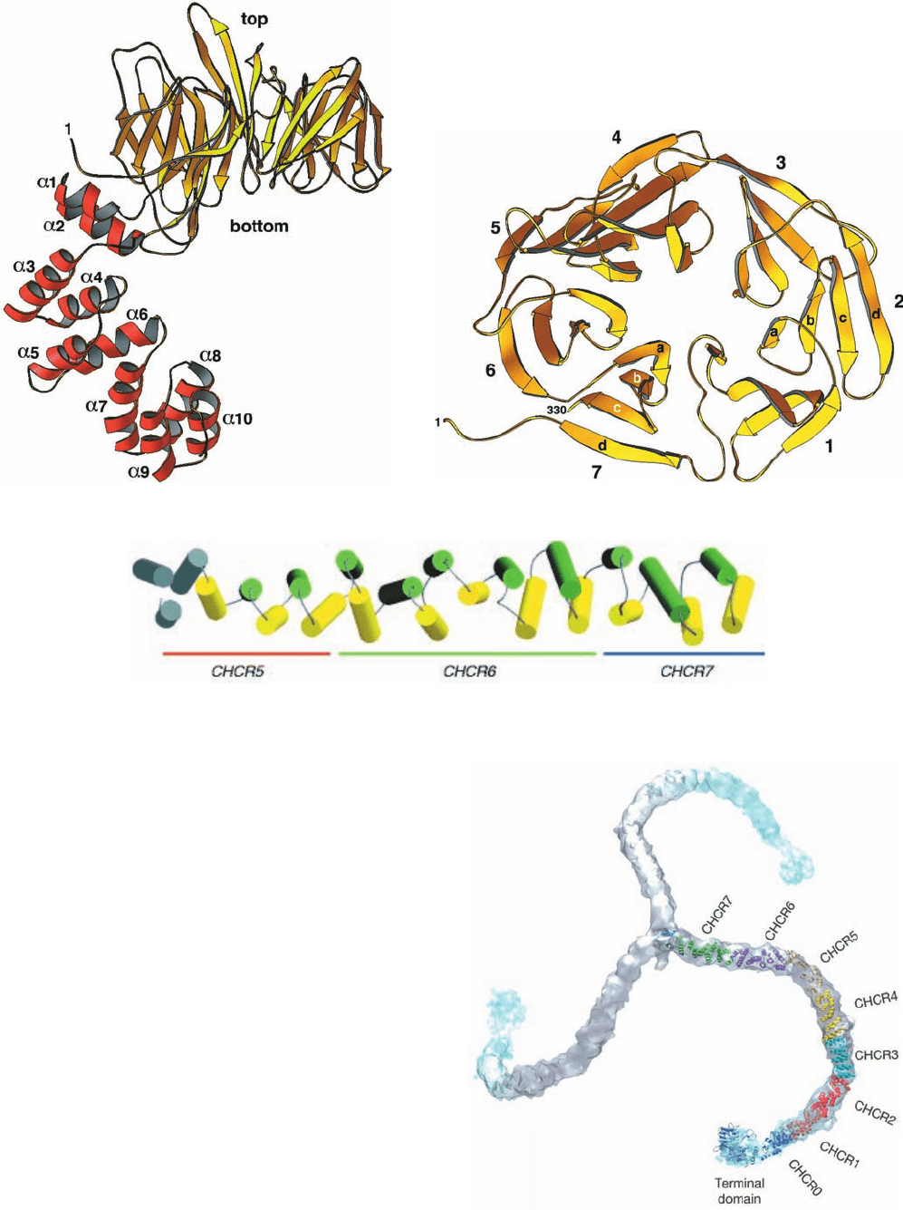
Figure 12-63 Structure of the clathrin heavy chain. (a) The
X-ray structure of the N-terminal domain and part of the linker
of rat HC.The N-terminal domain forms a seven-bladed 
propeller (yellow) that is seen here in side view, and the linker
(red) forms an ␣ solenoid. (b) The  propeller as viewed from the
top along its pseudo-7-fold axis. [Parts a and b courtesy of Tomas
Kirchhausen, Harvard Medical School. PDBid 1BPO.] (c) The
X-ray structure of bovine clathrin HC residues 1210 to 1516 as
viewed with its N-terminus on the left.The helices are alternately
colored yellow and green with the exception of the three
N-terminal helices, which are colored gray to indicate that they
are poorly resolved.The orange, green, and blue bars denote the
regions of CHCR5, CHCR6, and CHCR7, respectively. [Courtesy of
Peter Hwang, University of California at San Francisco. PDBid
1B89.] (d) A backbone model of a triskelion (residues 1–1597)
generated by docking the foregoing X-ray structures together with
homology models of the remaining CHCRs into the
cryo-EM–determined electron density of a heavy chain (Fig.
12-62a). [Courtesy of Stephen Harrison, Tomas Kirchhausen, and
Thomas Walz, Harvard Medical School. PDBid 1XI4.]
(a)
(b)
(c)
(d)
JWCL281_c12_386-466.qxd 6/9/10 12:06 PM Page 432
Although the X-ray structure of an entire HC has not
been determined, those of its N-terminal portion and a part
of its proximal segment have been elucidated:
1. The N-terminal segment (residues 1–494; Fig. 12-
63a,b), whose structure was determined by Harrison and
Kirchhausen, consists of two domains: (i) an N-terminal
seven-bladed  propeller in which each structurally simi-
lar propeller blade is formed by a four-stranded antiparal-
lel  sheet (Fig. 12-63b; the terminal domain) named the
WD40 sequence motif because it often contains the dipep-
tide WD and is ⬃40 residues long; and (ii) a C-terminal
linker that consists of 10 ␣ helices of variable lengths
(2–4 turns) connected by short loops and arranged in
an irregular right-handed helix (a helix of helices, that
is, a superhelix) named an ␣ solenoid (alternatively, an
␣-zigzag).
2. The proximal segment (residues 1210–1516; Fig.
12-63c), whose structure was determined by Peter Hwang
and Robert Fletterick, consists of 24 linked ␣ helices that
are arranged similarly but more regularly than the above ␣
solenoid to form a rod-shaped right-handed superhelix.
The rigidity of this motif is attributed to its continuous hy-
drophobic core together with the efficient interdigitation
of its side chains where its crossing antiparallel ␣ helices
come into contact (Section 8-3B).
Sequence and structural alignments indicate that HC
residues 537 to 1566 consist of seven homologous ⬃145-
residue clathrin heavy chain repeats (CHCRs) that are
arranged in tandem and which each contain 10 helices
(the proximal segment consists of all of CHRC6 together
with the C- and N-terminal portions of CHRC5 and
CHRC7; Fig. 12-63c).This has permitted the generation of
a backbone model of a triskelion by docking the forego-
ing X-ray structures and homology models of the CHCRs
whose structures have not been experimentally deter-
mined in the cryo-EM–determined electron density (Fig.
12-63d; homology modeling is discussed in Section 9-3B).
Each HC leg consists of an extended superhelix of linked
␣ helices. Nevertheless, triskelion legs exhibit consider-
able flexibility (Fig. 12-61), a functional necessity for the
formation of different sized vesicles as well as for the
budding of a vesicle from a membrane surface, which is
accompanied by a change in its curvature. The HC ap-
pears to flex mainly along its knee and ankle segments
(Fig. 12-62b).
The proximal segment bears extensive hydrophobic sur-
face patches that follow the grooves between adjacent he-
lices. Apparently, the lengthwise association of two proxi-
mal segments in a clathrin cage (Fig. 12-62a) is stabilized by
the burial of these hydrophobic patches through the com-
plementary packing of the helices of one proximal leg in
the grooves on another.
Light chains (LCs) are not required for clathrin cage as-
sembly. Indeed, LCs inhibit heavy chain polymerization in
vitro, which suggests that they have a regulatory role in
preventing inappropriate clathrin cage assembly in the
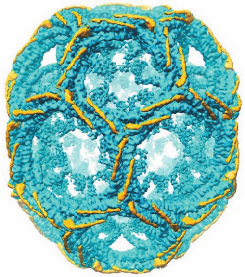
cytosol. Comparison of the cryo-EM structures of intact
and LC-free hexagonal barrels reveals that the central por-
tion of an LC consists of a 71-residue helix that binds to a
surface formed by the interhelical loops along the HC
proximal segment with the C-terminus of the LC closest to
the triskelion hub (Fig. 12-64). The segments of the 60%
identical LCa and LCb that differ in sequence are largely
confined to their N- and C-terminal regions, which do not
participate in HC binding and hence are likely to contain
sites for the attachment of cytosolic factors that regulate
vesicle uncoating.
c. Clathrin-Coated Vesicles Also
Participate in Endocytosis
CCVs, as we have seen, transport TM and secretory
proteins from the trans Golgi network (TGN) to the
plasma membrane (Fig. 12-58). In addition, through a
process known as endocytosis (discussed in Section 12-
5Bc), they act to engulf specific proteins from the extra-
cellular medium by the invagination of a portion of the
plasma membrane and to transport them to intracellular
destinations.
Section 12-4. Membrane Assembly and Protein Targeting 433
Figure 12-64 Arrangement of light chains on a clathrin cage.
The differences between the cryo-EM–determined electron
densities of a hexagonal barrel with and without light chains are
shown in yellow with the light chain–free electron density shown
in blue. [Courtesy of Stephen Harrison,Tomas Kirchhausen, and
Thomas Walz, Harvard Medical School.]
JWCL281_c12_386-466.qxd 6/9/10 12:06 PM Page 433
Membrane
Golgi
AP
Triskelions
Soluble cargo
proteins
Membrane
cargo protein
Clathrin cage
Membrane
receptor protein
Dynamin
Dynamin
Hsc70
GDP
GTP
GTP
1. Priming
2. Assembly
3. Release
4. Uncoating
Cytosol
GDP
ARF1
ARNO
GDP
GTP
GTP
GTP
GTP
d. The Formation of CCVs Is a Complex Process
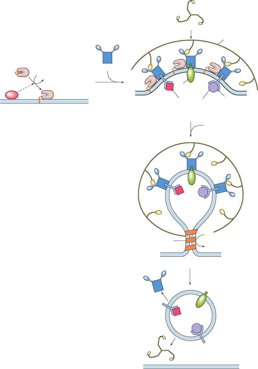
The formation of CCVs involves four stages (Fig. 12-65):
(1) priming,(2) assembly, (3) release, and (4) uncoating.We
outline these processes below.
1. Priming: The Activation of ARF1. Vesicle formation
begins with the binding to the membrane of the myristoyl-
ated small (181-residue) GTPase named ARF1 (ARF for
ADP-ribosylation factor, because it was first described as a
cofactor in the cholera toxin–catalyzed ADP-ribosylation of
the GTPases known as heterotrimeric G proteins; Section
19-2). ARFs, which are members of the Ras superfamily
(Ras is a small GTPase that participates in intracellular sig-
naling; Section 19-3C), are water-soluble cytosolic proteins
when binding GDP, but when binding GTP they associate
with membranes through the insertion of their N-terminal
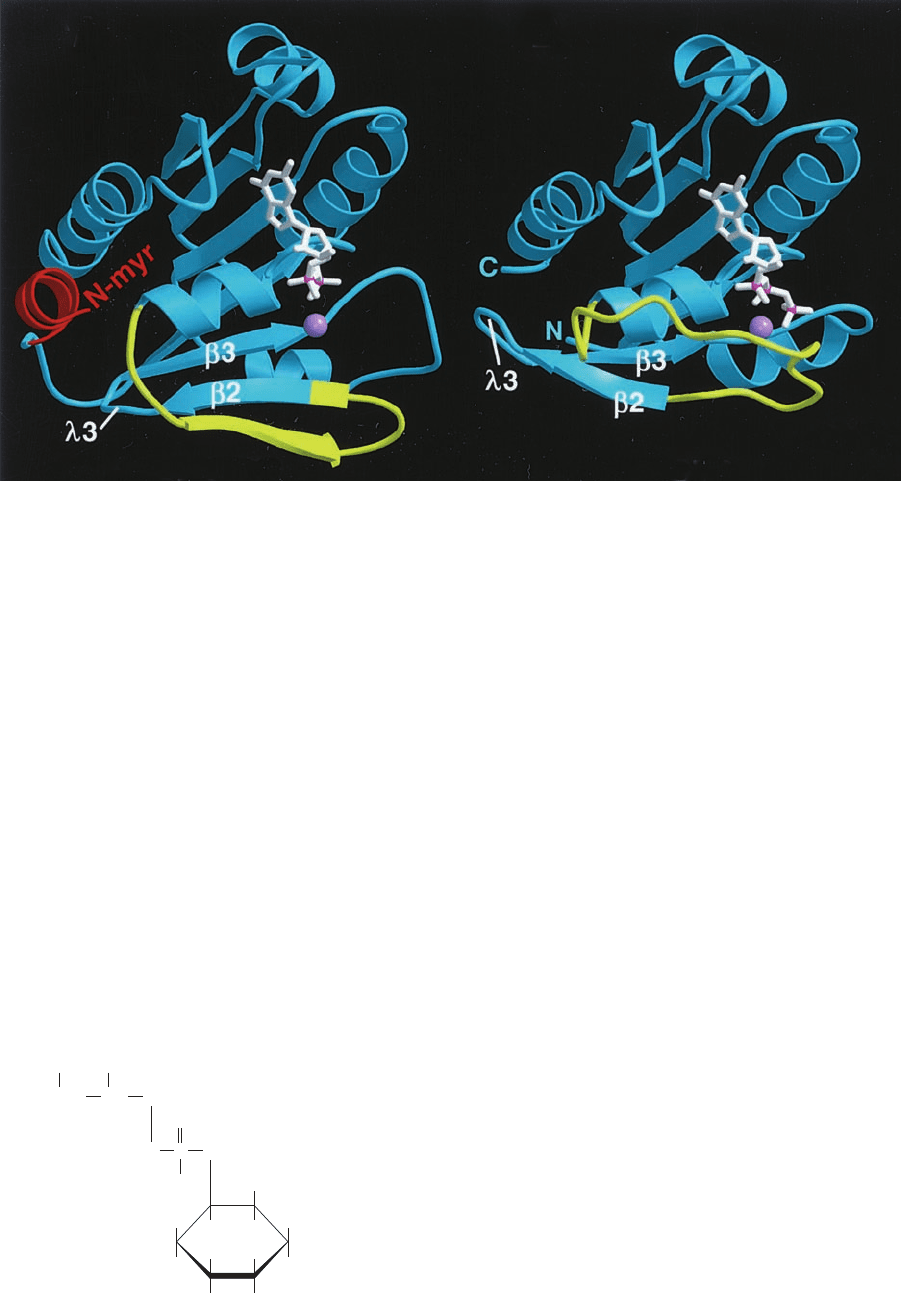
myristoyl groups into the bilayer (Section 12-3Bb).The com-
parison of X-ray structures of ARF1 ⴢ GDP and ARF1 ⴢ
GTP, determined by Dagmar Ringe and by Jonathan Gold-
berg, indicate that this occurs because the N-terminal helix
of ARF1 ⴢ GDP together with its appended myristoyl group
are bound in a shallow groove in the protein (Fig. 12-66a)
that is absent in ARF1 ⴢ GTP (Fig. 12-66b).
The guanine nucleotide exchange factor (GEF) for
ARF1, which in humans is called ARNO (for ARF nu-
cleotide-binding site opener; 399 residues), contains an
434 Chapter 12. Lipids and Membranes
Figure 12-65 Formation of clathrin-coated vesicles.
(1) The ARNO-stimulated exchange of ARF1’s bound GDP for
GTP frees ARF1 ⴢ GDP’s protein-bound N-terminal myristoyl
group for insertion into the membrane. (2) Membrane-bound
ARF1 ⴢ GTP recruits adapter proteins (APs). These, in turn, bind
triskelions, thereby promoting the formation of a clathrin coat,
which causes the vesicle to bud out from the membrane. In
addition,APs bind the transmembrane receptors of cargo
proteins as well as transmembrane cargo proteins. (3) The vesicle
is released from the membrane through the action of the GTPase
dynamin. (4) Shortly after the vesicle is released from the
membrane, the clathrin coat and the APs dissociate from the
vesicle.
JWCL281_c12_386-466.qxd 6/9/10 12:06 PM Page 434
⬃200-residue domain similar to the highly conserved yeast
protein Sec7. When ARNO or its isolated Sec7 domain is
incubated with myristoylated ARF1 ⴢ GDP, it fails to cat-
alyze nucleotide exchange unless lipid micelles are also
present, thereby suggesting that ARNO is activated only
when localized to a membrane surface. Indeed, ARNO
contains a pleckstrin homology (PH) domain, an ⬃100-
residue module occurring in numerous proteins (Section
19-3Ce) that binds the minor membrane phospholipid
phosphatidylinositol-4,5-bisphosphate (PIP
2
),
Phosphatidylinositol-4,5-bisphosphate (PIP
2
)
O
H
H
HO
OH
H
H
H
HO
H
OPO
2
3
–
OPO
2
3
–
O
O
–
P
O
CH
2
OR
2
OR
1
CH
CH
2
which is also a precursor of compounds that participate in
intracellular signaling (Section 19-4A).
2. Assembly: Adaptor Proteins Link Cargo Proteins to
the Clathrin Coat. Membrane-bound ARF1 ⴢ GTP acts to
recruit adapter proteins (APs) to the membrane surface.
APs bind clathrin HC together with TM proteins that are
either receptors that selectively bind soluble cargo proteins
inside the budding vesicle or are cargo proteins themselves.
APs comprise the central cores of CCVs and, in fact, are
the scaffolding on which clathrin cages form.The APs bind
clathrin via its N-terminal  propeller domain (Fig. 12-63a),
which forms the knobs that project inward from clathrin
cages (Fig. 12-62a). The grooves between the propeller
blades on the top face of the  propeller (Fig. 12-63b) prob-
ably form the AP binding sites.
AP1 is the most common AP contained in the coated
vesicles originating from the TGN, whereas the homolo-
gous AP2 predominates in endocytotic vesicles. Both APs
are heterotetramers: AP1 consists of the subunits ␥, 1
(⬃110 kD each), 1 (⬃50 kD), and 1 (⬃17 kD), whereas
the corresponding subunits of the better characterized AP2
are named ␣, 2, 2, and 2 (Fig. 12-67). Electron mi-
croscopy and X-ray studies indicate that the large subunits
each consist of a trunk and an appendage domain joined by
Section 12-4. Membrane Assembly and Protein Targeting 435
Figure 12-66 X-ray structures of (a) ARF1 ⭈ GDP and (b)
ARF1 ⭈ GMPPNP. (GMPPNP is a nonhydrolyzable GTP analog
in which the O atom linking GTP’s - and ␥-phosphorus atoms is
replaced by an NH group.) The bound nucleotides are drawn in
stick form in white with their phosphorus atoms magenta and
their bound Mg
2⫹
ions shown as lavender spheres. In ARF1 ⴢ
GDP, the protein’s N-terminal helix (red) together with its
covalently linked myristoyl group (not present in the X-ray
structures) are bound in a shallow hydrophobic groove on the
surface of the protein formed in part by the residues of loop 3.
However, the replacement of GDP by GMPPNP (and presumably
GTP) induces a conformational change in residues 37 to 53
(yellow) that displaces strand 2 by two residues along strand 3,
a shift of 7 Å.The resulting movement of loop 3 eliminates the
binding site for the N-terminus, thereby making the myristoyl
group available for membrane insertion (residues 1–17 of the
GMPPNP complex are disordered). [Courtesy of Jonathan
Goldberg, Memorial Sloan-Kettering Cancer Center, New York.
The X-ray structure of ARF1 ⴢ GDP was determined by Dagmar
Ringe, Brandeis University. PDBid 1HUR.]
(a)
(b)
JWCL281_c12_386-466.qxd 6/9/10 12:06 PM Page 435
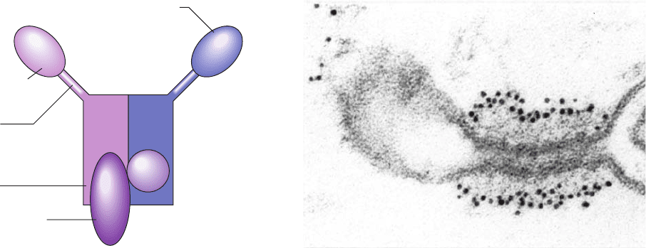
a flexible and proteolytically sensitive hinge region (Fig.
12-67). The AP2 hinge region of 2 binds to the clathrin 
propeller, whereas the cytoplasmic domains of target pro-
teins bind most commonly to 2 via YXX sequences
(where is a bulky hydrophobic residue), but in some
cases to its ␣ and 2 subunits via [D/E]XXXL[L/I] se-
quences, which are known as dileucine motifs. This explains
why the proteolytic excision of AP2’s appendage domain
prevents the assembly of clathrin coats, although the re-
maining AP2 trunk can still bind to membranes that contain
proteins bearing a YXX internalization signal. In addi-
tion, both AP1 and AP2 bind PIP
2
and mutating their PIP
2
-
binding sites prevents them from localizing to their target
membranes.
Mammals have two additional heterotetrameric APs,
AP3 and AP4, both of which function in the TGN. More-
over, database searches for AP homologs have identified a
family of monomeric clathrin adapters named GGAs (for
Golgi-localized ␥-ear-containing ARF-binding proteins),
whose C-terminal domain is homologous to the appendage
or “ear” domain of AP1’s ␥ subunit (and AP2’s ␣ subunit;
Fig.12-67).These various adapter proteins participate in the
transport of their target proteins between different pairs of
membanes so that CCVs are multifunctional entities.
3. Release: Vesicle Scission Is Mediated by Dynamin.
The budding of a CCV from its parent membrane appears
to be mechanically driven by the formation of the clathrin
cage. However, the actual scission of the coated bud from
its parent membrane to form a coated vesicle requires the
participation of dynamin, an ⬃870-residue GTPase. Dy-
namin contains a PIP
2
-binding PH domain, which recruits
dynamin to the membrane. On binding GTP, dynamin
forms a helical oligomer that wraps about the base of the
budding vesicle so as to squeeze this region down to a thin
tube (Fig. 12-68). The oligomerization together with the
presence of PIP
2
stimulates dynamin to hydrolyze its
bound GTP (dynamin also contains a GAP domain), caus-
ing the helical oligomer to lengthen its pitch. However, the
way in which this process releases the vesicle from the
membrane is not well understood.
4. Uncoating: The Recycling of Clathrin and Adapter
Proteins. Shortly after the formation of a CCV, the clathrin
is released as triskelions, thereby recycling them for partic-
ipation in the formation of additional coated vesicles. This
process is mediated by the ATPase Hsc70 (Hsc for heat
shock cognate), an ⬃650-residue homolog of the chaper-
one Hsp70 (Section 9-2C) present in all eukaryotic cells,
which on ATP hydroysis forms a complex with clathrin.
Hsc70 is recruited to the appropriate sites on the clathrin
lattice by the ⬃910-residue cochaperone auxilin, which
binds to specific sites on the clathrin heavy chains. Auxilin
contains a J-domain that induces Hsc70 to hydrolyze its
bound ATP to ADP, thereby causing Hsc70 to bind to and
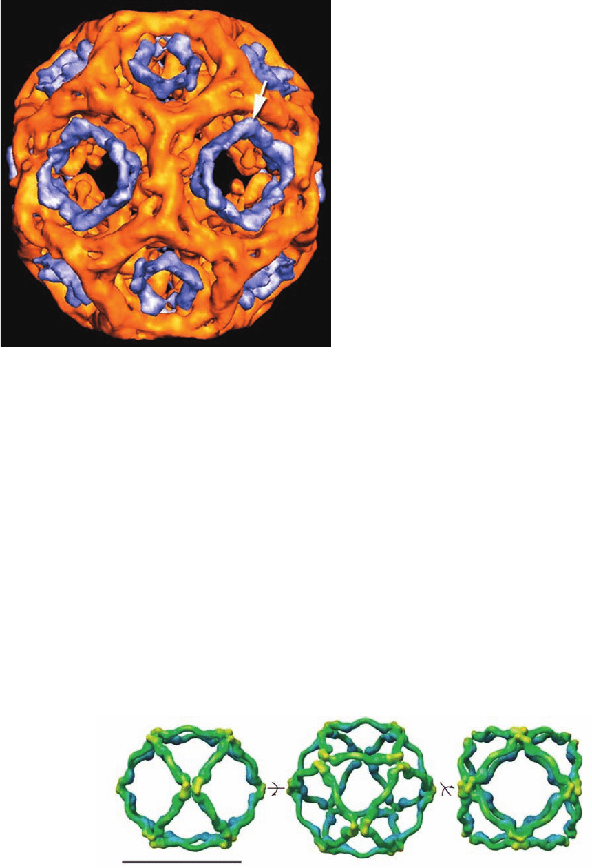
dismantle the clathrin lattice.The cryo-EM structure of the
clathrin “hexagonal barrel” in complex with Hsc70 and a J-
domain-containing fragment of auxilin at 28 Å resolution,
determined by Alasdair Steven, indicates that Hsc70 is lo-
cated within diffuse rings inside the clathrin cage’s pentag-
onal and hexagonal rings (Fig. 12-69). This suggests that
triskelions are pried out of the clathrin lattice by the con-
certed action of up to six Hsc70 molecules. This may occur
by a simple clockwise rotation of a triskelion as viewed in
Fig. 12-62a. On the subsequent exchange of its bound ADP
for ATP, the Hsc70 releases its bound triskelions.
Following clathrin release from newly formed vesicles,
the APs are also released. This process may be initiated by
the hydrolysis of ARF1’s bound GTP to GDP, which would
release ARF1 from the membrane and, presumably, from
binding an AP. In any case, the coating and uncoating of
vesicles by clathrin must be closely regulated processes
since both occur simultaneously.
436 Chapter 12. Lipids and Membranes
Figure 12-68 Electron micrograph of a budding coated
vesicle. The vesicle was incubated with the nonhydrolyzable GTP
analog GTP␥S (in which a terminal O atom on the ␥-phosphorus
of GTP is replaced by S) and then treated with gold-tagged
anti-dynamin antibodies (black dots). Note that the dynamin
surrounds a long narrow tube at the base of the budding vesicle
that has not pinched off from the membrane. [Courtesy of Pietro
De Camilli,Yale University School of Medicine.]
Figure 12-67 Schematic diagram of the AP2 heterotetramer.
AP1 has a similar structure. [After Pearse, B.M., Smith, C.J., and
Owen, D.J., Curr. Opin. Struct. Biol. 10, 223 (2000).]
Trunk
YXXφ endocytic
motifs
Hinge
DPF/W
motifs
Appendage
μ2
αβ2
σ2
JWCL281_c12_386-466.qxd 6/9/10 12:06 PM Page 436
A variety of regulatory and accessory proteins of
largely unknown function have also been implicated in
CCV formation. Moreover, many of the proteins de-
scribed above are each present in several isoforms. Hence
it is clear that our understanding of this process is far from
complete.
e. The Assembly of COPI- and COPII-Coated Vesicles
Resembles That of Clathrin-Coated Vesicles
COPI- and COPII-coated vesicles are both assembled
in processes, elucidated in large part by Randy Schekman,
that resemble CCV assembly:
1. Priming: COPI-coated vesicles are primed identi-
cally to CCVs: ARF1 is recruited to the membrane by the
ARNO-promoted exchange of its bound GDP for GTP
(Fig. 12-65, Step 1). COPII-coated vesicle assembly is simi-
larly primed but by different proteins: Sar1 (for secretion-
associated and Ras-related protein-1) is the small ARF
family GTPase that carries out this process, and the ex-
change of its GDP for GTP is mediated by the transmem-
brane GEF Sec12.
2. Assembly: ARF1 ⴢ GTP stoichiometrically recruits
intact coatomers to form COPI-coated vesicles. Most of the
seven COPI coatomer subunits have homologs in the
clathrin system and function accordingly:The -, ␥-, ␦-, and
-COPs correspond to the 2, ␣, 2, and 2 subunits of
AP2, respectively (Fig. 12-67), and the ␣- and ε-COPs cor-
respond to the clathrin heavy and light chains. In COPII
coat formation, Sar1 ⴢ GTP recruits the TM complex
Sec23/24, which in turn recruits cargo proteins and
Sec13/31, which forms the budding vesicle’s polyhedral
outer shell (see below).
3. Release: Both COPI- and COPII-coated vesicles
spontaneously bud off from their parent membranes; these
processes appear to have no requirement for an analog of
dynamin as does CCV release.
4. Uncoating: As is the case for CCVs, COPI- and
COPII-coated vesicles uncoat shortly after being released
from their parent membranes. These processes appear to
be initiated by the hydrolysis of the GTPs bound to ARF1
and Sar1, which thereby weaken the attachment of COPI
and COPII to their respective vesicles.The GTPase activat-
ing protein (GAP) for COPI vesicles, a 415-residue protein
named ARF GAP, appears to be a component of the COPI
coat. In COPII vesicles, Sec23 is the GAP for Sar1.
f. The Components of COPII and Clathrin Cages Are
Structurally Similar but Functionally Different
Cryo-EM studies of the COPII component Sec13/31 by
Bridget Carragher and William Balch reveal that, in vitro,
this heterodimer forms a 600-Å-diameter cuboctahedral
cage (Fig. 12-70). A cuboctahedron has O symmetry (the
symmetry of a cube; Section 8-5B) and has 24 edges of
equal length; 12 vertices, each of which is formed by the
Section 12-4. Membrane Assembly and Protein Targeting 437
Figure 12-69 Cryo-EM–based image of a clathrin hexagonal
barrel in complex with Hsc70 and a J-domain-containing
fragment of auxilin at 28 Å resolution. The clathrin cage is gold
and electron density attributable to the Hsc70 is blue. The white
arrow indicates the position at which the Hsc70 most closely
approaches the clathrin lattice. [Courtesy of Alasdair Steven, NIH,
Bethesda, Maryland.]
Figure 12-70 Cryo-EM structure of the human Sec13/31
COPII cage at 30 Å resolution. The views are along the
cuboctahedral cage’s 2-fold axis (left), it 3-fold axis (middle), and
its 4-fold axis (right).The surfaces of the cage elements are
colored according to their distance from the center of the cage
with blue nearest and yellow farthest.The scale bar is 500 Å long.
[Courtesy of Bridget Carragher and William Balch, The Scripps
Research Institute, La Jolla, California.]
JWCL281_c12_386-466.qxd 6/9/10 12:06 PM Page 437
interection of four edges (in contrast to clathrin cages
whose vertices are each formed by the insection of three
edges; Fig. 12-62c); and 14 faces, of which 8 are equilateral
triangles and 6 are squares. In vivo, COPII vesicles are often
larger than 600 Å in diameter. However, several larger
polyhedra are known whose vertices are each formed by
the intersection of four equal-length edges.
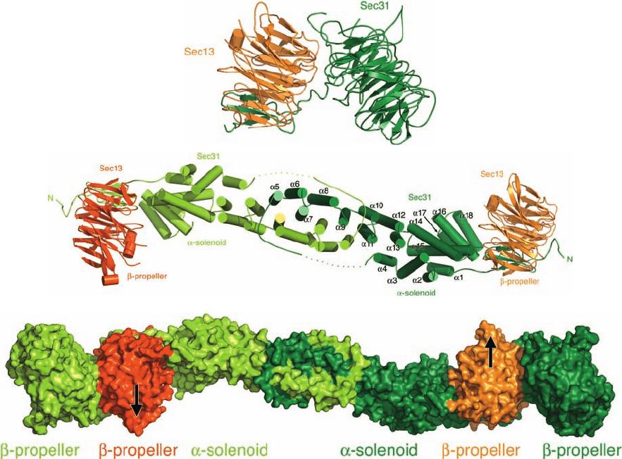
Although the full-length Sec13/31 complex has not been
crystallized, its limited proteolysis led to two X-ray struc-
tures determined by Jonathan Goldberg:
1. That of the 297-residue Sec13 in complex with
residues 1 to 411 of the 1297-residue Sec31 (Fig. 12-71a).
Sec13 forms six blades of a  propeller and the Sec31 frag-
ment forms a seven-bladed  propeller with its C-terminal
segment contributing a seventh blade to the Sec13  pro-
peller. Each blade of these propellers consists of a WD40
repeat as do the blades of the clathrin  propeller.
2. That of the Sec13/31 edge element (Fig. 12-71b),
which is a 2-fold symmetric heterotetramer that contains
the full length Sec13 in complex with residues 370–763 of
Sec31. As in the previous structure, Sec13 forms six blades
of a  propeller with a seventh blade contributed by the
here N-terminal segment of the Sec31 fragment. The re-
mainder of the Sec31 fragment consists of an ␣ solenoid
with its N-terminal end folded back over itself and its C-
terminal end overlapping the C-terminal end of another
Sec31 fragment to form an interlocked dimer. Thus, the
central portion of the complex consists of a double layer of
␣ solenoids.
Since the same segment of Sec31 passes through Sec13 in
both complexes and their Sec13 subunits are superimpos-
able, this strongly suggests that the Sec13/31 complex con-
tains the assembly unit shown in Fig. 12-71c. This assembly
unit has been docked into the cryo-EM–determined struc-
438 Chapter 12. Lipids and Membranes
Figure 12-71 X-ray structures of portions of the Sec13/31
complex from yeast. (a) The Sec13/31 vertex element, which
consists of Sec13 (orange) in complex with residues 1 to 411 of
Sec31 (green).The complex forms two seven-bladed  propellers
with one blade of the mainly Sec13  propeller contributed by
the C-terminal portion of the Sec31 fragment. (b) The Sec13/31
edge element, which is a heterotetramer composed of two
molecules each of Sec13 (red and orange) and residues 370 to
763 of Sec31 (light and dark green).The complex is viewed along
its 2-fold axis and oriented as in Part a. Here Sec13 forms 
propellers as in Part a and the Sec31 fragment forms a 215-Å-long
double layered ␣ solenoid. (c) Molecular model of the Sec13/31
assembly unit drawn as a surface diagram that is colored and
oriented as in Parts a and b. [Courtesy of Jonathan Goldberg,
Memorial Sloan-Kettering Cancer Center, New York, New York.
PDBids 2PM6 and 2PM9.]
(a)
(b)
(c)
JWCL281_c12_386-466.qxd 6/9/10 12:06 PM Page 438
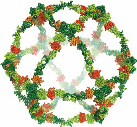
ture of the Sec13/31 cage (Fig. 12-70) to yield the model for
the COPII cage drawn in Fig. 12-72.
It is instructive to consider the similarities and differ-
ences between COPII and clathrin cages. Both consist of
seven-bladed  propellers and ␣ solenoids. In COPII cages,
all such motifs participate in forming its edges with four
Sec31  propellers associating to form each of its vertices.
In contrast, clathrin cages are constructed entirely from
their ␣ solenoidal segments with three such segments asso-
ciating to form each of its vertices and with their  propeller
motifs located in the interior of the cage, where they inter-
act with adapter proteins. Moreover, the ⬃40-Å-diameter
edges of COPII cages each consist of a double layer of
␣ solenoids, whereas the ⬃120-Å-diameter edges of clathrin
cages each consist of the interdigitated ␣ solenoidal seg-
ments from four triskelions. Evidently, evolution has
molded the similar components of these cages to different
functions. Sequence analysis of COPI coat proteins have
identified ␣ solenoid and  propeller motifs, which suggests
that the clathrin-, COPI-, and COPII-coated vesicles arose
from the same proto-coatamer.
The C-terminal segment of Sec31, which is not present
in the forgoing X-ray structures, contains an apparently
unstructured Pro-rich segment (residues 770–1110 are
20% Pro) that has been implicated in binding the
Sar1–Sec23/24 complex (which initiates vesicle budding
by binding to the cytoplasmic regions of cargo TM pro-
teins). Based on the X-ray structure of the Sar1–Sec23/24
complex and the fact that a cuboctahedral Sec13/31 cage
has has 48 binding sites for this complex, it would appear
that the Sar1–Sec23/24 complex forms a 50-Å-thick layer
beneath the surface of the COPII cage. Indeed, cryo-EM
studies on COPII vesicles assembled from purified
Sec13/31 and Sec23/24 complexes reveal that the Sec23/24
complexes form a cage that is concentric to and inside the
Sec13/31 cage.
g. Proteins Are Directed to the Lysosome by
Carbohydrate Recognition Markers
How are proteins in the ER selected for transport to the
Golgi apparatus and from there to their respective mem-
branous destinations? A clue as to the nature of this
process is provided by the human hereditary defect known
as I-cell disease (alternatively, mucolipidosis II), which in
homozygotes is characterized by severe progressive psy-
chomotor retardation, skeletal deformities, and death by
age 10.The lysosomes in the connective tissue of I-cell dis-
ease victims contain large inclusions (after which the dis-
ease is named) of glycosaminoglycans and glycolipids as a
result of the absence of several lysosomal hydrolases.These
enzymes are synthesized on the RER with their correct
amino acid sequences but, rather than being dispatched to
the lysosomes, are secreted into the extracellular medium.
This misdirection results from the absence of a mannose-6-
phosphate recognition marker on the carbohydrate moi-
eties of these hydrolases because an enzyme required for
mannose phosphorylation fails to recognize the lysosomal
proteins. The mannose-6-phosphate residues are normally
bound by a receptor in the coated vesicles that transport
lysosomal hydrolases from the Golgi apparatus to the lyso-
somes (Section 23-3Bj). Other glycoproteins are directed
to their intracellular destinations by similar carbohydrate
markers.
h. ER-Resident Proteins Have the C-Terminal
Sequence KDEL
Most soluble ER-resident proteins in mammals have
the C-terminal sequences KDEL (HDEL in yeast),
KKXX, or KXKXXX (where X represents any amino acid
residue), whose alteration results in the secretion of the
resulting protein. By what means are these proteins selec-
tively retained in the ER? Since many ER-resident pro-
teins freely diffuse within the ER, it seems unlikely that
they are immobilized by membrane-bound receptors
within the ER. Rather, it has been shown that ER-resident
proteins, as do secretory and lysosomal proteins, readily
leave the ER via COPII-coated vesicles but that ER-resident
proteins are promptly retrieved from the Golgi and returned
to the ER in COPI-coated vesicles. Indeed, coatomer binds
the Lys residues in the C-terminal KKXX motif of trans-
membrane proteins, which presumably permits it to gather
these proteins into COPI-coated vesicles. Furthermore,
genetically appending KDEL to the lysosomal protease
cathepsin D causes it to accumulate in the ER, but it nev-
ertheless acquires an N-acetylglucosaminyl-1-phosphate
group, a modification that is made in an early Golgi com-
partment. Presumably, a membrane-bound receptor in a
post-ER compartment binds the KDEL signal and the
Section 12-4. Membrane Assembly and Protein Targeting 439
Figure 12-72 Molecular model of the COPII cage viewed
approximately along its 3-fold axis. Its 48 Sec13/31 subunits are
drawn in worm form colored as in Fig. 12-71. Four Sec31 
propellers associate to form each vertex of the cuboctahedral
cage with the remaining portions of the heterotetrameric
Sec13/31 assembly units forming its edges.The inner diameter of
the cage is ⬃520 Å. [Courtesy of Jonathan Goldberg, Memorial
Sloan-Kettering Cancer Center, New York, New York.]
JWCL281_c12_386-466.qxd 6/9/10 12:06 PM Page 439
resulting complex is returned to the ER in a COPI-coated
vesicle. KDEL receptors have, in fact, been identified in
yeast and humans. However, the observation that former
KDEL proteins whose KDEL sequences have been deleted
are, nevertheless, secreted relatively slowly suggests that
there are mechanisms for retaining these proteins in the
ER by actively withholding them from the bulk flow of
proteins through the secretory pathway.
D. Vesicle Fusion
Vesicles that travel only short distances (⬍1 m) between
their parent and target membranes (e.g., between neigh-
boring Golgi cisternae) do so via simple diffusion, a
process that typically takes from one to several minutes.
However, vesicles that have longer distances to commute
(e.g., from the TGN to the plasma membrane) are actively
transported along cytoskeletal microtubules (Section 1-2A)
by the motor proteins dynein and kinesin, which unidirec-
tionally crawl along microtubule “tracks” in an ATP-driven
process (Section 35-3H).
a. Vesicle Fusion Is Most Easily Studied in Yeast
and in Synapses
On arriving at its target membrane, a vesicle fuses with
it, thereby releasing its contents on the opposite side of the
target membrane (Fig. 12-60). How do vesicles fuse and
why do they fuse only with their target membranes and not
other membranes? Progress in answering these questions
has been made mainly by using two experimental ap-
proaches, the genetic dissection of this process in yeast and
its biochemical analysis in synapses, the junctions between
neurons (nerve cells) and between neurons and muscles
(Fig. 12-73).
When a nerve impulse in the presynaptic cell reaches a
synapse, it triggers the fusion of neurotransmitter-containing
synaptic vesicles with the presynaptic membrane (a spe-
cialized section of the neuron’s plasma membrane),
thereby releasing the neurotransmitter (a small molecule)
into the ⬃200-Å-wide synaptic cleft (the process whereby
membranous vesicles fuse with the plasma membrane to
release their contents outside the cell is called exocytosis).
The neurotransmitter rapidly diffuses across the synaptic
cleft to the postsynaptic membrane, where it binds to spe-
cific receptors that then trigger the continuation of the
nerve impulse in the postsynaptic cell (Section 20-5C).The
homogenization of nerve tissue causes its presynaptic end-
ings to pinch off and reseal to form synaptosomes, which
can be readily isolated by density gradient ultracentrifuga-
tion for subsequent study.
b. Vesicle Fusion Requires the Coordinated Actions
of Many Proteins
Biological membranes do not spontaneously fuse. In-
deed, being negatively charged, they strongly repel one an-
other at short distances. These repulsive forces must be
overcome if biological membranes are to fuse. As we shall
see below, we are just beginning to understand how this
complicated process occurs.
Studies of the mechanism of vesicle fusion were pio-
neered by Rothman, who demonstrated that the fusion
process is blocked by low concentrations of the cysteine-
alkylating agent N-ethylmaleimide (NEM),
NH
CO
CH
CH
2
S
NH
CO
CH
CH
2
HS
⫹
O
O
Cys
N
-Ethylmaleimide (NEM)
CH
2
CH
3
N
O
O
CH
2
CH
3
N
440 Chapter 12. Lipids and Membranes
Synaptic cleft
(b)
Synaptic
vesicles
Neurotransmitter
molecules
Postsynaptic membrane
Presynaptic
membrane
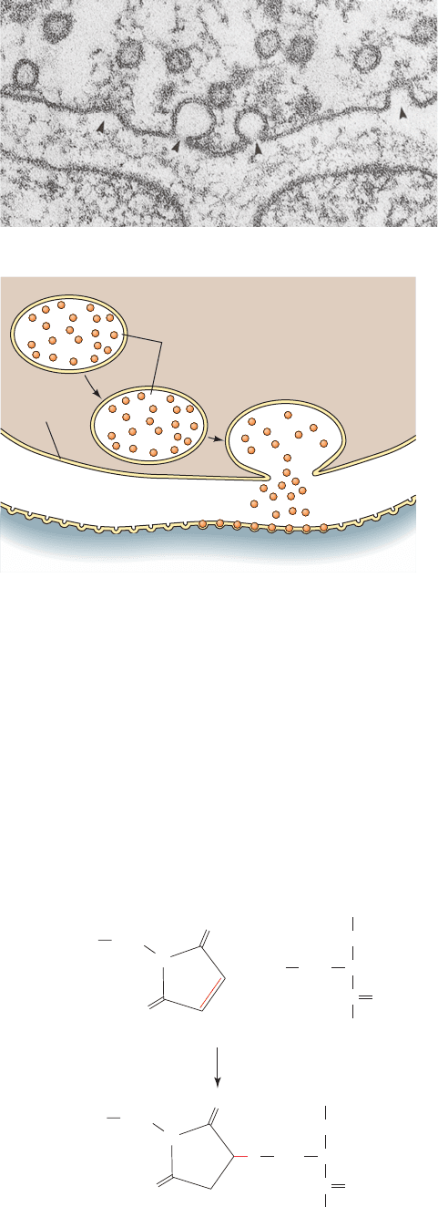
Figure 12-73 Transmission of nerve impulses across a synaptic
cleft. (a) Electron micrograph of a frog neuromuscular junction
in which the synaptic vesicles are undergoing exocytosis (arrows)
with the presynaptic membrane (top). [Courtesy of John Heuser,
Washington University School of Medicine, St. Louis, Missouri.]
(b) The neurotransmitter, which is thereby discharged into the
synaptic cleft, rapidly (in ⬍0.1 ms) diffuses to the postsynaptic
membrane, where it binds to transmembrane receptors,
triggering a new nerve impulse.
(a)
JWCL281_c12_386-466.qxd 6/9/10 12:06 PM Page 440
