Voet D., Voet Ju.G. Biochemistry
Подождите немного. Документ загружается.

indicating the presence of an NEM-sensitive fusion (NSF)
protein. NSF is a cytosolic ATPase that does not bind to
membranes unless a soluble NSF attachment protein
(SNAP) is also present. SNAPs bind to membranes in the
absence of NSF, demonstrating that SNAPs bind before
NSF. SNAPs bind to alkali-extracted membranes, which in-
dicates that SNAP receptors (SNAREs) are integral or
lipid-linked proteins.
Three classes of proteins appear to participate in all
vesicle fusion reactions:
1. Rab proteins, which are small (20–29 kD) GTPases
of the Ras superfamily that play a central role in directing
vesicle transport. Cells express numerous Rab isoforms, 11
in yeast and 63 in humans, each localized to a specific mem-
brane compartment. Rab proteins have two tandem Cys
residues at their C-termini, both of which are geranylger-
anylated (Section 12-3Ba). A soluble protein named GDP
dissociation inhibitor (GDI) binds to Rab ⴢ GDP so as to
mask its geranylgeranyl groups and thus maintain it in the
cytoplasm. However, when Rab ⴢ GDP interacts with a
cognate Rab-GEF on the surface of its target vesicle, the
geranylgeranyl groups on the resulting Rab ⴢ GTP are un-
masked and insert into the vesicle membrane—much like
the anchoring of ARF1 ⴢ GTP to the Golgi membrane (Fig.
12-65). Rab ⴢ GTP then binds to rodlike proteins emanat-
ing from the vesicle’s target membrane known as tethering
factors to form a relatively loose association between the
two membranes. After vesicle fusion, Rab hydrolyzes its
bound GTP to GDP in a process induced by a specific Rab-
GAP and the resulting Rab ⴢ GDP is extracted from the
membrane by GDI, thereby recycling the system. Rab pro-
teins are also implicated in initiating the actual membrane
fusion step (see below) as well as in the vesicle interactions
with the cytoskeleton that function in transporting vesicles
to their proper destinations.
2. SNAREs, which form cognate combinations of mem-
brane-associated proteins known as R-SNAREs and Q-
SNAREs (because they contain conserved Arg and Gln
residues in their cytoplasmic domains; they were originally
named v-SNAREs and t-SNAREs, respectively, because
they are mainly associated with the vesicle and target mem-
branes).The best characterized SNAREs are those function-
ing at neuronal synapses: Synaptobrevin (alteratively,
VAMP for vesicle associated membrane protein) is an R-
SNARE, whereas syntaxin and SNAP-25 (for synaptosome
associated protein of 25 kD) are Q-SNAREs. R-SNAREs
and Q-SNAREs associate to firmly anchor the vesicle to its
previously loosely tethered target membrane, a process called
“docking.” The docked complexes, which are described be-
low, are eventually disassembled by NSF in association with
a SNAP protein. (Note that SNAP-25 is not a SNAP protein;
by curious coincidence, the two independently characterized
proteins were assigned the same acronym before it was real-
ized that they are functionally associated.)
3. The SM proteins (so called because they are named
Sec1 in yeast and Munc18 in mammals), which in synapses
bind to syntaxin so as to prevent synaptobrevin and SNAP-
25 from binding to it. Mutational studies indicate that these
65- to 70-kD hydrophilic proteins are essential for vesicle
fusion.
c. SNAREs Form a Stable Four-Helix Bundle
The R-SNARE synaptobrevin and the Q-SNAREs syn-
taxin and SNAP-25 form a highly stable complex; boiling
SDS solution is required to dissociate it. Synaptobrevin and
syntaxin each have a C-terminal TM helix, and SNAP-25 is
anchored to the membrane via palmitoyl groups that are
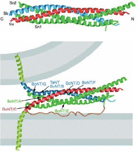
linked to Cys residues in its central region. The X-ray struc-
ture of the associating portions of this complex (Fig. 12-74a),
determined by Reinhard Jahn and Axel Brünger, reveals it
to be a bundle of four parallel ⬃65-residue ␣ helices with
two of the helices formed by the N- and C-terminal seg-
ments of SNAP-25. Since synaptobrevin is anchored in the
vesicle membrane and syntaxin and SNAP-25 are an-
chored in the target membrane, this so-called core complex
firmly ties together the two membranes (Fig. 12-74b).
Section 12-4. Membrane Assembly and Protein Targeting 441
Figure 12-74 X-ray structure of the syntaxin–synaptobrevin–
SNAP-25 core complex. (a) Ribbon diagram showing the
syntaxin helix (Sx) in red, the synaptobrevin helix (Sb) in blue,
and the N- and C-terminal helices of SNAP-25 (Sn1 and Sn2) in
green. (b) Model of the synaptic fusion complex linking two
membranes (gray).The helices of the core complex are colored
as in Part a.The transmembrane C-terminal extensions of
syntaxin and synaptobrevin are modeled as helices (light green).
The loop connecting the N- and C-terminal helices of SNAP-25
is speculatively represented as an unstructured loop (brown).
Recall that this loop is anchored to the membrane via Cys-linked
palmitoyl groups (not shown).The cleavage sites for the various
clostridial neurotoxins are indicated by the arrows. [Courtesy of
Axel Brünger,Yale University. PDBid 1SFC.]
(a)
(b)
JWCL281_c12_386-466.qxd 6/9/10 12:06 PM Page 441
The four helices of the core complex wrap around each
other with a gentle left-handed twist.For the most part, the se-
quence of each helix has the expected 7-residue repeat, (a-b-
c-d-e-f-g)
n
, with residues a and d hydrophobic (Section 8-2A;
note that this property is characteristic of 4- and 3-helix bun-
dles as well as of coiled coils). However, the central layer of
side chains along the length of the 4-helix bundle consists of
an Arg residue from synaptobrevin that is hydrogen bonded
to three Gln side chains, one from syntaxin and one from each
of the SNAP-25 helices. These highly conserved polar
residues are sealed off from the aqueous environment such
that their interactions are enhanced by the low dielectric con-
stant of their environment. It therefore appears that these in-
teractions serve to bring the four helices into proper register.
Since cells contain large numbers of different R-
SNAREs and Q-SNAREs (25 in yeast and 36 in humans),it
would seem likely that their interactions are at least par-
tially responsible for the specificity that vesicles exhibit in
fusing with their target membranes. Indeed, Rothman has
shown this to be the case by determining,in vitro, the rate of
fusion of liposomes bearing different SNAREs. In testing
all the R-SNAREs in the yeast genome against Q-SNAREs
known to be localized to the yeast Golgi, vacuole, and
plasma membranes, he found that liposome fusion only oc-
curs when the combinations of R- and Q-SNAREs corre-
spond to those mediating membrane flow in vivo. Never-
theless, it seems likely that the in vivo specificity of vesicle
fusion is augmented by other mechanisms such as the local-
ization of cognate R- and Q-SNAREs to particular regions
in the cell and by the actions of regulatory proteins includ-
ing,as is indicated above and discussed below, Rab proteins.
d. Tetanus and Botulinus Toxins Specifically
Cleave SNAREs
The frequently fatal infectious diseases tetanus (which
arises from wound contamination) and botulism (a type of
food poisoning) are caused by certain anaerobic bacteria of
the genus Clostridium. These bacteria produce extremely
potent protein neurotoxins that inhibit the release of neuro-
transmitters into synapses. In fact, botulinal toxins are the
most powerful known toxins;they are ⬃10 millionfold more
toxic than cyanide (10
⫺10
g ⴢ kg
⫺1
will kill a mouse).
There are seven serologically distinct types of botulinal
neurotoxins, designated BoNT/A through BoNT/G, and
one type of tetanus neurotoxin, TeTx. Each of these ho-
mologous proteins is synthesized as a single ⬃150-kD
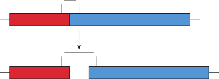
polypeptide chain that is cleaved by host proteases to yield
an ⬃50-kD L chain that remains disulfide-linked to the
⬃100-kD H chain (Fig. 12-75). The H chains bind to spe-
cific types of neurons (via gangliosides and protein recep-
tors), where they facilitate the uptake of the L chain by en-
docytosis. The L chains are proteases, and each cleaves its
target SNARE at a specific site (Fig. 12-74b). This prevents
the formation of the core complex and thereby halts the
exocytosis of synaptic vesicles. The H chain of TeTx specif-
ically binds to inhibitory neurons (which function to mod-
erate excitory nerve impulses) and is thereby responsible
for the spastic paralysis characteristic of tetanus. The H
chains of the BoNTs instead bind to motor neurons (which
innervate muscles) and thus cause the flaccid paralysis
characteristic of botulism.
The administration of carefully controlled quantities of
botulinal toxin (trade name Botox) is medically useful in
relieving the symptoms of certain types of chronic muscle
spasms. Moreover, this toxin is used cosmetically: Its injec-
tion into the skin relaxes the small muscles causing wrin-
kles and hence these wrinkles disappear for ⬃3 months.
e. Bilayer Fusion Is Mechanically Induced
The association of Q-SNAREs on a vesicle with an R-
SNARE on its target membrane brings the two bilayers into
close proximity, yielding a so-called trans-SNARE complex.
But what induces the fusion of the juxtaposed bilayers? The
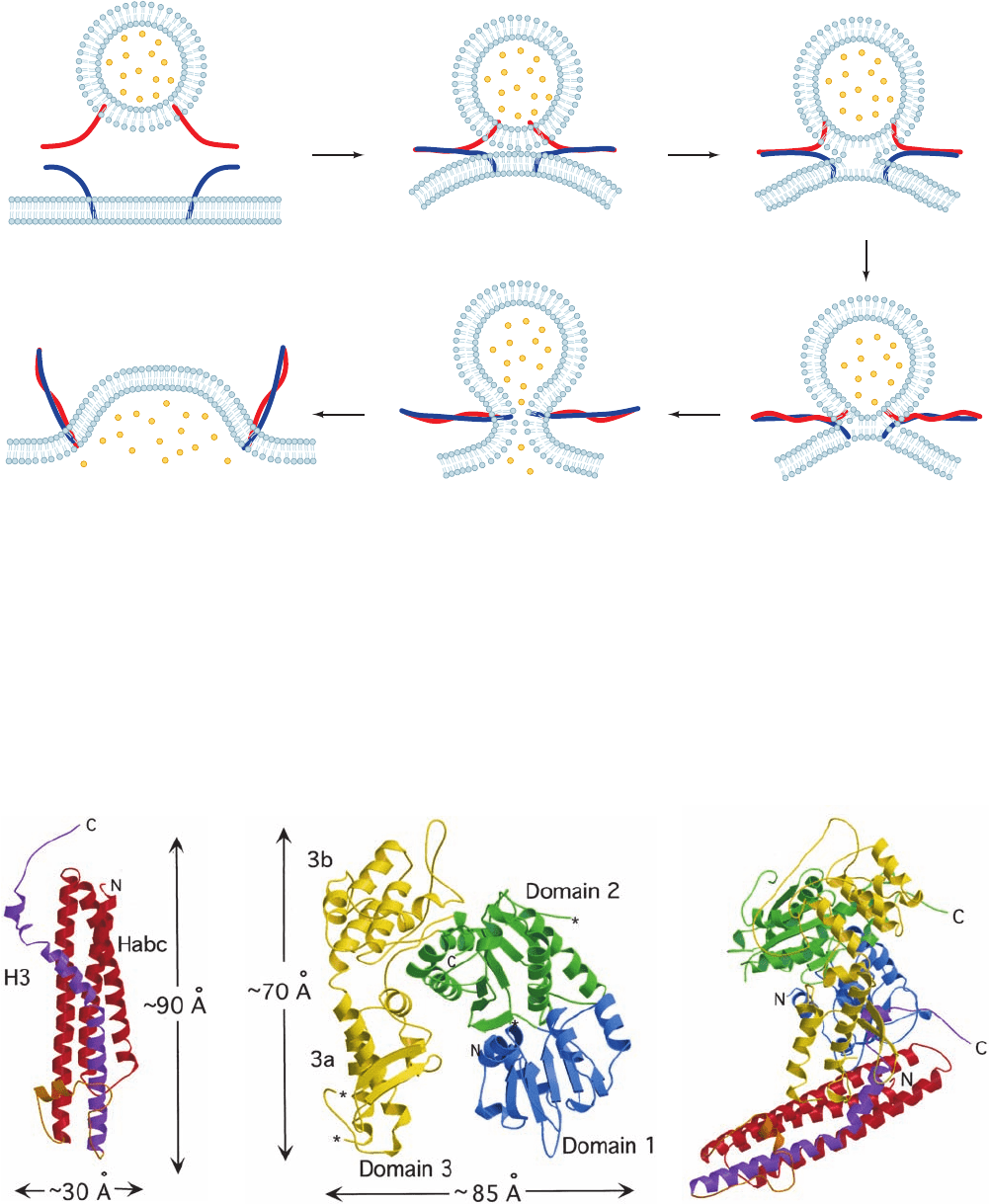
answer,which is diagrammed in Fig. 12-76, is that the mechan-
ical forces arising from the formation of a ring of several (es-
timated to be 5–10) trans-SNARE complexes pulls together
apposing bilayers. This expels the contacting lipids between
them so as to join their outer leaflets, a process known as
hemifusion. Indeed, the pressure (force/area) within the ring
of trans-SNARE complexes is estimated to be 100 to 1000
atm. In the resulting transient structure, no aqueous contact
between the two membrane systems has yet been established.
However, as the fusion process proceeds (the trans-SNAREs
continue zipping up), the two inner leaflets of the now par-
tially joined membranes come together to form a new bilayer,
whose component lipids are subsequently similarly expelled
to yield a fusion pore. The fusion pore then rapidly expands,
thereby fully joining the two membranes as well as their con-
tents. Thus, vesicle fusion is driven by the protein folding
forming the trans-SNARE complexes.
As we discussed above, liposomes containing the corre-
sponding Q- and R-SNAREs spontaneously fuse. How-
ever, this in vitro process takes 30 to 40 minutes whereas,
for example, the in vivo fusion of a synaptic vesicle with the
presynaptic membrane takes ⬍0.3 ms (Section 20-5C).This
suggests that other proteins such as Rab proteins and/or
their effectors (proteins with which they interact) partici-
pate in mediating the bilayer fusion process.
f. The Structure of the nSec1–Syntaxin Complex
Suggests a Function for Rab Protein
The neuronal SM protein, which is named nSec1, binds
to syntaxin with high affinity to form a complex that is
mutually exclusive with the formation of the syntaxin–
442 Chapter 12. Lipids and Membranes
Figure 12-75 Model of clostridial neurotoxins and their
activation by host proteases. The disulfide bond linking the L
and H segments is cleaved after the neurotoxin is taken up by its
target neuron.
SS
N
N
C
SS
C
Proteolytic activation
H chainL chain
JWCL281_c12_386-466.qxd 6/9/10 12:06 PM Page 442
synaptobrevin–SNAP-25 complex. The X-ray structure of
nSec1 in complex with the cytoplasmic domain of syntaxin
(Fig. 12-77), determined by William Weis, reveals that this
portion of the 288-residue syntaxin forms an N-terminal
up–down–up–down four-helix bundle. Syntaxin’s C-termi-
nal helix (but lacking its TM portion) adopts a bent and
somewhat irregular conformation, which differs from that
in the core complex displayed in Fig. 12-74. In contrast, the
Section 12-4. Membrane Assembly and Protein Targeting 443
Figure 12-76 Model for SNARE-mediated vesicle fusion.
Here the R-SNARE and the Q-SNAREs are schematically
Figure 12-77 X-ray structure of the complex between nSec1
and syntaxin. (a) Ribbon diagram of syntaxin with its N-terminal
3-helix bundle (Habc) red and the cytoplasmic portion, its
C-terminal helix (H3; the segment that forms a component of the
core complex), purple. (b) Ribbon diagram of nSec1 with its
1 Zipping: As the vesicle
approaches its target
membrane, the SNAREs begin
zipping together (docking) from
their N-termini, which draws
the two membranes toward
each other to form
trans-SNARE complexes.
2 Hemifusion: As docking
proceeds, the increased
curvature and lateral
tension induce the
approaching bilayer
leaflets to fuse, thereby
exposing the bilayer
interior.
3 The two bilayer leaflets that were
originally farthest apart are brought
together to form a new bilayer.
5 The fusion pore expands as the
now fused membrane relaxes
yielding cis-SNARE complexes.
4 Fusion pore formation: The continuing
SNARE-induced lateral tension causes
membrane breakdown, resulting in the
formation of a fusion pore.
represented by red and blue worms. [After a drawing by Chen,
Y.A. and Scheller, R.H., Nature Rev. Mol. Cell Biol. 2, 98 (2001).]
three domains differently colored. (c) The nSec1–syntaxin
complex colored as in Parts a and b and viewed such that the
nSec1 is rotated by 90º about the vertical axis relative to Part b.
[Courtesy of William Weis, Stanford University School of
Medicine. PDBid 1DN1.]
(a)
(b)
(c)
JWCL281_c12_386-466.qxd 6/9/10 12:06 PM Page 443
remaining N-terminal 3-helix bundle is closely superimpos-
able on the NMR structure of this segment alone. The 594-
residue nSec1 is an arch-shaped molecule that binds syn-
taxin, and in particular its C-terminal helix, in the cleft of
the arch (Fig. 12-77c).
The formation of the syntaxin–synaptobrevin–SNAP-25
complex that mediates vesicle fusion requires that the
nSec1–syntaxin complex dissociate and that syntaxin’s N-
terminal 3-helix bundle release the C-terminal helix. Muta-
tional studies indicate that a Rab protein and/or its effec-
tors mediate this process. It has therefore been proposed
that the binding of Rab and/or its effectors to the
nSec1–syntaxin complex causes nSec1 to change confor-
mation, which in turn induces syntaxin’s N-terminal 3-helix
bundle to release the C-terminal helix, thereby permitting
the SNARE complex to form. Thus Rab controls the avail-
ability of syntaxin.
g. NSF Mediates Core Complex Disassembly
The SNARE complex in the fused membranes, the so-
called cis-SNARE complex, must eventually be dissoci-
ated in order for its component proteins to participate in a
new round of vesicle fusion. This process is mediated by
NSF, an ATP-dependent cytosolic protein that binds to
SNAREs (SNAP receptors) through the intermediacy of
adaptor proteins called SNAPs (soluble NSF attachment
proteins).Although it was initially proposed that the NSF-
mediated disassembly of the cis-SNARE complex some-
how directly drove membrane fusion, it is now clear that
NSF functions to recycle SNAREs after their participation
in membrane fusion, that is, NSF functions as an ATP-
driven molecular chaperone. However, since trans-SNARE
complexes form spontaneously, membrane fusion is indi-
rectly driven by NSF-mediated ATP hydrolysis.
NSF is a hexamer of identical 752-residue subunits. Se-
quence analysis and limited proteolysis studies indicate
that each subunit consists of three domains:
1. An N-terminal so-called N domain (residues 1–205),
which mediates NSF’s interactions with SNAPs and
SNAREs.
2. A D1 domain (206–487), which binds ATP and cat-
alyzes its hydrolysis in a process that drives the disassembly
of the cis-SNARE complex.
3. A C-terminal D2 domain (488–752), which is homol-
ogous to D1. D2 binds ATP with a much higher affinity
than does D1 but hydrolyzes it very slowly, if at all.
D2 ⴢ ATP mediates the hexamerization of NSF, which is re-
quired for NSF activity.
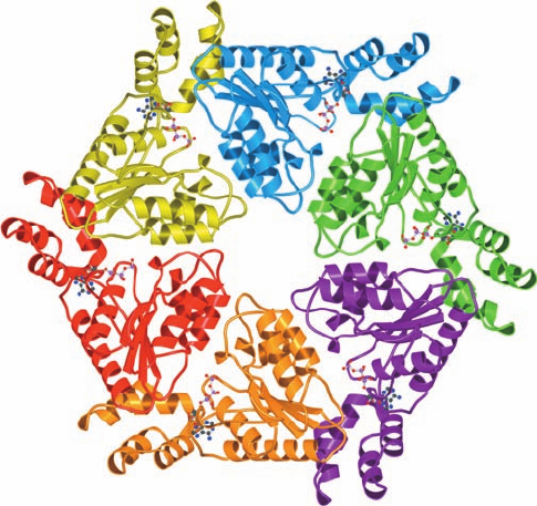
The X-ray structure of the D2 domain of NSF was inde-
pendently determined by Weis and by Jahn and Brünger. Its
wedge-shaped subunits associate to form a 116-Å-diameter
and 40-Å-high disk-shaped hexamer that has an ⬃18-Å-
diameter central pore (Fig.12-78).The ATP is bound near the
interface between two subunits, where it presumably helps
stabilize their association.
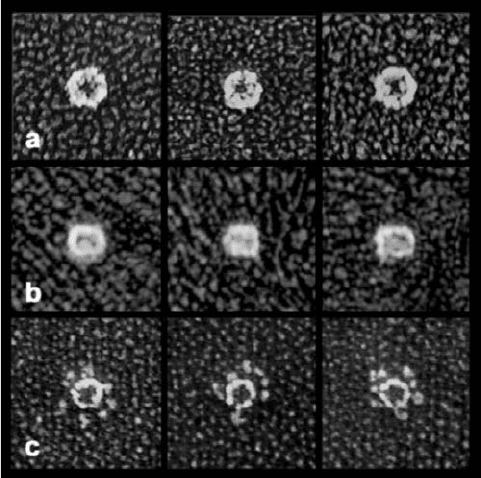
Electron micrographs by Jahn and John Heuser of intact
NSF in the presence of ATP have the appearance of an
⬃120-Å-diameter hexagonal ring with a 30- to 50-Å central
opening when seen in top view (Fig. 12-79a) and of a 120-Å
by 150-Å rectangle when seen in side view (Fig. 12-79b).
The length of the rectangle is about twice the height of the
D2 disk, which suggests that D1 forms a D2-like hexagonal
disk that stacks on D2. In the presence of ADP, NSF has an
identical appearance, which suggests that D1 rapidly hy-
drolyzes its bound ATP to form ADP. However,in the pres-
ence of the nonhydrolyzable ATP analog ATP␥S (in which
a terminal O atom on the ␥-phosphorus atom of ATP is re-
placed by S), NSF displays six globular feet that are tightly
packed around the somewhat smaller hexagonal ring (Fig.
12-79c). Since the hexagonal rings but not the globules are
seen when D1–D2 constructs are imaged in the presence of
ATP␥S, the globules must be the N domains. Evidently, the
N domains are held tightly around the central disk of
stacked D1 and D2 hexamers when D1 binds ADP but are
released when D1 binds ATP.
The mechanism whereby NSF disassembles the cis-
SNARE complex is largely unknown. The rod-shaped
SNARE core complex (Fig. 12-74a), which is 20 to 25 Å in
diameter,is too wide to fit inside the 18-Å-diameter central
pore of the D2 hexamer (and presumably the similarly
shaped D1 hexamer) without significant structural
changes. It is therefore unlikely that the core complex
binds inside NSF’s central cavity in a manner similar to the
way that the GroEL–GroES chaperonin system binds its
substrate proteins (Section 9-2Ca). Moreover, electron mi-
crographs indicate that the complex of SNAP and the
444 Chapter 12. Lipids and Membranes
Figure 12-78 X-ray structure of the NSF D2 hexamer as
viewed from its N-terminal end along its 6-fold axis. Each
subunit is differently colored.The bound ATPs are drawn in
ball-and-stick form. [Courtesy of Axel Brünger,Yale University.
PDBid 1NSF.]
JWCL281_c12_386-466.qxd 6/9/10 12:06 PM Page 444
three SNARE proteins binds to one end of NSF in the
presence of ATP␥S (but not at all in the presence of ADP).
Since NSF oligomers containing mixtures of active and in-
active D1 domains are unable to disassemble SNARE
complexes, it appears that the NSF subunits function in a
cooperative manner.
E. Protein Targeting to Mitochondria
Although mitochondria contain functioning genetic and
protein synthesizing systems, their genomes encode only a
handful of inner membrane proteins (13 in humans; 8 in
yeast). The vast majority of mitochondrial proteins
(⬃99%), which comprise 10 to 20% of intracellular pro-
teins, are encoded by nuclear genes and are synthesized by
cytosolic ribosomes. They must therefore traverse one or
both mitochondrial membranes (Section 1-2Ac) to reach
their final destinations. In this subsection, we discuss how
proteins are imported into mitochondria and are directed
to their correct destinations [outer membrane, inner mem-
brane, intermembrane space, or matrix (the space enclosed
by the inner membrane)]. Our rapidly developing knowl-
edge of this process was elucidated in large part through in-
vestigations in yeast and in the pink bread mold Neu-
rospora crassa by Walter Neupert, Nikolaus Pfanner,
Trevor Lithgow, and Gottfried Schatz. However, there is
considerable evidence that this process is well conserved
among all eukaryotes. The transport systems we describe
here and in Section 12-4B resemble those that mediate the
import of proteins into chloroplasts (in which proteins
must cross up to three membranes; Section 1-2Ag) and
peroxisomes (Section 1-2Ad).
a. Proteins Must Be Unfolded for
Import Into Mitochondria
Most nuclear-encoded mitochondrial proteins are fully
synthesized by cytosolic ribosomes before they are imported
into mitochondria; that is, they are post-translationally
imported. One might expect, therefore, that mitochondrial
proteins, many of which are integral proteins, would at least
partially fold and/or nonspecifically aggregate in the cy-
tosol before encountering the mitochondrial import sys-
tem. Yet a variety of evidence indicates that only unfolded
proteins can pass through mitochondrial membranes. For
example, dihydrofolate reductase (DHFR), a normally cy-
tosolic enzyme, is imported into yeast mitochondria when
it is preceded by the targeting sequence (see below) of a cy-
tosolically synthesized mitochondrial protein. However,
the importation of this chimeric protein is arrested by the
presence of methotrexate, an analog of DHFR’s normal
substrate dihydrofolate (Section 28-3Be), which binds to
DHFR with such high affinity that it stabilizes the protein’s
native conformation.
The import competence of mitochondrially destined
proteins is maintained in the cytosol by a variety of ATP-
dependent molecular chaperones. These include members
of the Hsp70 family (Section 9-2C) and, in mammals, a pro-
tein named mitochondrial import stimulation factor
(MSF). Consequently, the genetically engineered shut-
down of Hsp70 production in yeast causes the cells to cy-
tosolically accumulate proteins that would otherwise be
imported into the mitochondria. Moreover, the rate of the
Hsp70-facilitated mitochondrial import of a protein is en-
hanced by its prior denaturation by urea. Evidently,
Hsp70 functions in this process as an ATP-driven “protein
unfoldase.”
b. Translocation of Proteins Across the Outer
Mitochondrial Membrane
Most cytosolically synthesized matrix proteins have
cleavable N-terminal signal sequences that do not interact
with the SRP. These presequences consist of 10 to 80
residues that form amphipathic helices with one face posi-
tively charged. However, many mitochondrial proteins, in-
cluding most metabolite carrier proteins of the inner mem-
brane (see below), have poorly characterized internal
targeting sequences.
The protein subunits that participate in importing pro-
teins across the outer mitochondrial membrane are called
TOM proteins (for translocase of the outer mitochondrial
membrane) and are named Tomxx, where xx is the molec-
ular mass of the subunit in kilodaltons. Likewise, many of
the proteins involved in translocating proteins across the
inner mitochondrial membrane are called TIM proteins
(for translocase of the inner mitochondrial membrane) and
are named Timxx.
Section 12-4. Membrane Assembly and Protein Targeting 445
Figure 12-79 Quick-freeze/deep-etch electron micrographs of
NSF hexamers. (a) Top and (b) side views in the presence of
ATP. (c) Top view in the presence of ATP␥S. [Courtesy of John
Heuser,Washington University School of Medicine, St. Louis,
Missouri.]
JWCL281_c12_386-466.qxd 6/9/10 12:06 PM Page 445
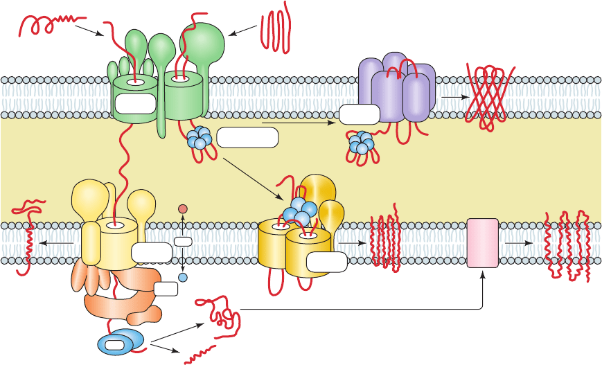
The TOM complex, the machinery that imports all mito-
chondrial proteins through the outer mitochondrial mem-
brane, does so as follows (Fig. 12-80, top left—green):
1. The signal sequences of unfolded preproteins associ-
ate with the cytoplasmic domains of mitochondrial receptor
proteins: N-terminal signal sequences interact mainly with
Tom20 in complex with Tom22, whereas internal signal se-
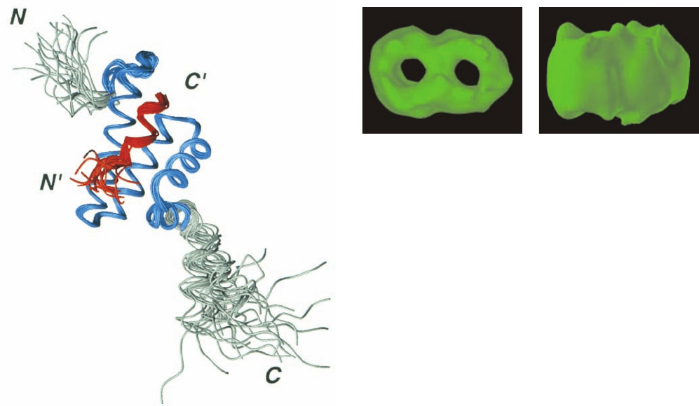
quences interact mainly with Tom70. The NMR structure of
a portion of Tom20’s cytosolic domain in complex with an
11-residue segment of a presequence peptide (Fig. 12-81),
determined by Toshiya Endo and Daisuke Kohda, reveals
that the Tom20 domain consists mainly of five helices. Its
two N-terminal helices form a nonpolar surface groove in
which the helical presequence binds, mainly via hydropho-
bic interactions rather than ionic interactions. Evidently,
Tom20 recognizes the presequence’s amphipathic helix but
not its positive charges.These positive charges, which are re-
quired for mitochondrial import, interact with Tom22.
2. Tom20 and Tom70 deliver preproteins to the general
import pore (GIP), so called because all nuclear-encoded
mitochondrial proteins must pass through it. The GIP is
formed by Tom40, a polytopic TM protein, which CD meas-
urements indicate consists mainly of  sheets and hence has
a TM  barrel structure that presumably resembles that of
bacterial porins (Fig. 12-27). Electrophysiological measure-
ments demonstrate that Tom40 contains a cation-selective
hydrophilic channel through which precursor proteins are
transported. Tom40 is closely associated with three small
single pass TM subunits, Tom5, Tom6, and Tom7, to form
the TOM core complex. The deletion of any one of these
small subunits has only minor effects but the deletion of all
three is lethal. They appear to stabilize the TOM complex
but their individual functions are largely unknown. Elec-
tron micrographs of the Neurospora TOM core complex
(Fig. 12-82) reveal an ⬃70-Å-high (⬃20 Å larger than the
thickness of the lipid bilayer) and ⬃120-Å-wide particle
containing two ⬃21-Å-diameter pores that presumably are
the protein-conducting channels. This agrees with perme-
ability experiments using cations of various sizes, which in-
dicate that the Tom40 pore is ⬃22 Å in diameter.
3. The forces driving the translocation of polypeptides
through the TOM complex remain largely enigmatic.A pro-
posed mechanism, the acid chain hypothesis, is that the pos-
itively charged presequence is sequentially transferred be-
tween acidic (negatively charged) patches to which it binds
with successively higher affinities. Such patches are present
on the cytoplasmic faces of Tom20, Tom22, and Tom5, as
well as on the intermembrane faces of Tom40 and Tom22.
At this stage, the import pathway for mitochondrial pro-
teins splits several ways.We discuss these various pathways
below.
c. Translocation of Proteins Into the Matrix
Polypeptides with N-terminal signal sequences, which
include the precursors of all matrix-destined proteins, most
inner membrane proteins, and many proteins that occupy
the intermembrane space (IMS), are translocated across
the inner mitochondrial membrane by the TIM23 complex
446 Chapter 12. Lipids and Membranes
Figure 12-80 Schematic diagram of the mitochondrial protein
import machinery in yeast. See the text for a description. The
subunit compositions of these complexes in mitochondria from
7
6
20
5
Cytosol
C
N
Protein with
presequence
Protein with internal
targeting signals
Outer
membrane
Intermembrane
space
Inner
membrane
inner membrane
protein
50
21
17
23
Pam
18
Tim44
Metabolic carrier protein
TM protein
Matrix
protein
PAM
16
17
ATP
Matrix
70
40
Sam
37
Sam
50
Mdm
10
Sam
35
Sam
complex
Tim9-Tim10
complex
+++
+++
+
–
TIM23
complex
TIM22
complex
β-barrel
protein
mtHsp70
Mge1
Oxa1
22
TOM
complex
MPP
54
18
22
9
10
9
12
ΔΨ
other organisms are similar. [After Bolender, N., Sickmann, A.,
Wagner, R., Meisinger, C., and Pfanner, N., EMBO Rep. 9, 42–49
(2008).]
JWCL281_c12_386-466.qxd 6/9/10 12:06 PM Page 446
(Fig. 12-80, bottom left—yellow). This complex contains a
protein channel formed by Tim23, which is closely associ-
ated with Tim17. The peripheral protein Tim50 binds the
polypeptide emerging from the Tom40 channel and passes
it to Tim23. Electron microscopy studies indicate that the
TOM and TIM23 complexes are in apposition at sites
where the inner and outer mitochondrial membranes ap-
proach each other most closely. Indeed, Tim21 transiently
associates with Tom22 across this contact site by displacing
the emerging signal sequence.
In the presence of methotrexate, the above DHFR
chimera becomes stuck in the membrane with the spacer
that linked the enzyme to its N-terminal presequence si-
multaneously spanning the TOM and TIM23 complexes.
The N-terminal end of the spacer is presumably trapped in
the matrix through its association with mtHsp70 (see be-
low). Consequently, if the spacer is so short that it cannot
span both membranes (less than ⬃40 residues), no stable
translocation intermediate is formed. Thus, it appears that
presequences make their way between the TOM and
TIM23 complexes without the aid of chaperones.
The translocation of a protein across the inner mito-
chondrial membrane requires energy in the form of both
ATP and an electrostatic potential across the inner mito-
chondrial membrane. This so-called membrane potential
(Section 20-1), ⌬⌿, which is metabolically generated (Sec-
tion 22-3Ba), apparently functions to electrophoretically
transport the positively charged N-terminal signal se-
quence into the matrix (the matrix is negatively charged
with respect to the cytosol).
The ATP is utilized by matrix Hsp70 (mtHsp70; alterna-
tively, mHsp70), the central component of the presequence
translocase-associated motor (PAM; Fig. 12-80, bottom
left—orange). This molecular chaperone binds to Tim44 on
the inner face of the inner mitochondrial membrane, where
it is thought to mechanically pull the protein through the
Tim23 pore via a Brownian ratchet mechanism (Section
12-4Bg). Pam 18 (alternatively, Tim14), which associates
with Tim44, has a J-domain that presumably recruits
mtHsp70 and induces it to hydrolyze its bound ATP to
ADP, thus activating it to bind the incoming polypeptide.
Pam16 (alternatively Tim16), which binds Tim14, is thought
to act as a negative regulator of Tim14 by physically block-
ing its access to mtHsp70. Pam17 is required for the assem-
bly of the Pam18–Pam16 module.The matrix protein Mge1
stimulates mtHsp70 to exchange its bound ADP for ATP,
thus permitting it to participate in another cycle of the
Brownian ratchet.
Once a preprotein, or at least its N-terminal segment,
has entered the matrix, its N-terminal signal sequence is
excised by matrix processing peptidase (MPP), an essen-
tial protein. The imported protein then folds/assembles to
its native state, a process that is facilitated by a battery of
ATP-dependent chaperone proteins including mtHsp70
(only about 10% of which is associated with Tim44) and
Hsp60/Hsp10 (homologs of the GroEL/ES system; Sec-
tion 9-2C).
Some of the polypeptides that are translocated by the
TIM23 complex have a stop-transfer anchor sequence. The
TIM23 complex laterally inserts the resulting TM helix into
the inner mitochondrial membrane (Fig. 12-80, bottom, far
left) such that its N-terminal portion occupies the matrix,
where MPP excises its N-terminal signal sequence.
Section 12-4. Membrane Assembly and Protein Targeting 447
Figure 12-81 NMR structure of the cytoplasmic domain of rat
Tom20 in complex with the C-terminal 11-residue segment
(GPRLSRLLSYA) of the 22-residue presequence of the rat mi-
tochondrial enzyme aldehyde dehydrogenase. The diagram is a
superposition of the 20 final structures in the NMR analysis (Sec-
tion 8-3A) in which the residues used to make the superposition
are blue (Tom20) and red (presequence) and the remaining
residues are gray (Tom20) and orange (presequence). [Courtesy
of Toshiya Endo, Nagoya University, Nagoya, Japan, and Daisuke
Kohda, Biomolecular Engineering Research Institute, Osaka,
Japan. PDBid 1OM2.]
Figure 12-82 Electron microscopy–based image of the TOM
core complex particles from Neurospora. The particles, which are
shown in top view (left) and side view (right), contain two
openings that presumably represent the mitochondrial outer
membrane’s protein-conducting channels. [Courtesy of Stephan
Nussberger and Walter Neupert, Universität München, Germany.]
JWCL281_c12_386-466.qxd 6/9/10 12:06 PM Page 447
d. Insertion of Metabolite Carrier Proteins Into
the Inner Mitochondrial Membrane
The mitochondrial inner membrane is impermeable to
nearly all polar substances and hence contains numerous (35
in yeast) metabolite carrier proteins to permit the acquisi-
tion of reactants and the delivery of products. The most
abundant members of this family are the ATP–ADP
translocator (which exchanges the ATP synthesized in the
matrix for the ADP product of cytosolic ATP hydrolysis;
Section 20-4C) and the phosphate carrier (which returns the
phosphate product of cytosolic ATP hydrolysis to the ma-
trix;Section 22-1Ba).All metabolite carrier proteins have six
TM helices with both their N- and C-termini in the IMS.
Most members of the metabolite carrier family lack N-
terminal signal sequences and are therefore translocated
through the TOM complex via interactions with its Tom70
receptor. Curiously, however, the Tom20–Tom22 complex
is the receptor for most other outer membrane proteins
that have internal signal sequences. Metabolite carrier
proteins are escorted across the IMS by a hexameric
complex of the homologous proteins Tim9 and Tim10,
(Tim9)
3
(Tim10)
3
, which is thought to shield the hydropho-
bic domains of the metabolite carrier proteins (Fig. 12-80,
middle—blue). Metabolite carrier proteins in a preparation
of mitochondria depleted of Tim9 and Tim10 are not in-
serted into the GIP, as indicated by their failure to reach a
protease-resistant state. This suggests that it is the binding
of the Tim9–Tim10 complex to an unfolded metabolite car-
rier protein that drives its translocation across the outer
mitochondrial membrane.
The Tim9–Tim10 complex delivers the metabolite car-
rier protein to the peripheral protein Tim12 (a homolog of
Tim9 and Tim10), which is associated with the integral
proteins Tim22 (which is homologous to Tim 23), Tim54,
and Tim18 to form the TIM22 complex (Fig. 12-80, bottom
middle—gold). Tim22 then mediates the lateral insertion of
the metabolite carrier protein into the inner mitochondrial
membrane, where it assembles to form homodimers. This
process occurs via an unknown but membrane potential–
dependent mechanism. The functions of Tim54 and Tim18
are unknown.
e. Soluble Proteins Occupying the Intermembrane
Space Are Imported via Three Mechanisms
Despite the fact that its width is around that of a mem-
brane bilayer, the IMS contains a large collection of essen-
tial proteins. The precursors of some of these proteins are
imported, as described above, such that they become
anchored to the IMS by a single TM helix that has its N-
terminal end in the matrix (Fig.12-80, bottom,far left). Such
a protein is then cleaved by an inner membrane protease on
the C-terminal side of its TM helix, thereby releasing it into
the IMS, where it folds to its native conformation. Since
the mature protein lacks a signal sequence, it is no longer
subject to importation into the matrix and hence remains in
the IMS. Coproporphyrinogen oxidase, which participates
in heme biosynthesis (Section 26-4Ae), is such a protein.
Many small proteins that lack N-terminal signal se-
quences are imported, via the TOM complex, into the IMS.
There they assume their native fold, thus trapping them in
the IMS—the so-called folding-trap mechanism. Such pro-
teins have conserved patterns of Cys and/or His residues
that enable them to bind metal ion–containing cofactors in
the IMS or to form disulfide bonds, both of which stabilize
their native structures. [Note that the latter proteins are
among the few intracellular proteins that have disulfide
bonds (Section 8-4D). Evidently, the IMS has an oxidative
environment.] For example, apocytochrome c (cytochrome
c without its covalently attached heme group; Fig. 9-39)
folds when the IMS-resident enzyme cytochrome c heme
lyase (CCHL) catalyzes the attachment of its heme group,
whereas Tim9, Tim10, and Tim12 each contain twin CX
3
C
motifs that form disulfide bonds.
A third class of IMS-resident proteins remain in the
IMS through their association with the inner membrane,
that is, they are peripheral proteins. CCHL is a member of
this class of proteins.
f. Many Polytopic Inner Membrane Proteins Are First
Imported to the Matrix
Many cytosolically synthesized polytopic proteins des-
tined for insertion into the mitochondrion’s inner mem-
brane are first imported into the matrix as described above
and then inserted into the inner membrane, an indirect
routing that reflects the mitochondrion’s bacterial origin
[the primordial mitochondrion, being a gram-negative bac-
terium, synthesized all of its proteins in its cytoplasm (the
primordial matrix) so that membrane-bound or intermem-
brane proteins had to be exported to these destinations].
These proteins, for the most part, are synthesized with bi-
partite N-terminal targeting sequences whose inner (more
C-terminal) segments, once exposed by the removal of the
above-described N-terminal presequence, direct the pro-
teins to the inner membrane. The insertion of several such
proteins into the inner mitochondrial membrane is medi-
ated by the TM protein Oxa1, which also occupies the in-
ner mitochondrial membrane (Fig. 12-80, bottom right,
pink). Oxa1, which binds mitochondrial ribosomes on its
matrix side, also inserts mitochondrially synthesized pro-
teins into the inner mitochondrial membrane. As might be
expected, Oxa1 is related to a protein that inserts proteins
into the inner membrane of gram-negative bacteria.
g. Insertion of  Barrel Proteins Into the Outer
Mitochondrial Membrane
The outer membranes of mitochondria and chloroplasts
contain proteins, such as porins (Section 12-3Ad) and
Tom40, that have TM  barrels.These are the only places in
eukaryotic cells that TM  barrels occur, which also reflects
the bacterial origins of these organelles (Sections 1-2Ac
and 1-2Ag).
 barrel proteins are imported into the IMS by the TOM
complex. There they are bound by the Tim9–Tim10 com-
plex, which escorts them to the SAM complex (for sorting
and assembly machinery; alternatively TOB complex for
topogenesis of mitochondrial outer membrane  barrel),
which in turn inserts them into the outer mitochondrial
membrane (Fig. 12-80, top right—purple).The SAM complex
448 Chapter 12. Lipids and Membranes
JWCL281_c12_386-466.qxd 6/9/10 12:06 PM Page 448
is formed by the TM  barrel–containing protein Sam50
(alternatively, Tob55 or Tom55) in association with Sam37
(Mas37/Tom37), Sam35 (Tob35/Tom38), and Mdm10
(mitochondrial distribution and morphology 10).  barrel
proteins are inserted into the outer membrane from its
inner side, which presumably is also an evolutionary conse-
quence of the mitochondrion’s bacterial origin.Nevertheless,
the TOM and SAM complexes are functionally coupled as
indicated by the observation that when  barrel proteins
are imported into mitochondria lacking Sam50, they accu-
mulate in the TOM complex rather than in the IMS. Sam50
is homologous to the bacterial outer membrane protein
Omp85, which participates in inserting  barrel proteins
into the bacterial outer membrane.
5 LIPOPROTEINS
Lipids and proteins associate noncovalently to form
lipoproteins, which function in the blood plasma as trans-
port vehicles for triacylglycerols and cholesterol. In this sec-
tion, we discuss the structure, function, and dysfunction of
lipoproteins, and how eukaryotic cells take up lipoproteins
and other specific proteins from their external medium
through receptor-mediated endocytosis.
A. Lipoprotein Structure
Lipids, such as phospholipids, triacylglycerols, and choles-
terol, are but sparingly soluble in aqueous solution. Hence,
they are transported by the circulation as components of
lipoproteins, globular micellelike particles that consist of a
nonpolar core of triacylglycerols and cholesteryl esters sur-
rounded by an amphiphilic coating of protein, phospho-
lipid, and cholesterol. Lipoproteins have been classified
into five broad categories on the basis of their functional
and physical properties (Table 12-6):
1. Chylomicrons, which transport exogenous (exter-
nally supplied; in this case, dietary) triacylglycerols and
cholesterol from the intestines to the tissues.
2–4. Very low density lipoproteins (VLDL), intermedi-
ate density lipoproteins (IDL), and low density lipoproteins
(LDL), a group of related particles that transport endoge-
nous (internally produced) triacylglycerols and cholesterol
from the liver to the tissues (the liver synthesizes triacyl-
glycerols from excess carbohydrates; Section 25-4).
5. High density lipoproteins (HDL), which transport
endogenous cholesterol from the tissues to the liver.
Lipoprotein particles undergo continuous metabolic
processing, so that they have variable properties and com-
positions (Table 12-6). Each contains just enough protein,
phospholipid, and cholesterol to form an ⬃20-Å-thick
monolayer of these substances on the particle surface
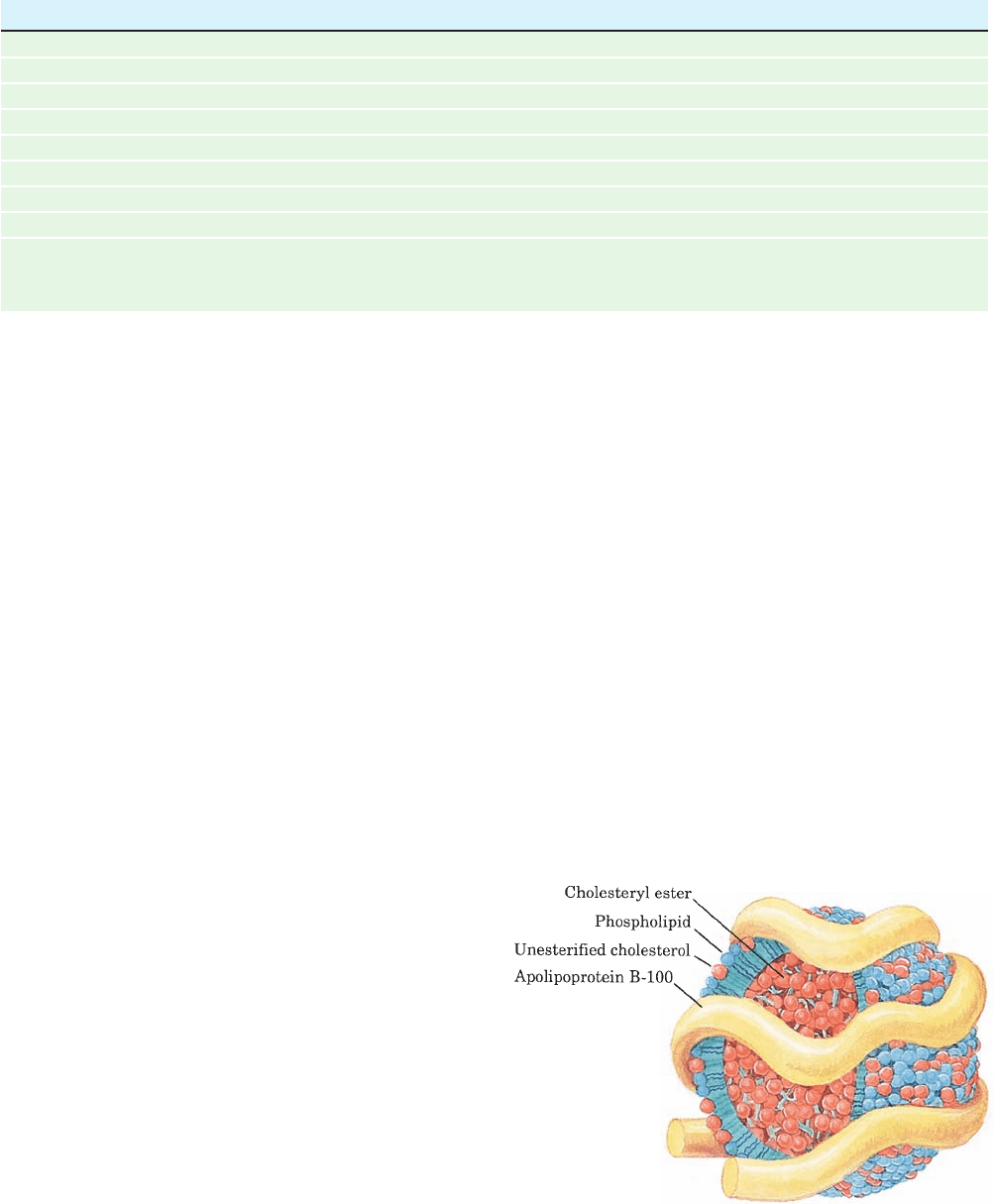
(Fig. 12-83). Lipoprotein densities increase with decreasing
particle diameter because the density of their outer coating
is greater than that of their inner core.
Section 12-5. Lipoproteins 449
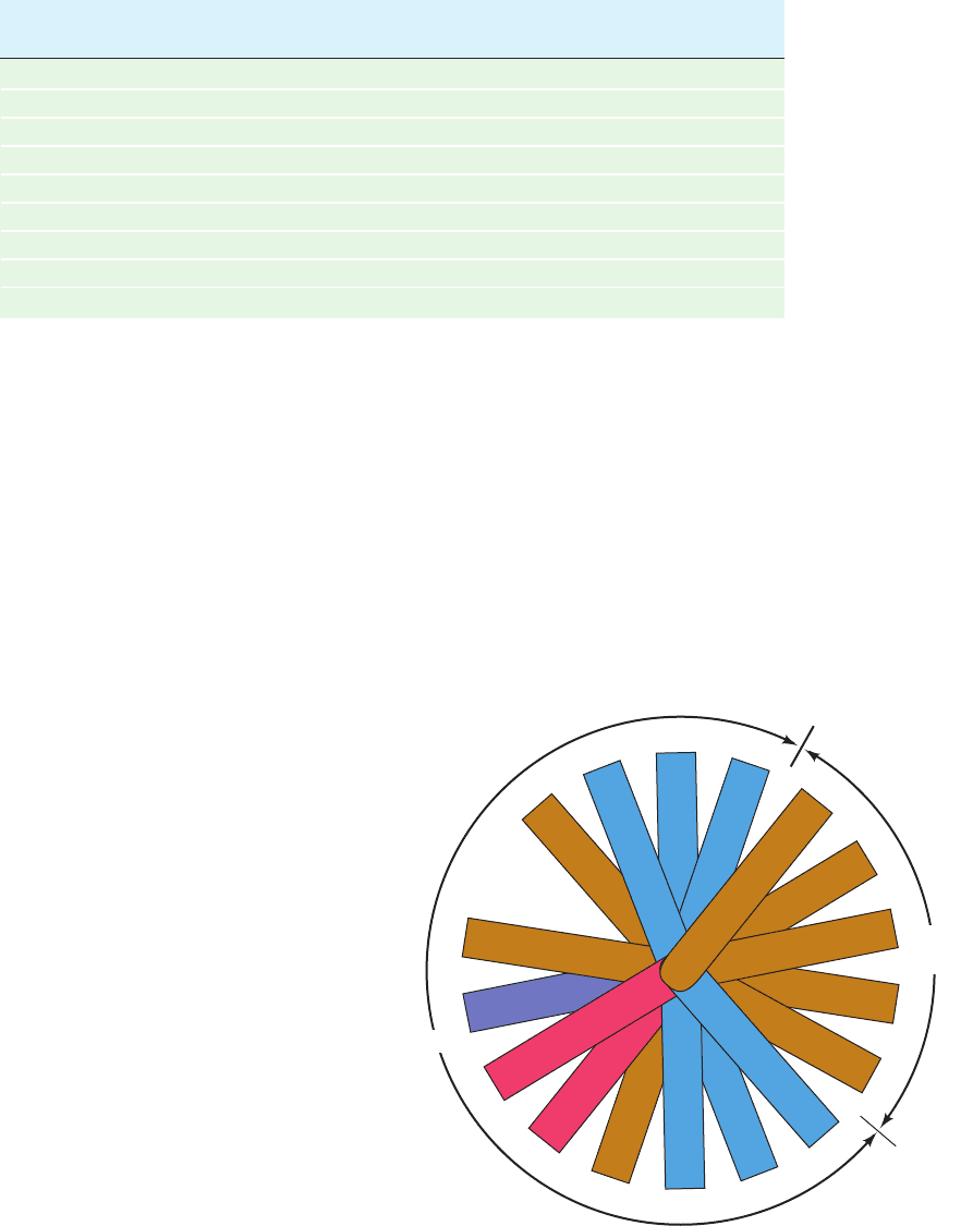
Figure 12-83 LDL, the major cholesterol carrier of the
bloodstream. This spheroidal particle consists of some 1500
cholesteryl ester molecules surrounded by an amphiphilic coat of
800 phospholipid molecules, 500 cholesterol molecules, and a
single 4536-residue molecule of apolipoprotein B-100.
Table 12-6 Characteristics of the Major Classes of Lipoproteins in Human Plasma
Chylomicrons VLDL IDL LDL HDL
Density (g ⴢ cm
⫺3
) ⬍0.95 ⬍1.006 1.006–1.019 1.019–1.063 1.063–1.210
Particle diameter (Å) 750–12,000 300–800 250–350 180–250 50–120
Particle mass (kD) 400,000 10,000–80,000 5000–10,000 2300 175–360
% Protein
a
1.5–2.5 5–10 15–20 20–25 40–55
% Phospholipids
a
7–9 15–20 22 15–20 20–35
% Free cholesterol
a
1–3 5–10 8 7–10 3–4
% Triacylglycerols
b
84–89 50–65 22 7–10 3–5
% Cholesteryl esters
b
3–5 10–15 30 35–40 12
Major
apolipoproteins A-I, A-II, B-48, C-I, B-100, C-I, C-II, B-100, C-I, C-II, B-100 A-I, A-II, C-I,
C-II, C-III, E C-III, E C-III, E C-II, C-III, D, E
a
Surface components.
b
Core lipids.
JWCL281_c12_386-466.qxd 6/10/10 11:16 AM Page 449
a. Apolipoproteins Have Amphipathic Helices That
Coat Lipoprotein Surfaces
The protein components of lipoproteins are known as
apolipoproteins or just apoproteins. At least nine
apolipoproteins are distributed in significant amounts in
the different human lipoproteins (Tables 12-6 and 12-7).
Most of them are water-soluble and associate rather
weakly with lipoproteins. Hence, they readily transfer be-
tween lipoprotein particles via the aqueous phase. CD
measurements indicate that apolipoproteins have a high
helix content, which increases when they are incorporated in
lipoproteins. Apparently, the helices are stabilized by a
lipid environment, presumably because helices fully satisfy
the polypeptide backbone’s hydrogen bonding potential in
a lipoprotein’s water-free interior.
b. The X-ray Structure of ApoA-I Mimics That in HDL
Apolipoprotein A-I (apoA-I) is HDL’s major apopro-
tein. Sequence analysis indicates that apoA-I consists
mainly of repeated amphipathic ␣ helices of 11 or 22
residues that provide the protein’s lipid-binding regions.
These putative ␣ helices, as well as similar helices that occur
in most other apolipoproteins, have their hydrophobic and
hydrophilic residues on opposite sides of the helical cylin-
ders (Fig. 12-84). Furthermore, the polar helix face has a
zwitterionic character in that its negatively charged
residues project from the center of this face, whereas its
positively charged residues are located at its edges. Indeed,
a synthetic 22-residue polypeptide of high helix-forming
propensity, which was designed by E. Thomas Kaiser to
have this polarity distribution but to otherwise have mini-
mal similarity to the repeating apoA-I sequences, behaves
much like apoA-I in binding to egg lecithin liposomes.
Evidently, the structural role of apoA-I, and probably most
other apolipoproteins, is fulfilled by its helical segments
rather than by any organized tertiary structure. This sug-
gests that lipoprotein ␣ helices float on phospholipid sur-
faces, much like logs on water. The phospholipids are pre-
sumably arrayed with their charged groups bound to
oppositely charged residues on the polar face of the helix
and with the first few methylene groups of their fatty acid
residues in hydrophobic association with the nonpolar face
of the helix.
A variety of criteria indicate that apoA-I undergoes sig-
nificant secondary structural changes on binding lipid.
However, apo ⌬(1–43)A-I, a truncation mutant that lacks
residues 1 to 43 of the 243-residue human apoA-I, has a
450 Chapter 12. Lipids and Membranes
Table 12-7 Properties of the Major Species of Human Apolipoproteins
Number of Molecular Mass
a
Apolipoprotein Residues (kD) Function
A-I 243 29 Activates LCAT
b
A-II 77 17 Inhibits LCAT, activates hepatic lipase
B-48 2152 241 Cholesterol clearance
B-100 4536 513 Cholesterol clearance
C-I 56 6.6 Activates LCAT?
C-II 79 8.9 Activates LPL
c
C-III 79 8.8 Inhibits LPL, activates LCAT?
D 169 19 Unknown
E 299 34 Cholesterol clearance
a
All apolipoproteins are monomers but apoA-II, which is a disulfide-linked dimer.
b
LCAT ⫽ lecithin–cholesterol acyltransferase.
c
LPL ⫽ lipoprotein lipase.
Figure 12-84 A helical wheel projection of the amphipathic ␣
helix constituting residues 148 to 164 of apolipoprotein A-I. (In
a helical wheel representation, the side chain positions are
projected down the helix axis onto a plane.) Note the segregation
of nonpolar, acidic, and basic residues to different sides of the
helix. Other apolipoprotein helices have similar polarity
distributions. [After Kaiser, E.T., in Oxender, D.L. and Fox, C.F.
(Eds.), Protein Engineering, p. 194, Liss (1987).]
Ala
158
Arg
151
His
162
His
155
Met
148
Leu
159
Ala
152
Leu
163
Val
156
Arg
149
Arg
160
Arg
153
Ala
164
Asp
157
Asp
150
Thr
161
Ala
154
Non-
polar
Polar
JWCL281_c12_386-466.qxd 6/9/10 12:06 PM Page 450
