Voet D., Voet Ju.G. Biochemistry
Подождите немного. Документ загружается.

from the ribosome–translocon complex. This permits the
bound ribosome to resume polypeptide synthesis such that
the growing polypeptide’s N-terminus passes through the
translocon into the lumen of the ER. Most ribosomal
processes, as we shall see in Section 32-3, are driven by
GTP hydrolysis.
Section 12-4. Membrane Assembly and Protein Targeting 421
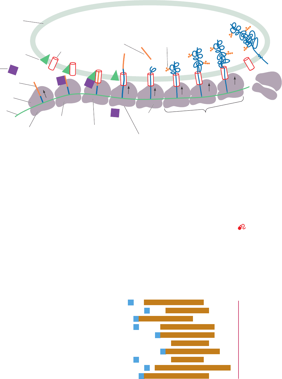
Figure 12-46 The ribosomal synthesis, membrane insertion,
and initial glycosylation of an integral protein via the secretory
pathway. (1) Protein synthesis is initiated at the N-terminus of
the polypeptide, which consists of a 13- to 36-residue signal
sequence. (2) A signal recognition particle (SRP) binds to the
ribosome and the signal sequence emerging from it, thereby
arresting polypeptide synthesis. (3) The SRP is bound by the
transmembrane SRP receptor (SR) in complex with the
translocon, thereby bringing together the ribosome and the
translocon. (4) The SRP and SR hydrolyze their bound GTPs,
causing them to dissociate from the ribosome–translocon
complex.The ribosome then resumes the synthesis of the
polypeptide, which passes through the translocon into the lumen
Figure 12-47 N-Terminal sequences of some eukaryotic
secretory preproteins. The hydrophobic cores (brown) of most
Lumen
N
H
3
+
NH
3
+
Carbohydrate
Signal
sequence
removed
Translocon
COO
–
3
′
SRP receptor
SRP
Rough
endoplasmic
reticulum
Signal
sequence
Ribosome
Messenger
RNA
5
′
Polypeptide
synthesis
initiated
Signal
sequence
excised
Protein extrudes, folds,
and is anchored to
membrane
Ribosome
dissociates
NH
3
+
N
H
3
+
N
H
3
+
GDP
GDP
NH
3
+
Polypeptide
synthesis
inhibited
2
GTP
GTP
GTP
GTP
GDP
GDP
SRP and SRP receptor
dissociate and polypeptide
synthesis resumes
1
SRP docks to
the SRP receptor
to form the ribosome-
translocon complex.
3
4
5
6
7
Bovine growth hormone
Bovine proalbumin
Human proinsulin
Human interferon
γ
Z
ea mays rein protein 22.1
Human
α-fibrinogen
Human IgG heavy chain
Rat amylase
Murine
α-fetoprotein
Chicken lysozyme
P
W
M
R
M
M
M
A
T
R
K
M
M
K
T
S
M
L
Y
F
E
W
M
K
L
K
L
T
S
F
I
R
I
L
W
P
S
M
G
T
S
L
L
V
L
Y
R
L
M
P
L
A
A
T
L
I
I
S
K
A
L
L
F
F
A
L
V
W
F
S
I
L
A
I
L
A
C
L
V
L
L
A
L
S
L
F
L
F
L
I
V
L
L
L
A
Q
V
L
L
L
L
L
C
L
L
L
L
V
L
L
C
A
L
L
W
C
S
A
S
L
F
L
P
L
G
I
V
I
L
L
L
L
W
F
P
Y
V
L
I
H
P
V
T
S
D
L
G
K
G
F
L
S
Q
S
P
G
T
G
F
A
A
A
V
A
A
S
A
V
C
A
A
T
V
Y
A
L
W
Q
W
S
L
N
G
S
A
G
T
C
A
K
G
A
A
R
F
C
A
E
Q
A
K
F
F
G
V
Y
D
V
Y
L
V
I
P
V
N
C
S
Q
D
H
F
I
MAAGM
MA L
Signal
peptidase
cleavage
site
of the ER. (5) Shortly after the entrance of the signal sequence
into the lumen of the endoplasmic reticulum, it is proteolytically
excised. (6) As the growing polypeptide chain passes into the
lumen, it commences folding into its native conformation, a
process that is facilitated by its interaction with the chaperone
protein BiP (not shown). Simultaneously, enzymes initiate the
polypeptide’s specific glycosylation. Once the protein has folded,
it cannot be pulled out of the membrane. At points determined by
its sequence, the polypeptide becomes anchored in the membrane
(proteins destined for secretion pass completely into the ER
lumen). (7) Once polypeptide synthesis is completed, the
ribosome dissociates into its two subunits.
See the Animated
Figures
signal peptides are preceded by basic residues (blue). [After
Watson, M.E.E., Nucleic Acids Res. 12, 5147–5156 (1984).]
JWCL281_c12_386-466.qxd 6/9/10 12:06 PM Page 421
5. Shortly after the signal peptide enters the ER lumen,
it is specifically cleaved from the growing polypeptide by a
membrane-bound signal peptidase (polypeptide chains
with their signal peptide still attached are known as prepro-
teins; signal peptides are alternatively called presequences).
6. The nascent (growing) polypeptide starts to fold to
its native conformation, a process that is facilitated by its
interaction with an ER-resident chaperone protein Hsp70
(Section 9-2C). Enzymes in the ER lumen then initiate
post-translational modification of the polypeptide, such as
the specific attachments of “core” carbohydrates to form
glycoproteins (Section 23-3B); the formation of disulfide
bonds as facilitated by protein disulfide isomerase (Section
9-2A), an ER-resident protein; and the attachment of GPI
anchors (Section 23-3Bk).
7. When polypeptide synthesis is completed, the pro-
tein is released from both the ribosome and the translocon,
and the ribosome dissociates from the RER. Secretory,
ER-resident, and lysosomal proteins pass completely
through the RER membrane into the lumen. TM proteins,
in contrast, contain one or more hydrophobic ⬃22-residue
TM sequences that remain embedded in the membrane.
The secretory pathway also occurs in prokaryotes for the
insertion of certain proteins into the cell membrane
(whose exterior is equivalent to the ER lumen). Indeed, all
forms of life yet tested have homologous SRPs and SRs.
However, in bacteria, the binding of the SRP to the ribo-
some does not arrest translation.
b. The Cryo-Electron Microscopy Structure of the
SRP in Complex with a Translating Ribosome
Reveals How the SRP Binds Signal Peptide and
Arrests Translation
Mammalian SRPs consist of six polypeptides known as
SRP9, SRP14, SRP19, SRP54, SRP68, and SRP72 (where
the numbers are their molecular masses in kilodaltons) and
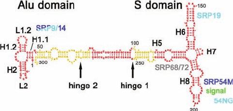
an ⬃300-nucleotide (nt) 7S RNA [Fig. 12-48; RNAs are of-
ten classified according to their sedimentation rate in Sved-
berg units (S), which increases with their molecular mass
(Section 6-5Aa)]. Many prokaryotic SRPs are much sim-
pler; that in E. coli consists of a single polypeptide named
Ffh that is homologous to SRP54 (Ffh for Fifty-four ho-
molog) and a 4.5S RNA (114 nt) that, in part, is predicted to
have a secondary structure similar to that portion of the 7S
RNA to which SRP54 binds. Indeed, replacing SRP54 with
Ffh or vice versa yields functional SRPs, at least in vitro,
thereby suggesting that the Ffh–4.5S RNA complex is a
structurally minimized version of the eukaryotic SRP.
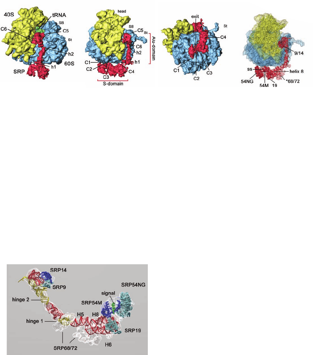
The 12-Å resolution cryo-electron microscopy (cryo-
EM)–based structure of canine SRP in complex with a
wheat germ ribosome containing a nascent (growing)
polypeptide chain was determined by Joachim Frank and
Roland Beckmann.The structure reveals that the so-called
S domain of the ⬃270-Å-long SRP binds at the base of the
large (60S) ribosomal subunit next to the exit of the tunnel
through which newly synthesized polypeptide emerges,
whereas the Alu domain bends around the large subunit to
contact the ribosome at the interface between its large and
small (40S) subunits (Fig. 12-49).
The ribosome–SRP structure was modeled by fitting the
much higher resolution X-ray structures of the yeast ribo-
some (Section 32-3Af) and various SRP fragments to the
cryo-EM–derived electron density (Figs. 12-49d and 12-50).
The model indicates that the 7S RNA consists mainly of a
long double helical rod that is bent at two positions named
hinge 1 and hinge 2 (RNA, as does DNA, can form a base-
paired double helix, although its conformation is distinctly
different from that of B-DNA; Section 29-1Bc). The signal
sequence exiting the ribosome, which was modeled as an ␣
helix, binds to SRP54, which contacts the ribosome near the
mouth of its peptide exit tunnel.
SRP54 consists of three domains: the N-terminal N do-
main; the central G domain, which contains the SRP’s
GTPase function and together with the N domain mediates
the SRP’s interaction with the SRP receptor; and the C-
terminal M domain, which is rich in methionine (25 of its
209 residues in humans). The N domain forms a bundle of
four antiparallel ␣ helices that closely associates with the
G domain, which consists of an open  sheet (Section 8-3Bi)
that structurally resembles those of other GTPases. The
M domain contains a deep groove that binds the helical
signal sequence. The groove is lined almost entirely with
hydrophobic residues including many of SRP54’s Met
residues (the Met side chain has physical properties similar
to that of an n-butyl group). Its flexible unbranched Met
side chain “bristles” presumably provide the groove with
the plasticity to bind a variety of different signal sequences
so long as they are hydrophobic and form an ␣ helix.
Ribosomes, as we shall see in Section 32-3Dk, employ
protein elongation factors to deliver aminoacyl-tRNAs
and to motivate the sequence of reactions that appends an
amino acid residue to the growing polypeptide chain. The
eukaryotic SRP’s Alu domain, which is required for trans-
lational arrest, contacts the ribosome’s intersubunit region
at the same positions to which the ribosomal elongation
factors bind. This suggests that the Alu domain arrests
translation by binding to the ribosome with sufficient affin-
ity to block the binding of the ribosome’s required elonga-
tion factors. This is corroborated by the observation that
422 Chapter 12. Lipids and Membranes
Figure 12-48 Sequence and secondary structure of canine 7S
RNA. Its various double helical segments (denoted H1 through
H8) and loops (denoted L1 and L1.2), are drawn in red and
yellow with Watson–Crick base pairs represented by connecting
lines and non-Watson–Crick base pairs indicated by dots. The
positions at which the various SRP proteins bind to the 7S RNA
are indicated in cyan, blue, and gray. [Courtesy of Roland
Beckmann, Humboldt University of Berlin, Germany.]
JWCL281_c12_386-466.qxd 6/9/10 12:06 PM Page 422
bacterial SRP’s, which do not arrest translation on binding
to a ribosome, lack Alu domains.
c. Secretory Pathway Initiation Is Driven
by GTP Hydrolysis
In eukaryotes, the SRP receptor is a heterodimer of sub-
units named SR␣ and SR. SR is a 271-residue integral
protein that has an N-terminal TM segment, whereas SR␣
is a 638-residue peripheral protein that is apparently
membrane-bound through the association of its N-terminal
segment with SR. Both SR␣ and SR are GTPases.
In E. coli, the SR consists of a single 497-residue subunit
named FtsY, whose C-terminal portion is homologous to
that of SR␣, although their N-terminal portions have no
sequence similarity. Curiously, the X-ray structure of the C-
terminal portion of FtsY closely resembles that of the N and
G domains of SRP54, with which it shares ⬃34% identity.
The targeting of the SRP–ribosome complex to the ER
membrane is mediated by the GTPase functions of SRP54,
SR␣, and SR. In numerous biological systems, mainly
those mediating translation (Section 32-3), vesicle trans-
port (Sections 12-4C and 12-4D), and signal transduction
(Section 19-2), GTPases function as molecular switches that
endow the system with unidirectionality and specificity.
These so-called G proteins have at least two stable confor-
mations: GDP-bound and GTP-bound. Interconversion
between these states only occurs in a unidirectional cycle
due to the irreversibility of GTP hydrolysis. In most cases,
a G protein must interact with other proteins in order to
change conformational states. Thus, GTP hydrolysis often
requires stimulation by a specific GTPase activating pro-
tein (GAP), and the exchange of bound GDP for GTP may
require the assistance of a specific guanine nucleotide ex-
change factor (GEF; Section 19-2Ca). The need for these
particular factors confers specificity on the system.
The GEF for the SRP is the complex of the newly
emerged signal sequence with the M domain of SRP54,
which induces the adjoining G domain to exchange its
bound GDP for GTP (Fig. 12-46, Stage 2).The formation of
the resulting SRP ⴢ GTP complex results in a conforma-
tional change that locks the SRP to the ribosome, which, in
turn, induces translational arrest. The GEF for the SR ap-
pears to be an empty translocon, which thereby associates
with the resulting SR ⴢ GTP complex to which the SRP ⴢ
GTP–ribosome complex then binds (Fig. 12-46, Stage 3).
Evidently, the SRP and SR, both in their GTP forms, act as
“molecular matchmakers” to bring together an empty
translocon with a ribosome synthesizing a signal sequence–
bearing polypeptide.The SRP and the SR then reciprocally
stimulate each other’s GTPase functions (act as mutual
GAPs; neither protein alone has significant GTPase activ-
ity) followed by their dissociation, yielding free SRP ⴢ GDP
and SR ⴢ GDP complexes ready to participate in a new
Section 12-4. Membrane Assembly and Protein Targeting 423
Figure 12-49 Cryo-EM structure of a translating wheat germ
ribosome in complex with canine SRP at 12 Å resolution.
(a) Surface diagram showing the small (40S) ribosomal subunit
in yellow, the large (60S) ribosomal subunit in blue, the SRP in
red, and the tRNA occupying the ribosomal P-site (to which the
growing polypeptide chain is covalently linked; Fig. 5-28) in
green. C1 to C6 indicate the six positions at which the SRP
Figure 12-50 Molecular model of the SRP. The transparent
cryo-EM–based electron density is shown in white and the rib-
bon diagrams of the X-ray structures of SRP proteins and RNA
fragments that have been docked into it are colored as is indi-
cated in Fig. 12-48. The signal sequence, modeled as an ␣ helix, is
green. Note that no atomic resolution structure of the SRP68/72
heterodimer is available. [Courtesy of Roland Beckmann,
Humboldt University of Berlin, Germany. PDBid 2G05.]
(c)
(d)
(c)
(a)
(b)
(d)
contacts the ribosome and h1 and h2 indicate the 7S RNA’s
hinge positions. St and SB stand for stalk and stalk base. (b) As in
Part a but rotated 70° about the vertical axis. (c) As in Part a but
rotated 90° about the horizontal axis. (d) As in Part b but with
transparent surfaces showing the molecular models of the
ribosome and SRP. [Courtesy of Roland Beckmann, Humboldt
University of Berlin, Germany.]
JWCL281_c12_386-466.qxd 6/9/10 12:06 PM Page 423
round of the secretory pathway (Fig. 12-46, Stage 4). The
release of the SRP and SR permits the now translocon-
associated ribosome to recommence translation, thereby
extruding the polypeptide it is synthesizing into or through
the ER membrane as described below.
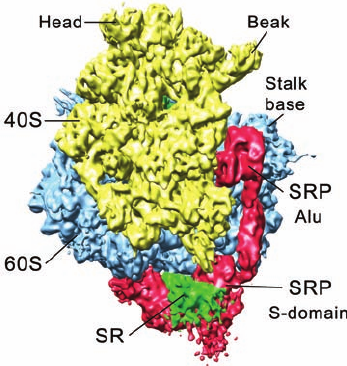
In the structure of the SRP–ribosome complex (Fig.
12-49), the S domain of the SRP blocks the binding of
the translocon at the mouth of the peptide exit tunnel.
However, the cryo-EM–based structure of the SR–SRP–
ribosome complex (but lacking SR’s TM segment), deter-
mined by Irmgard Sinning and Beckmann, reveals that the
SR contacts both the large ribosomal subunit and the S do-
main of the SRP (Fig. 12-51) in a way that pushes the SRP S
domain in the SRP–ribosome complex away from the pep-
tide exit site. This allows the translocon access to its riboso-
mal binding site and presumably positions it such that the M
domain–bound signal sequence can readily be transferred
to it. The position of the eukaryotic SRP’s Alu domain on
the ribosome is unaffected by the binding of the SR.
d. The Translocon Forms a Transmembrane Pore
How are preproteins transported across or inserted into
the RER membrane? In 1975, Blobel postulated that these
processes are mediated by a TM channel. However, it was
not until 1991 that he was able to show that these channels
actually exist through electrophysiological measurements
indicating that the RER membrane contains ion-conducting
channels. These increase in number when the ribosome-
bearing side of the RER is treated with puromycin (an an-
tibiotic that causes the ribosome to prematurely release
the growing polypeptide; Section 32-3Df), thereby suggest-
ing that the channels are usually plugged by the presence
of the polypeptides. By linking fluorescent dyes whose flu-
orescence is sensitive to the polarity of their environment
to a nascent polypeptide, Arthur Johnson demonstrated
that these channels, now called translocons, enclose aque-
ous pores that completely span the ER membrane.
The various ER transmembrane proteins that comprise
the translocon were identified through the use of photoac-
tivatable groups that were attached to signal sequences
and mature regions of preproteins. On exposure to light of
the proper wavelength, the photoactivatable groups react
with nearby proteins to form covalent cross-links, thereby
permitting the identification of these proteins. The central
component of the translocon, named Sec61 (Sec for secre-
tion) in eukaryotes and the SecY complex in prokaryotes,
is a heterotrimeric protein. Its ␣ and ␥ subunits, but not its
 subunit, are essential for channel function and are con-
served across all kingdoms of life (these subunits are re-
spectively named Sec61␣, Sec61, and Sec61␥ in eukary-
otes and SecY, SecE, and SecG in bacteria).
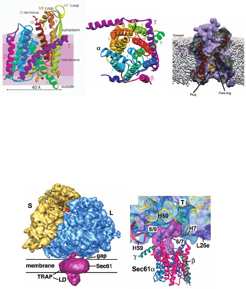
The X-ray structure of the SecY complex from the
archaeon Methanococcus jannaschii, determined by Stephen
Harrison and Tom Rapoport, reveals that its ␣, ,and ␥ sub-
units, respectively, have 10, 1, and 1 TM ␣ helices (Fig. 12-
52a,b). The ␣ subunit’s TM helices are wrapped around an
hourglass-shaped channel whose minimum diameter is ⬃3
Å (Fig. 12-52c). The channel is blocked at its extracellular
end by a short, relatively hydrophilic helix (blue unnum-
bered helix in Fig. 12-52a,b and yellow helix in Fig. 12-52c).A
variety of evidence indicates that this helix functions as a plug
to prevent small molecules and ions from leaking across the
membrane in the absence of a translocating polypeptide and
that an incoming signal peptide pushes this helix aside.The ␥
subunit extends diagonally across the back of the ␣ subunit
so as to buttress it. The  subunit makes relatively tenuous
contacts with the ␣ subunit, which likely explains why the 
subunit is dispensable for translocon function.
A cryo-EM–based structure of a mammalian ribosome–
Sec61 complex (Fig. 12-53a), determined by Rapoport and
Christopher Akey at 11 Å resolution, reveals that a single
Sec61 channel is positioned over the ribosome’s peptide
exit tunnel with Sec61’s 6/7 and 8/9 loops extending into
the peptide exit tunnel (Fig. 12-53b). Indeed, mutating the
positively charged residues of the 6/7 and 8/9 loops, which
presumably bind to negatively charged ribosomal RNA,
abolishes ribosome binding.
How wide is the SecY complex’s protein-conducting
channel (PCC) when it is translocating a polypeptide? At
minimum, it would have to be ⬃7 Å across (the diameter of
an extended anhydrous polypeptide), although if a TM se-
quence assumed its helical conformation while still in the
PCC, the PCC would have to be at least ⬃12 Å wide. Such
widening could be accomplished by movements of the he-
lices from which the pore ring side chains emanate. The
maximum dimensions of the PCC,as estimated from a con-
sideration of the SecY structure (Fig. 12-52), are 15 ⫻ 20 Å.
Such movements are supported by molecular dynamics
calculations (Section 9-4a). Despite the large pore size of
an active translocon, the ER membrane’s permeability bar-
rier is largely maintained. Evidently, the pore ring fits
around the translocating polypeptide chain like a gasket,
thereby preventing the passage of small molecules and ions
during polypeptide translocation.
424 Chapter 12. Lipids and Membranes
Figure 12-51 Cryo-EM structure of the eukaryotic
SR–SRP–ribosome complex at 8 Å resolution. The complex is
oriented and colored as in Fig. 12-49b with the SR colored green.
[Courtesy of Roland Beckmann, Humboldt University of Berlin,
Germany. ]
JWCL281_c12_386-466.qxd 6/9/10 12:06 PM Page 424
Section 12-4. Membrane Assembly and Protein Targeting 425
Figure 12-52 X-ray structure of the M. jannaschii SecY
complex. (a) X-ray structure of the complex, with shading
indicating the positions of membrane phospholipid head groups
(violet) and hydrocarbon tails (pink).The ␣ subunit of SecY
(436 residues) is colored in rainbow order and its helices are
numbered from its N-terminus (dark blue) to its C-terminus
(red), the  subunit (74 residues) is magenta, and the ␥ subunit
(53 residues) is purple. (b) View of SecY from the cytosol. The
(c)
(b)(a)
translocon’s putative lateral gate is on the left between helices
2 and 7. (c) Cross-section of the protein-conducting channel as
viewed from the bottom of Part b.The helix that plugs the
channel is yellow and the six hydrophobic side chains that form
the narrowest part of the channel, the so-called pore ring, are
green. [Courtesy of Stephen Harrison and Tom Rapoport,
Harvard Medical School. PDBid 1RH5.]
Figure 12-53 Cryo-EM structure of a canine
ribosome–Sec61–TRAP complex at 11 Å resolution. (a) A
surface diagram viewed parallel to the ER membrane. The
ribosome’s small (S) and large (L) subunits are yellow and blue,
a tRNA occupying the ribosome’s exit site (Section 32-3Bd) is
red, and the Sec61–TRAP complex is magenta. LD is TRAP’s
lumenal domain. (b) A thin slab showing the interface between
the ribosome and Sec61.The modeled structures of the ribosome
and Sec61, shown as ribbons, are embedded in their transparent
surface diagrams, which are colored as in Part a. Note how loops
6/7 and 8/9 of Sec61 are inserted into the ribosome’s peptide exit
tunnel (T), where they interact with RNA helices H7 and H50.
L26e is a protein subunit. [Courtesy of Tom Rappoport, Harvard
Medical School; and Christopher Akey, Boston University
School of Medicine. PDBid 3DKN.]
(a)
(b)
JWCL281_c12_386-466.qxd 6/9/10 12:06 PM Page 425
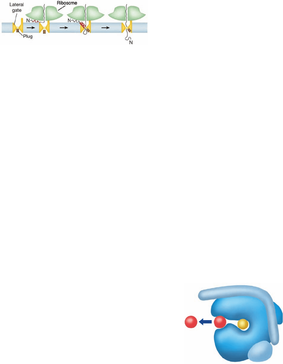
The translocation of a secretory protein begins with its in-
sertion as a loop into the PCC (Fig. 12-54, left). This was es-
tablished by using a mutant protein whose signal sequence is
not excised by the signal protease and showing,through pro-
teolysis experiments, that the protein’s N- and C-termini
both remained on the ER membrane’s cytoplasmic side.
Subsequently, as was shown by photo-cross-linking experi-
ments, the signal sequence forms an ⬃2-turn helix that in-
serts between TM helices 2 and 7 of Sec61␣ (Fig. 12-54, mid-
dle). The separation of helices 2 and 7 (the lateral gate; Fig.
12-52b) helps displace the plug helix, which following signal
sequence excision, allows the nascent polypeptide pass
through the PCC into the ER (Fig. 12-54, right).
Additional components of the mammalian translocon
are named translocating chain-associated membrane pro-
tein (TRAM, ⬃375 residues; predicted to have 8 TM he-
lices with both its N- and C-termini in the cytosol) and
translocon-associated membrane protein (TRAP; an
⬃800-residue heterotetramer with its ␣, , and ␦ subunits
each having one TM helix and its ␥ subunit having four TM
helices). Through the use of Sec61-containing liposomes
that either did or did not also contain TRAM, Rapoport
demonstrated that TRAM is required for the translocation
and membrane integration of most but not all preproteins
into the liposome. Whether or not a given preprotein re-
quires TRAM for translocation depends on its signal
sequence, although no particular characteristic of this se-
quence appears to be critical for TRAM dependence.
TRAP, which is seen in Fig. 12-53a, functions similarly to
increase the translocational efficiency of proteins with cer-
tain signal sequences.
e. The Translocon Laterally Inserts Transmembrane
Helices Into the ER Membrane
In addition to forming a conduit for soluble proteins to
enter the ER, the translocon must insert an integral protein’s
TM segments into the ER membrane.The translocon,in con-
cert with the ribosome, recognizes these TM segments and
installs them into the lipid bilayer via a lateral gate between
helices 2 and 7 in the SecY/Sec61 ␣ subunit (Fig. 12-55).
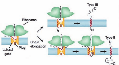
Monotopic (alternatively, single-pass) TM proteins fall
into one of three classes:
1. Type I proteins have cleavable N-terminal signal se-
quences. They are inserted into the membrane much like
secretory proteins (Fig. 12-54) but have an ⬃22-residue hy-
drophobic stop-transfer anchor sequence that the translo-
con laterally inserts into the membrane as a helix. Hence a
type I protein has its N-terminus in the ER.
2. Type II proteins lack a cleavable N-terminal signal
sequence. However, they have an ⬃22-residue hydropho-
bic signal-anchor sequence, not necessarily near the pro-
tein’s N-terminus, that is recognized by the SRP. The SRP
then passes the nascent polypeptide to the translocon,
which laterally inserts the signal-anchor sequence into the
membrane oriented such that the protein’s N-terminus is in
the cytoplasm. This requires the polypeptide to loop
around inside the translocon before being inserted into the
membrane (Fig. 12-56, lower portion). The C-terminal seg-
ment of a type II protein is presumably extruded into the
gap between the ribosome and the translocon (Fig. 12-53a)
before being passed through the translocon.
3. Type III proteins, like type II proteins, lack a cleav-
able N-terminal signal sequence and have a signal-anchor
sequence that is not necessarily near the protein’s N-terminus.
However, the orientation of these signal-anchor sequences
in the membrane is opposite to that of type II proteins.
Hence the way that type III proteins are inserted into the
membrane resembles that of type I proteins (Fig. 12-56,
upper portion).They differ, however, in that the transfer of
a type III protein’s N-terminal segment across the membrane
cannot be initiated until after its signal-anchor sequence has
been synthesized.
For polytopic (alternatively, multipass or type IV) TM
proteins, looping must occur each time an additional helix is
to be installed in the membrane. Evidently, the PCC of the
426 Chapter 12. Lipids and Membranes
α
β
γ
Figure 12-55 Model for the insertion of a TM helix into a
membrane. The translocon (blue) is viewed as in Fig. 12-52b.A
polypeptide chain (yellow) is shown bound in the translocon’s
pore during its translocation through the membrane, and a TM
helix (red) is shown passing through the translocon’s lateral gate
and being released into the membrane (arrow). [Based on a
drawing by Dobberstein, B. and Sinning, I., Science 303, 320
(2004).]
Figure 12-54 The stages of polypeptide translocation of a
secretory protein. The red line represents the hydrophobic
portion of the signal sequence. The process begins with the
insertion of the nascent peptide as a loop into the PCC (left).
The signal sequence then binds as an ⬃2-turn helix between the
SecY/Sec61 ␣ subunit’s helices 2 and 7, which helps displace
the plug helix (middle). Finally, the signal sequence is excised by
the signal protease (not shown) and the nascent peptide enters
the ER through the PCC (right). [Courtesy of Tom Rapoport,
Harvard Medical School.]
JWCL281_c12_386-466.qxd 6/9/10 12:06 PM Page 426
active translocon has sufficient room for successive TM seg-
ments to reverse their direction prior to being inserted into
the ER membrane. Helices may be inserted into the mem-
brane either singly or in pairs, depending on their hydropho-
bicity and their ability to form stable helix–helix interactions.
What controls the orientations of the helices in a TM
protein, that is, its topogenesis? Most TM proteins, as
Gunnar von Heijne pointed out, adopt an orientation such
that their cytoplasmically exposed ends, those that are not
translocated across the membrane, are more positively
charged (have more Arg and Lys residues) than their lume-
nally exposed ends—the positive-inside rule. This appears
mainly due to the charge distribution within the translo-
con, which is oriented with its more positive face on the cy-
toplasmic side of the membrane (in accordance with the
positive-inside rule). In fact, mutating certain charged
residues of Sec61␣ so as to reverse their charge (e.g.,
changing an Arg to Glu), inverts the orientation of the TM
helices it installs in the membrane. Another important in-
fluence on the orientation of a TM helix is its hydrophobic-
ity gradient: The more hydrophobic end of a TM helix is
preferentially translocated across the membrane.
Despite the foregoing,one might reasonably expect that
the membrane orientation of the N-terminal TM helix of a
polytopic TM protein dictates the orientations of the suc-
ceeding TM helices (many, if not all, of which have yet to be
synthesized at the time the N-terminal helix is inserted into
the membrane). However, the deletion or insertion of a
TM helix from/into a polypeptide does not necessarily
change the membrane orientations of the succeeding TM
helices: When two successive TM helices have the same
preferred orientation,one of them may be forced out of the
membrane. Moreover, the topological organization of TM
proteins is influenced by the membrane lipid composition.
This suggests that the translocon’s lateral gate frequently
opens and closes so as to allow its transiting peptide to
sample the outside lipid environment and only inserts a
peptide segment into the lipid bilayer if it is thermodynam-
ically favorable to do so; that is, helix insertion may be con-
sidered as a partitioning between the aqueous environ-
ment in the translocon and that of the membrane.
Polytopic TM proteins can fold to their native confor-
mations only after all their TM helices have been inserted
into the membrane.This process is guided by packing inter-
actions between helices as well as specific interactions with
membrane lipids. Thus, although a TM protein’s sequence
determines its topology, it does so for a specific membrane
lipid environment. Evidently, the lipid composition of a
membrane and the topologies of its embedded TM pro-
teins have coevolved.
f. Protein Folding in the ER Is Monitored by
Molecular Chaperones
The ER, as does the cytosol,contains a battery of molec-
ular chaperones that assist in protein folding and act as
agents of quality control.The best characterized of these is
the Hsp70 homolog (Section 9-2C) BiP (for binding pro-
tein). BiP associates with many secretory and TM proteins
although, if folding proceeds normally, these interactions
are weak and short-lived. However, proteins that are im-
properly folded, incorrectly glycosylated, or improperly as-
sembled form stable complexes with BiP that are often ex-
ported, via a poorly understood process involving the
translocon called retrotranslocation, to the cytosol where
they are proteolytically degraded (Section 32-6).The entire
process is named ERAD (for ER-associated degradation).
Two other notable ER-resident chaperones are calreticulin
and calnexin, homologous proteins that participate in facil-
itating and monitoring the folding and assembly of glyco-
proteins (Section 23-3Bf). The ER also contains protein
disulfide isomerases (PDIs; Section 9-2A) and peptidyl
prolyl cis–trans isomerases (PPIs; Section 9-2B).
Abnormalities of protein folding and assembly are im-
portant mechanisms of disease (e.g., Section 9-5). For in-
stance, cystic fibrosis is the most common life-threatening
recessive genetic disease in the Caucasian population (af-
fecting one in ⬃2000 individuals). It occurs in homozygotes
for a defective cystic fibrosis transmembrane regulator
(CFTR) protein, a 1480-residue glycoprotein with 12 TM
helices that functions as a Cl
⫺
transporter in the plasma
membrane of epithelial cells. Individuals with cystic fibrosis
produce highly viscous mucus that, in its most damaging ef-
fects, blocks the small airways in the lungs.This leads to per-
sistent infections, which cause severe progressive lung de-
generation that is usually fatal by around age 30. Although
cystic fibrosis is caused by any of more than 1000 known mu-
tations in the CFTR gene, 70% of the cases arise from the
deletion of Phe 508 (⌬F508),which is located in a cytoplasmic
domain of the CFTR protein (which is initially inserted into
the ER membrane). Although this mutant domain in ⌬F508
folds to nearly its native conformation (⌬F508 retains almost
full biological activity), it does so much more slowly than in
the wild-type protein. This results in its retrotranslocation
Section 12-4. Membrane Assembly and Protein Targeting 427
Figure 12-56 The generation of types II and III proteins.
Here, the red line represents a signal-anchor sequence. The
N-terminus of a type III protein must pass through the translocon
before its succeeding TM helix is laterally installed in the
membrane (top.). However, for a type II protein, whose
N-terminus is retained in the cytoplasm (bottom), the
subsequently synthesized polypeptide (represented by the loop
between the ribosome and the translocon) must pass through the
PCC. For polytopic TM proteins, these two processes alternate.
[Based on a drawing by Tom Rapoport, Harvard Medical
School.]
JWCL281_c12_386-466.qxd 6/9/10 12:06 PM Page 427
and degradation by an, in this case, overly zealous prote-
olytic surveillance system (Section 32-6B).
g. Some Proteins Are Post-Translationally
Transported through Membranes
The secretory proteins we discussed pass through the
membrane as they are being synthesized by the ribosome,
that is, their membrane translocation occurs cotranslation-
ally. However, some secretory proteins are translocated only
after they have been fully synthesized in the cytoplasm,
that is, post-translationally. Nevertheless, both co- and post-
translational translocation is mediated by the translocon.
Yet, the translocon is a passive pore, that is, it does not pro-
vide the free energy that drives translocation. In cotransla-
tional translocation, it is the ribosomally mediated extension
of the polypeptide that pushes it through the translocon.
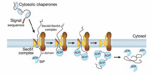
In eukaryotes, post-translationally translocated secre-
tory proteins have signal sequences that are only moder-
ately hydrophobic; they are not recognized by the SRP but
still bind to Sec61. These proteins are bound by cytoplas-
mic chaperones, which keep them in a loosely folded or un-
folded state that permits them to pass through the translo-
con. Their translocation is driven by a so-called Brownian
ratchet mechanism that is mediated by the partnering of
Sec61 with the tetrameric TM complex Sec62/Sec63 and
the lumenal Hsp70-like chaperone BiP (Fig. 12-57). The
polypeptide in the translocon randomly slides back and
forth via Brownian motion. However, in the ER, the so-
called J-domain (a homolog of the E. coli cochaperone
DnaJ;Section 9-2C) on the lumenal surface of Sec63 induces
BiP to hydrolyze its bound ATP to ADP. The resulting
BiP–ADP complex then binds the polypeptide emerging
from the translocon, which prevents it from sliding back to-
ward the cytoplasm.When the peptide again slides forward,
another BiP–ADP complex binds to it, etc., until the entire
polypeptide has entered the ER. BiP eventually exchanges
its ADP for ATP, which causes it to release the polypeptide,
which then folds to its native conformation.
In bacteria, the motor that drives post-translational
translocation is SecA, which binds to the cytoplasmic face
of the SecY complex and pushes the polypeptide through
the translocon via repeated cycles of ATP hydrolysis. SecA
is aided in doing so by the cytosolic chaperone SecB, which
prevents the polypeptide from folding in the cytoplasm.
C. Vesicle Formation
Shortly after their polypeptide synthesis is completed, the
partially processed transmembrane, secretory, and lysoso-
mal proteins appear in the Golgi apparatus (Fig. 1-5), a 0.5-
to 1.0-m-diameter organelle consisting of a stack of 3 to 6
or more (depending on the species) flattened and function-
ally distinct membranous sacs known as cisternae, where
further post-translational processing, mainly glycosylation,
occurs (Section 23-3Bg). The Golgi stack (Fig. 12-58) has
two distinct faces, each comprised of a network of intercon-
nected membranous tubules: the cis Golgi network (CGN),
which is opposite the ER and is the port through which
proteins enter the Golgi apparatus; and the trans Golgi
network (TGN), through which processed proteins exit to
their final destinations. The intervening Golgi stack con-
tains at least three different types of sacs, the cis, medial,
and trans cisternae, each of which contains different sets of
glycoprotein processing enzymes.
Proteins transit from one end of the Golgi stack to the
other while being modified in a stepwise manner, a process
that is described in Section 23-3Bg. These proteins are
transported via two mechanisms:
1. They are conveyed between successive Golgi com-
partments in the cis to trans direction as cargo within mem-
branous vesicles that bud off of one compartment and fuse
with a successive compartment, a process known as for-
ward or anterograde transport.
2. They are carried as passengers in Golgi compart-
ments that transit the Golgi stack, that is, the cis cisternae
eventually become trans cisternae, a process called cister-
nal progression or maturation. This process is mediated
through the backward or retrograde transport of Golgi-
resident proteins from one compartment to the preceeding
one via membranous vesicles.
The cisternal progression mechanism has been clearly
shown to occur but the significance of the anterograde
transport mechanism is as yet unclear. In any case, on
reaching the trans Golgi network, the now mature proteins
are sorted and sent to their final cellular destinations.
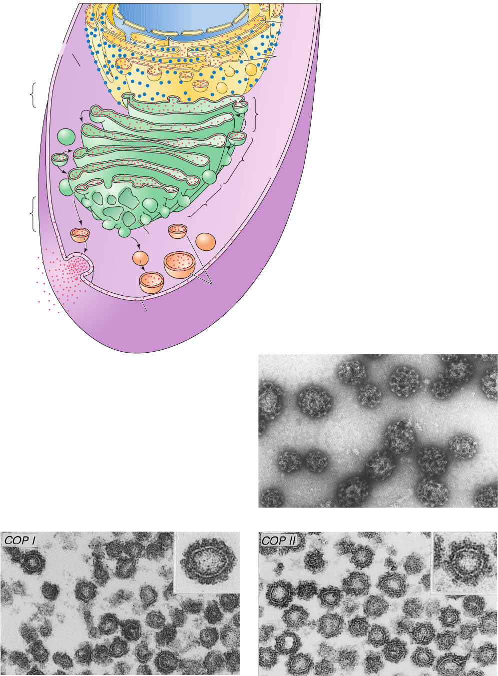
a. Membrane, Secretory, and Lysosomal Proteins
Are Transported in Coated Vesicles
The vehicles in which proteins are transported be-
tween the RER, the Golgi apparatus, and their final desti-
nations, as well as between the different compartments
of the Golgi apparatus, are known as coated vesicles (Fig.
12-59). This is because these 60- to 150-nm-diameter
428 Chapter 12. Lipids and Membranes
Figure 12-57 Scheme for post-translational translocation in
eukaryotes. As the translocating polypeptide enters the ER
through Sec61, the BiP–ATP complex binds to Sec63, whose
J-domain induces BiP to hydrolyze its bound ATP to ADP. The
resulting BiP–ADP complex binds to the emerging peptide so as
to prevent its backsliding.As additional peptide segments
emerge from Sec61, the process repeats until the entire protein
has entered the ER. BiP eventually exchanges its bound ADP for
ATP causing it to release the peptide, which then folds to its
native conformation. [Courtesy of Tom Rapoport, Harvard
Medical School.]
JWCL281_c12_386-466.qxd 6/9/10 12:06 PM Page 428
Section 12-4. Membrane Assembly and Protein Targeting 429
Figure 12-58 Post-translational processing
of proteins. Proteins destined for secretion,
insertion into the plasma membrane, or
transport to lysosomes are synthesized by
RER-associated ribosomes (blue dots; top).
As they are synthesized, the proteins (red
dots) are either translocated into the lumen
of the ER or inserted into its membrane.
After initial processing in the ER, the
proteins are encapsulated in vesicles that
bud off from the ER membrane and
subsequently fuse with the cis Golgi
network.The proteins are progressively
processed in the cis, medial, and trans
cisternae of the Golgi. Finally, in the trans
Golgi network (bottom), the completed
glycoproteins are sorted for delivery to their
final destinations, the plasma membrane,
secretory vesicles, or lysosomes, to which
they are transported by yet other vesicles.
Nucleus
Lumen
Cytoplasm
Golgi
apparatus
Cis
Golgi
network
Rough
ER
Cis
cisternae
cis face
Medial
cisternae
Lysosomes
Secretion
Plasma
membrane
Trans
face
Secretory
vesicle
Trans
cisternae
Trans
Golgi
network
Figure 12-59 Electron micrographs of coated vesicles.
(a) Clathrin-coated vesicles. Note their polyhedral character.
[Courtesy of Barbara Pearse, Medical Research Council,
Cambridge, U.K.] (b) COPI-coated vesicles. (c) COPII-coated
vesicles.The insets in Parts b and c show the respective vesicles
at higher magnification. [Courtesy of Lelio Orci, University of
Geneva, Switzerland.]
(b)
(c)
(a)
JWCL281_c12_386-466.qxd 6/9/10 12:06 PM Page 429
membranous sacs are initially encased on their outer (cy-
tosolic) faces by specific proteins that act as flexible scaf-
folding in promoting vesicle formation.A vesicle buds off
from its membrane of origin and later fuses to its target
membrane. This process preserves the orientation of the
transmembrane protein (Fig. 12-60), so that the lumens of
the ER and the Golgi cisternae are topologically equivalent
to the outside of the cell. This explains why the carbohy-
drate moieties of TM glycoproteins and the GPI anchors of
GPI-linked proteins occur only on the external surfaces of
plasma membranes.
The three best characterized types of coated vesicles are
distinguished by their protein coats:
1. Clathrin (Fig. 12-59a), a protein that forms a polyhe-
dral framework around vesicles that transport TM, GPI-
linked, and secreted proteins from the Golgi to the plasma
membrane. The clathrin cages, which were first character-
ized by Barbara Pearse, can be dissociated to flexible
three-legged proteins known as triskelions (Fig. 12-61) that
consist of three so-called heavy chains (HC, 1675 residues)
that each bind one of two homologous light chains, LCa or
LCb (⬃240 residues), at random.
2. COPI protein (Fig. 12-59b; COP for coat protein),
which forms what appears to be a fuzzy rather than a poly-
hedral coating about vesicles that carry out both the an-
terograde and retrograde transport of proteins between
successive Golgi compartments. In addition, COPI-coated
vesicles return escaped ER-resident proteins from the
Golgi to the ER (see below). COPI consists of seven differ-
ent subunits (␣, 160 kD; , 110 kD; ¿, 102 kD; ␥, 98 kD; ␦,
61 kD; ε, 31 kD; and , 20 kD). The soluble complex com-
prising the COPI protomer is named coatomer.
3. COPII protein (Fig. 12-59c), which transports pro-
teins from the ER to the Golgi.The COPII vesicle compo-
nents are then recycled by COPI-coated vesicles for partic-
ipation in another round of vesicle formation (the COPI
vesicle components entering the ER are presumably recy-
cled by COPII-coated vesicles).The COPII coat consists of
the GTPase Sar1, the heterodimer Sec23/24 in which Sec23
is a Sar1-specific GAP and Sec24 functions in cargo selection,
430 Chapter 12. Lipids and Membranes
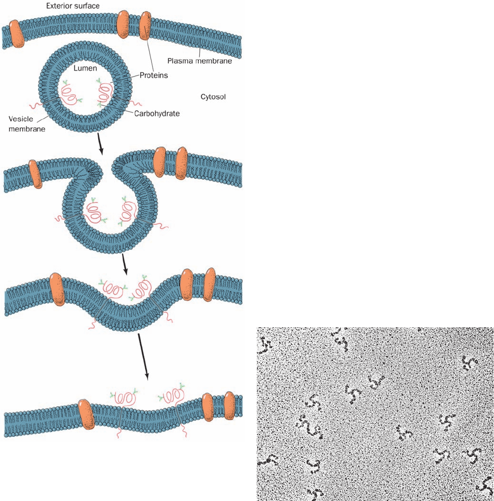
Figure 12-60 The fusion of a vesicle with the plasma
membrane preserves the orientation of the integral proteins
embedded in the vesicle bilayer. The inside of the vesicle and the
exterior of the cell are topologically equivalent because the same
side of the protein is always immersed in the cytosol. Note that
any soluble proteins contained within the vesicle would be
secreted. In fact, proteins destined for secretion are packaged in
membranous secretory vesicles that subsequently fuse with the
plasma membrane as shown.
Figure 12-61 Electron micrograph of triskelions. The variable
orientations of their legs are indicative of their flexibility.
[Courtesy of Daniel Branton, Harvard University.]
JWCL281_c12_386-466.qxd 6/9/10 12:06 PM Page 430
