Voet D., Voet Ju.G. Biochemistry
Подождите немного. Документ загружается.

4. Glycosphingolipids (which occur only in the outer
leaflet of the plasma membrane) and cholesterol pack to-
gether to form mobile rafts and ⬃75-nm-diameter flask-
shaped indentations named caveolae (Latin for small caves)
with which specific proteins preferentially associate. Gly-
cosphingolipids, by themselves, do not form bilayers be-
cause their large head groups prevent the requisite close
packing of their predominantly saturated hydrophobic tails.
Conversely, cholesterol by itself does not form a bilayer due
to its small head group. It therefore appears that the gly-
cosphingolipids in these microdomains associate laterally
via weak interactions between their carbohydrate head
groups with the voids between their tails filled in by choles-
terol. The sphingolipid–cholesterol rafts and caveolae are
not solublized at 4°C by uncharged detergents such as Tri-
ton X-100 (Fig. 12-19). The low density of the resulting de-
tergent-resistant membranes (DRMs) allows their isolation
by sucrose density gradient ultracentrifugation (Section 6-
5Ba), thereby permitting their associated proteins to be
identified. Many of the proteins that participate in trans-
membrane signaling processes (Chapter 19),including GPI-
linked proteins, preferentially associate with DRMs. Caveo-
lae, which appear to be rafts with which one or more
homologous proteins named caveolins are associated, are
likewise enriched with proteins that participate in signaling.
It should be noted that all of these aggregates are highly
dynamic structures that rapidly exchange both proteins
and lipids with their surrounding membrane as a conse-
quence of the weak and transient interactions between
membrane components and their interactions with the un-
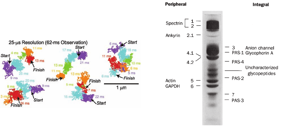
derlying cytoskeleton. In fact, single molecule tracking
techniques (Section 12-2Ca) have demonstrated that lipid
molecules in biological membranes undergo a series of
short random motions over short time periods (⬃10 ms) in-
terspersed by large hops (Fig. 12-36), a process called hop
diffusion. Evidently, biological membranes are partitioned
rather than continuous two-dimensional fluids.
D. The Erythrocyte Membrane
The erythrocyte membrane’s relative simplicity, availability,
and ease of isolation have made it the most extensively stud-
ied and best understood biological membrane. It is therefore
a model for the more complex membranes of other cell
types. A mature mammalian erythrocyte is devoid of or-
ganelles and carries out few metabolic processes; it is essen-
tially a membranous bag of hemoglobin. Erythrocyte mem-
branes can therefore be obtained by osmotic lysis, which
causes the cell contents to leak out. The resultant membra-
nous particles are known as erythrocyte ghosts because, on
return to physiological conditions, they reseal to form color-
less particles that retain their original shape. Indeed, by
transferring sealed ghosts to another medium, their contents
can be made to differ from the external solution.
a. Erythrocyte Membranes Contain a
Variety of Proteins
The erythrocyte membrane has a more or less typical
plasma membrane composition of about half protein, some-
what less lipid, and the remainder carbohydrate (Table 12-4).
Its proteins may be separated by SDS–polyacrylamide
gel electrophoresis (Section 6-4C) after first solubilizing
the membrane in a 1% SDS solution. The resulting elec-
trophoretogram for a human erythrocyte membrane ex-
hibits seven major and many minor bands when stained with
Coomassie brilliant blue (Fig. 12-37). If the electrophore-
togram is instead treated with periodic acid–Schiff’s reagent
(PAS), which stains carbohydrates, four so-called PAS
bands become evident. The polypeptides corresponding to
bands 1, 2, 4.1, 4.2, 5, and 6 are readily extracted from the
Section 12-3. Biological Membranes 411
Figure 12-36 Hop diffusion of individual colloidal
gold–tagged dioleoylphosphatidylethanolamine molecules in a
plasma membrane. The position of each particle was determined
at 25-s intervals (a video frame rate of 40,500 frames ⴢ s
⫺1
) over
a period of 62 ms (2500 steps). Colored lines connect successive
positions of the particle, with differently colored segments
representing the various plausible regions in the plasma
membrane to which the particle appears to have been transiently
confined (in the order purple, blue, green, orange, and red).
[Courtesy of Akihiro Kasumi, Nagoya University, Japan.]
Figure 12-37 SDS–PAGE electrophoretogram of human
erythrocyte membrane proteins as stained by Coomassie brilliant
blue. The bands designated 4.1 and 4.2 are not separated with the
1% SDS concentration used.The minor bands are not labeled for
the sake of clarity. The positions of the four sialoglycoproteins
that would be revealed by PAS staining are indicated. [Courtesy
of Vincent Marchesi, Yale University.]
JWCL281_c12_386-466.qxd 6/9/10 12:06 PM Page 411
membrane by changes in ionic strength or pH and hence are
peripheral proteins. These proteins are located on the inner
side of the membrane, as is indicated by the observation that
they are not altered by the incubation of intact erythrocytes
or sealed ghosts with proteolytic enzymes or membrane-
impermeable protein labeling reagents. These proteins are
altered, however, if “leaky” ghosts are so treated.
In contrast, bands 3, 7, and all four PAS bands correspond
to integral proteins; they can be released from the mem-
brane only by extraction with detergents or organic solvents.
Of these, band 3 and PAS bands 1 and 2 correspond to TM
proteins, as indicated by their different labeling patterns
when intact cells are treated with membrane-impermeable
protein-labeling reagents and when these reagents are intro-
duced inside sealed ghosts.The PAS band 1 is a dimer of gly-
cophorin A, which is formed through an SDS-resistant asso-
ciation between the TM helices of the polypeptide chains
(Fig. 12-21); this dimer is the protein’s native form.The PAS
band 2 protein is the monomeric form of glycophorin A.
The transport of CO
2
in blood (Section 10-1C) requires
that the erythrocyte membrane be permeable to HCO
⫺
3
and
Cl
⫺
(the maintenance of electroneutrality requires that for
every HCO
⫺
3
that enters a cell, a Cl
⫺
or some other anion
must leave the cell; Section 10-1Cb). The rapid transport of
these and other anions across the erythrocyte membrane is
mediated by a specific anion channel of which there are ⬃1
million/cell (comprising ⬎30% of the membrane protein).
Band 3 protein (929 residues and 5–8% carbohydrate)
specifically reacts with anionic protein-labeling reagents that
block the anion channel, thereby indicating that the anion
channel is composed of band 3 protein. Furthermore, cross-
linking studies with bifunctional reagents (Section 8-5Ca)
demonstrate that the anion channel is at least a dimer. He-
moglobin and the glycolytic (glucose metabolizing) enzymes
aldolase, phosphofructokinase (PFK), and the band 6 pro-
tein glyceraldehyde-3-phosphate dehydrogenase (GAPDH;
Section 17-2F) all specifically and reversibly bind to band 3
protein on the cytoplasmic side of the membrane. The func-
tional significance of this observation is unknown.
b. The Erythrocyte’s Cytoskeleton Is Responsible
for Its Shape and Flexibility
A normal erythrocyte’s biconcave disklike shape (Fig.
7-19a) assures the rapid diffusion of O
2
to its hemoglobin
molecules by placing them no farther than 1 m from the
cell surface. However, the rim and the dimple regions of an
erythrocyte do not occupy fixed positions on the cell mem-
brane. This can be demonstrated by anchoring an erythro-
cyte to a microscope slide by a small portion of its surface
and inducing the cell to move laterally with a gentle flow of
isotonic buffer.A point originally on the rim of the erythro-
cyte will move across the dimple to the rim on the opposite
side of the cell from where it began. Evidently, the mem-
brane rolls across the cell while maintaining its shape,
much like the tread of a tractor.This remarkable mechani-
cal property of the erythrocyte membrane results from the
presence of a submembranous network of proteins that
function as a membrane “skeleton”—the cell’s cytoskele-
ton. Indeed, this property is partially duplicated by a
mechanical model consisting of a geodesic sphere (a spher-
oidal cage) that is freely jointed at the intersections of its
struts but constrained from collapsing much beyond a flat
surface. When placed inside an evacuated plastic bag, this
cage also assumes a biconcave disklike shape.
The fluidity and flexibility imparted to an erythrocyte by
its cytoskeleton has important physiological consequences.
A slurry of solid particles of a size and concentration equal
to that of red cells in blood has the flow characteristics ap-
proximating that of sand. Consequently, in order for blood
to flow at all, much less for its erythrocytes to squeeze
through capillary blood vessels smaller in diameter than
they are, erythrocyte membranes, together with their cy-
toskeletons, must be fluidlike and easily deformable.
The protein spectrin, so called because it was discov-
ered in erythrocyte ghosts, accounts for ⬃75% of the ery-
throcyte cytoskeleton. It is composed of two similar
polypeptide chains, band 1 (␣ subunit; 2418 residues) and
band 2 ( subunit; 2137 residues), which sequence analysis
indicates each consist of repeating 106-residue segments
that are predicted to fold into triple-stranded ␣ helical
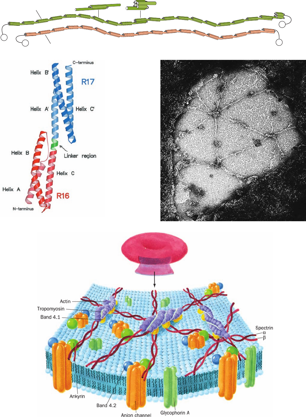
coiled coils (Fig. 12-38a,b). Electron microscopy indicates
that these large polypeptides are loosely intertwined to
form a flexible wormlike ␣ dimer that is ⬃1000 Å long
(Fig. 12-38c).Two such heterodimers further associate in a
412 Chapter 12. Lipids and Membranes
Figure 12-38 (Opposite) The human erythrocyte cytoskeleton.
(a) Structure of an ␣ dimer of spectrin. Both of these
antiparallel polypeptides contain multiple 106-residue repeats,
which are thought to form flexibly connected triple helical
bundles.Two of these heterodimers join, head to head, to form an
(␣)
2
heterotetramer. [After Speicher, D.W. and Marchesi, V.,
Nature 311, 177 (1984).] (b) X-ray structure of two consecutive
repeats of chicken brain ␣-spectrin. Each of these 106-residue
repeats consists of a down–up–down triple helical bundle in which
the C-terminal helix of first repeat (R16; red) is continuous, via a
5-residue helical linker (green), with the N-terminal helix of the
second repeat (R17; blue). The helices within each triple helical
bundle wrap around each other in a gentle left-handed supercoil
that is hydrophobically stabilized by the presence of nonpolar
residues at the a and d positions of heptad repeats on all three of
its component ␣ helices (Fig. 8-26). Despite the expected rigidity
of ␣ helices, there is considerable evidence that spectrin is a
flexible wormlike molecule. [Courtesy of Alfonso Mondragón,
Northwestern University. PDBid 1CUN.] (c) Electron
micrograph of an erythrocyte cytoskeleton that has been
stretched to an area 9 to 10 times greater than that of the native
membrane. Stretching makes it possible to obtain clear images of
the cytoskeleton, which in its native state is so densely packed
and irregularly flexed that it is difficult to pick out individual
molecules and to ascertain how they are interconnected. Note
the predominantly hexagonal network composed of spectrin
tetramers cross-linked by junctions containing actin and band 4.1
protein. [Courtesy of Daniel Branton, Harvard University.]
(d) Model of the erythrocyte cytoskeleton. The so-called
junctional complex, which is magnified in this drawing, contains
actin, tropomyosin (which, in muscle, also associates with actin;
Section 35-3Ac), and band 4.1 protein, as well as adducin,
dematin, and tropomodulin (not shown). [After Goodman, S.R.,
Krebs, K.E., Whitfield, C.F., Riederer, B.M., and Zagen, I.S., CRC
Crit. Rev. Biochem. 23, 196 (1988).]
JWCL281_c12_386-466.qxd 6/9/10 12:06 PM Page 412
Section 12-3. Biological Membranes 413
β chain
α chain
N
N
C
C
(a)
(b)
(c)
(d)
JWCL281_c12_386-466.qxd 6/9/10 12:06 PM Page 413
head-to-head manner to form an (␣)
2
heterotetramer.
These tetramers, of which there are ⬃100,000/cell, are
cross-linked at both ends by attachments to bands 4.1 and
5 to form a dense and irregular protein meshwork that un-
derlies the erythrocyte plasma membrane (Fig. 12-38c,d).
Band 5, a globular protein that forms filamentous
oligomers, has been identified as actin, a common cy-
toskeletal element in other cells (Section 1-2Ae) and a
major component of muscle (Section 35-3Ac). Spectrin
also associates with band 2.1, an 1880-residue monomer
known as ankyrin, which, in turn, binds to band 3, the an-
ion channel protein. This attachment anchors the cy-
toskeleton to the membrane. Indeed, on solubilization of
spectrin and actin by low ionic strength solutions, the ery-
throcyte ghosts’ biconcave shape is lost and their integral
proteins, which normally occupy fixed positions in the
membrane plane, become laterally mobile.
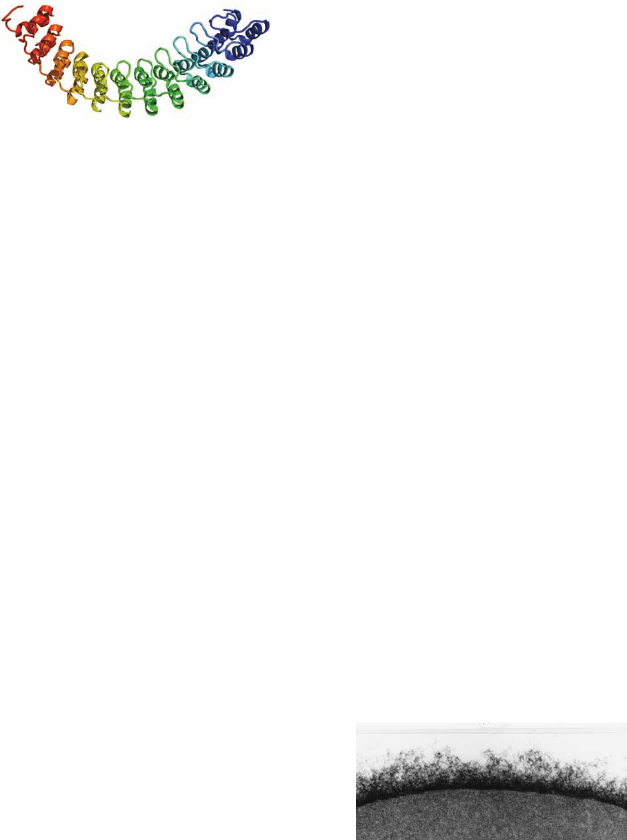
Ankyrin’s N-terminal 798-residue segment consists al-
most entirely of 24 tandem ⬃33-residue repeats known
as ankyrin repeats (Fig. 12-39), which also occur in a vari-
ety of other proteins. Each ankyrin repeat consists of two
short (8- or 9-residue) antiparallel ␣ helices followed by
a long loop. These structures are arranged in a right-
handed superhelical stack. The entire assembly forms an
elongated concave surface that is postulated to bind var-
ious integral proteins as well as spectrin. Immunochemi-
cal studies have revealed spectrinlike, ankyrinlike, and
band 4.1–like proteins in the cytoskeletons of a variety of
tissues.
c. Hereditary Spherocytosis and Elliptocytosis Arise
from Defects in the Erythrocyte Cytoskeleton
Individuals with hereditary spherocytosis have spher-
oidal erythrocytes that are relatively fragile and inflexible.
These individuals suffer from hemolytic anemia because
the spleen, a labyrinthine organ with narrow passages that
normally filters out aged erythrocytes (which lose flexibil-
ity toward the end of their ⬃120-day lifetime), prematurely
removes spherocytotic erythrocytes.The hemolytic anemia
may be alleviated by the spleen’s surgical removal. How-
ever, the primary defects in spherocytotic cells are reduced
synthesis of spectrin, the production of an abnormal spec-
trin that binds band 4.1 protein with reduced affinity, or the
absence of band 4.1 protein.
Hereditary elliptocytosis (having elongated or elliptical
red cells; also known as hereditary ovalcytosis), a condition
that is common in certain areas of Southeast Asia and
Melanesia, confers resistance to malaria in heterozygotes
(but apparently is lethal in homozygotes). This condition
arises from defects in the erythrocyte anion channel. A
common such defect consists of a 9-residue deletion that
inactivates this TM protein. The consequent reduced ca-
pacity of red cells to import phosphate or sulfate ions may
inhibit the intraerythrocytotic growth of rapidly develop-
ing malarial parasites.
The camel, the renowned “ship of the desert,” provides
a striking example of adaptation involving the erythrocyte
membrane. This remarkable animal is still active after a
loss of water constituting 30% of its body weight and, when
thus dehydrated, can drink sufficient water in a few min-
utes to become fully rehydrated. The rapid uptake of such
a large amount of water by the blood, which must deliver it
to the cells, would lyse the erythrocytes of most animals.
Yet camel erythrocytes, which have the shape of flattened
ellipsoids rather than biconcave disks, are resistant to os-
motic lysis. Camel spectrin binds to its membrane with par-
ticular tenacity, but on spectrin removal, which requires a
strong denaturing agent such as guanidinium chloride,
camel erythrocytes assume a spherical shape.
E. Blood Groups
The outer surfaces of erythrocytes and other eukaryotic
cells are covered with complex carbohydrates that are
components of plasma membrane glycoproteins and gly-
colipids. They form a thick, fuzzy cell coating, the glyco-
calyx (Fig.12-40),which contains numerous identity markers
that function in various recognition processes. Human ery-
throcytes have 30 genetically distinct blood group systems
comprised of ⬎600 known blood group determinants, al-
though many of these determinants are rare or occur only
in certain ethnic groups. Of these systems, only two—the
ABO blood group system (discovered in 1900 by Karl
414 Chapter 12. Lipids and Membranes
Figure 12-39 X-ray structure of human ankyrin repeats 13 to
24. The polypeptide is shown in ribbon form colored in rainbow
order from its N-terminus (blue, repeat 13) to its C-terminus
(red, repeat 24). [Based on an X-ray structure by Peter Michaely,
University of Texas Southwestern Medical Center, Dallas, Texas.
PDBid 1N11.]
Figure 12-40 The erythrocyte glycocalyx as revealed by electron
microscopy using special staining techniques. It is up to 1400 Å
thick and composed of closely packed, 12- to 25-Å-diameter
oligosaccharide filaments linked to plasma membrane–
associated proteins and lipids. [Courtesy of Harrison Latta, UCLA.]
JWCL281_c12_386-466.qxd 6/9/10 12:06 PM Page 414

Landsteiner) and the rhesus (Rh) blood group system—
have major clinical importance. The various blood groups
are identified by means of suitable antibodies or by specific
plant lectins.
a. ABO Blood Group Substances Are Carbohydrates
The ABO system consists of three blood group sub-
stances, the A, B, and H antigens, which are components of
erythrocyte surface sphingoglycolipids. [Antigens are char-
acteristic constellations of chemical groups on macromole-
cules that elicit the production of specific antibodies when
introduced into an animal (Section 35-2Aa). Each anti-
body molecule can specifically bind to at least two of its
corresponding antigen molecules, thereby cross-linking
them.] Individuals with type A cells have A antigens on
their cell surfaces and carry anti-B antibodies in their
serum; those with type B cells, which bear B antigens, carry
anti-A antibodies; those with type AB cells, which bear
both A and B antigens, carry neither anti-A nor anti-B an-
tibodies; and type O individuals, whose cells bear neither
antigen, carry both anti-A and anti-B antibodies. Conse-
quently, the transfusion of type A blood into a type B indi-
vidual, for example, causes an anti-A antibody–A antigen
reaction, which agglutinates (clumps together) the trans-
fused erythrocytes, resulting in an often fatal blockage of
blood vessels.The H antigen is discussed below.Anti-A and
anti-B antibodies, which are not present at birth, appear to
arise through an immune response to A-like and B-like
antigens in food and/or to the colonization of the infant gut
by bacteria that produce such antigens [the immune system
normally suppresses the production of antibodies directed
against the body’s own antigens (Section 35-2Ac) so, for
example, a type A individual does not produce anti-A anti-
bodies].
The ABO blood group substances are not confined to
erythrocytes but also occur in the plasma membranes of
many tissues as glycolipids of considerable diversity. In
fact, in the ⬃80% of the population known as secretors,
these antigens are secreted as O-linked components of gly-
coproteins into various body fluids, including saliva, milk,
seminal fluid, gastric juice, and urine. These diverse mole-
cules, which are 85% carbohydrate by weight and have mo-
lecular masses ranging up to thousands of kilodaltons, con-
sist of multiple oligosaccharides attached to a polypeptide
chain.
The A, B, and H antigens differ only in the sugar residues
at their nonreducing ends (Table 12-5). The H antigen oc-
curs in type O individuals; it is also the precursor oligosac-
charide of A and B antigens.Type A individuals have a 303-
residue glycosyltransferase that specifically adds an
N-acetylgalactosamine residue to the terminal position of
the H antigen, whereas in type B individuals, this enzyme,
which differs by four amino acid residues from that of type
A individuals, instead adds a galactose residue. In type O
individuals, the enzyme is inactive because its synthesis ter-
minates after its 115th residue.
Do the different blood groups confer any biological ad-
vantages or disadvantages? Epidemiological studies indicate
that type A and B individuals are less susceptible to
cholera infections than are type O individuals, with the rel-
atively rare type AB individuals being highly resistant to
this deadly disease. Apparently, type A and B oligosaccha-
rides block a receptor for the bacterium causing cholera,
Vibrio cholera (Section 19-2Cd). In addition, type O indi-
viduals, particularly nonsecretors, have a higher incidence
of peptic (stomach) ulcers. However, type A individuals
have a higher incidence of stomach cancer, heart disease,
and pernicious anemia (Section 25-2Ee).
F. Gap Junctions
Most cells in multicellular organisms are in metabolic and
electrical as well as physical contact with neighboring cells.
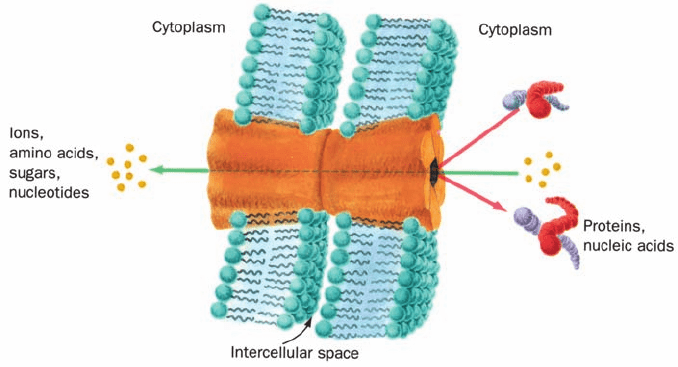
This contact is brought about by tubular particles, named
gap junctions, that join discrete regions of neighboring
plasma membranes much like hollow rivets (Fig. 12-41). In-
deed, these intercellular channels are so widespread that
many whole organs are continuous from within. Thus gap
junctions are important intercellular communication chan-
nels. For example, the synchronized contraction of heart
muscle is brought about by flows of ions through gap junc-
tions (heart muscle is not innervated as is skeletal muscle).
Likewise, gap junctions serve as conduits for some of the
substances that mediate embryonic development; blocking
gap junctions with antibodies that bind to them causes de-
velopmental abnormalities in species as diverse as hydras,
frogs, and mice.Gap junctions also function to nourish cells
that are distant from the blood supply, such as bone and
lens cells. Thus, it is not surprising that, in humans, defects
in gap junctions are associated with certain neurodegener-
ative diseases, cataracts, deafness, several skin diseases, and
developmental anomalies.
Gap junctions consist of a single sort of protein subunit
known as a connexin. A single gap junction consists of
two apposed hexagonal rings of connexins, called connex-
ons, one from each of the adjoining plasma membranes
Section 12-3. Biological Membranes 415
Table 12-5 Structures of the A, B, and H Antigenic
Determinants in Erythrocytes
Type Antigen
H Gal(1 S 4)GlcNAc
p
1,2
L-Fuc␣
A GalNAc␣(1 S 3)Gal(1 S 4)GlcNAc
p
1,2
L-Fuc␣
B Gal␣(1 S 3)Gal(1 S 4)GlcNAc
p
1,2
L-Fuc␣
¡¡¡
Abbreviations: Gal ⫽ galactose, GalNAc ⫽ N-acetylgalactosamine,
GlcNAc ⫽ N-acetylglucosamine,
L-Fuc ⫽ L-fucose.
JWCL281_c12_386-466.qxd 6/9/10 12:06 PM Page 415
(Fig. 12-41). Cells are normally connected by clusters of
hundreds to thousands of connexons. A given animal ex-
presses numerous genetically distinct connexins, with hu-
mans, for example, expressing 21 different connexins
ranging in molecular mass from 25 to 50 kD. Many types
of cells simultaneously express several different species
of connexins, and in cells that do so, there is considerable
evidence that at least some connexons may be formed
from two or more species of connexins. Moreover,the gap
junctions joining two cells may consist of two different
types of connexons. These various types of gap junctions
presumably differ in their selectivities for the substances
they transmit.
Mammalian gap junction channels are minimally 16 to
20 Å in diameter, which Werner Loewenstein established
by microinjecting single cells with fluorescent molecules of
various sizes and observing with a fluorescence microscope
whether the fluorescent probe passed into neighboring
cells. The molecules and ions that can pass freely between
neighboring cells are limited in molecular mass to a maxi-
mum of ⬃1000 D; macromolecules such as proteins and
nucleic acids cannot leave a cell via this route.
The diameter of a gap junction channel varies with Ca
2⫹
concentration: The channels are fully open when the Ca
2⫹
level is ⬍10
⫺7
M and narrow as the Ca
2⫹
concentration in-
creases until, above 5 ⫻ 10
⫺5
M, they close.This shutter sys-
tem is thought to protect communities of interconnected
cells from the otherwise catastrophic damage that would
result from the death of even one of their members. Cells
generally maintain very low cytosolic Ca
2⫹
concentrations
(⬍10
⫺7
M) by actively pumping Ca
2⫹
out of the cell as well
as into their mitochondria and endoplasmic reticulum
(Section 20-3B; Ca
2⫹
is an important intracellular messen-
ger whose cytosolic concentration is precisely regulated).
Ca
2⫹
floods back into leaky or metabolically depressed
cells, thereby inducing closure of their gap junctions and
sealing them off from their neighbors.
a. Connexins Contain Transmembrane
Four-Helix Bundles
The X-ray structure of the gap junction formed by the
226-residue human connexin 26 (Cx26), determined by
Tomitake Tsukihara, reveals a 12-mer with D
6
symmetry, a
height of 155 Å, and a maximal diameter of 92 Å that en-
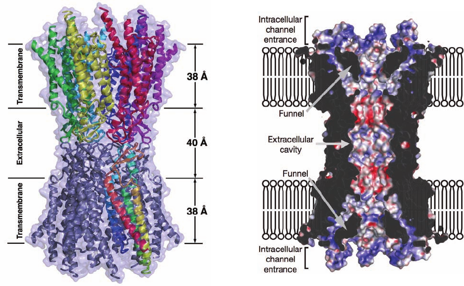
closes a central channel (Fig. 12-42a). The extracellular
portion of each connexon extends 23 Å from the extracel-
lular surface and interdigitates with the opposite connexon
by 6 Å to span an intercellular gap of 40 Å. Each Cx26 sub-
unit forms an up–down–up–down four-helix bundle in
which both the N- and C-termini occupy the cytosol. The
central channel has a diameter of ⬃40 Å at its cytosolic en-
trance that funnels down to 14 Å near the extracellular sur-
face of the membrane and then widens to 25 Å in the extra-
cellular space (Fig. 12-42b). The positively charged funnel
entrance would attract negatively charged molecules.
However, the region of maximal channel constriction is
negatively charged, which should also affect the channel’s
charge selectivity.
G. Channel-Forming Proteins
A number of bacterial toxins are synthesized as water-
soluble monomers that, on interacting with their target
membrane via a specific receptor protein, spontaneously
insert into the membrane as a TM pore.This process, which
for many such channel-forming toxins (CFTs) requires
their oligomerization, causes the leakage of small ions and
molecules from the target cell, thereby killing it through
loss of osmotic balance. The formation of only one CFT-
based pore is often sufficient to kill a cell.
416 Chapter 12. Lipids and Membranes
Figure 12-41 Model of a gap junction. Gap junctions between
adjacent cells consist of two apposed plasma membrane–
embedded hexagonal studs that bridge the gap between the cells.
Gap junctions hold cells a fixed distance apart—the gap. Small
molecules and ions, but not macromolecules, can pass between
cells via the gap junction’s central channel.
JWCL281_c12_386-466.qxd 10/14/10 6:21 PM Page 416
One of the best characterized CFTs is ␣-hemolysin,
which the human pathogen Staphylococcus aureus secretes
as a water-soluble 293-residue monomer and which sponta-
neously inserts into the membranes of erythrocytes and
several other types of cells in the form of heptameric pores.
Even though the ␣-hemolysin monomer is water-soluble
and lacks clearly hydrophobic segments, the heptamer acts
as a typical TM protein in that it is not released from the
membrane by treatment with high salt, low pH, or
chaotropic agents but, instead, requires treatment with de-
tergents for this to occur.
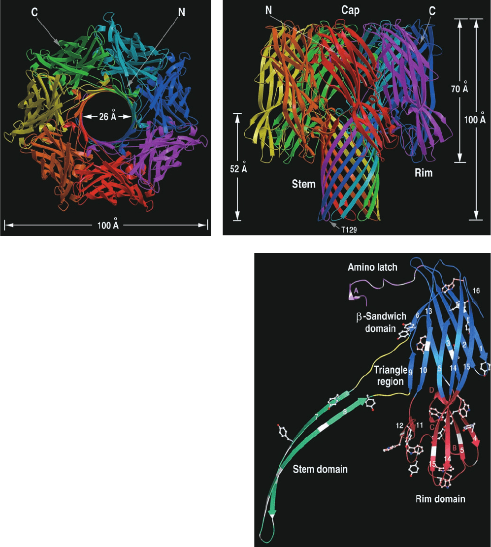
The X-ray structure of detergent-solublized ␣-hemolysin,
determined by Eric Gouaux, reveals a striking mushroom-
shaped heptameric complex that is 100 Å in height and
100 Å in diameter (Fig. 12-43a,b). A 14- to 46-Å-diameter
solvent-filled channel, which runs along the protein’s 7-fold
axis, forms a TM pore. The stem of the mushroom, the
protein’s TM segment, consists of a 52-Å-high and 26-Å-
diameter, porinlike, 14-stranded, antiparallel  barrel com-
posed of seven 2-stranded antiparallel  sheets, one from
each subunit (Fig. 12-43b). The remainder of each subunit
consists of a  sandwich domain and a rim domain, which
together form a 70-Å-long ellipsoid (Fig. 12-43c). Seven of
these ellipsoids are distributed in a ring, thereby forming
the mushroom’s cap and rim. The rim domain projects
toward and probably interacts with the membrane’s phos-
pholipid head groups via the basic and aromatic residues
that extend from the crevice between the top of the stem
and rim.
A variety of experimental evidence indicates that the
spontaneous formation of the heptameric TM pore occurs
via several discrete steps: (1) the binding of the aqueous
monomer to the membrane surface, probably through the
interaction of the protein’s polypeptide loops with the sur-
face groups of the lipid bilayer; (2) the formation of the
heptamer on the surface of the membrane; and (3) the in-
sertion of the 14-stranded  barrel through the membrane
to form the TM pore. The structural details of this process
are as yet unknown, although it seems clear that there is lit-
tle change in the monomers’ secondary structure on their
assembly to form the heptameric TM pore.The reason why
monomers do not form heptamers in aqueous solution is
Section 12-3. Biological Membranes 417
Figure 12-42 X-ray structure of the connexin 26 gap junction.
(a) View perpendicular to the protein’s 6-fold axis (parallel to
the planes of the membranes) in which the protein is drawn in
ribbon form embedded in its semitransparent molecular surface.
Each connexin of the upper connexon has a different color,
whereas one connexin in the lower connexon is colored in
rainbow order from its N-terminus (blue) to its C-terminus (red)
with the remaining connexins purple. The extent of the
transmembrane region was deduced from the distribution of
hydrophobic and aromatic residues (Section 12-3Af).
(b) Cutaway drawing through the surface diagram of the gap
junction channel.The channel surface is colored according to its
electrostatic potential with red positive, blue negative, and white
neutral. [Part a based on an X-ray structure by, and Part b
courtesy of Tomitake Tsukihara, University of Osaka, Japan.
PDBid 2ZW3.]
(a)
(b)
JWCL281_c12_386-466.qxd 6/9/10 12:06 PM Page 417
probably due to differences between the strengths of the
intrasubunit interactions in the monomer in aqueous solu-
tion and the intersubunit interactions in the heptamer in
the membrane.
Not all CFTs form pores using  barrels. Rather, a vari-
ety of CFTs, notably several E. coli proteins known as col-
icins, form pores that are lined with ␣ helices. Most such
pores consist of monomers.
4 MEMBRANE ASSEMBLY AND
PROTEIN TARGETING
As cells grow and divide, they synthesize new membranes.
How are such asymmetric membranes generated? One
way in which this might occur is through self-assembly. In-
deed, when the detergent used to disperse a biological
membrane is removed, liposomes form in which functional
integral proteins are embedded. In most cases, however,
these model membranes are symmetrical, both in their
lipid distribution between the inner and outer leaflets of
the bilayer and in the orientations of their embedded pro-
teins. An alternative hypothesis of membrane assembly is
that it occurs on the scaffolding of preexisting membranes;
that is, membranes are generated by the expansion of old
ones rather than by the creation of new ones. In this section
we shall see that this is, in fact, how biological membranes
are generated. In doing so, we shall consider how proteins
are inserted into and passed through membranes as well as
how portions of membranes in the form of vesicles pinch
off from one membrane and fuse with another, thereby
transporting proteins and lipids between these membranes.
These highly complex processes are indicative of the intri-
cacies of biological processes in general.
418 Chapter 12. Lipids and Membranes
Figure 12-43 X-ray structure of ␣-hemolysin. Views (a) along
and (b) perpendicular to the heptameric transmembrane pore’s
7-fold axis. Each subunit is drawn with a different color. (c) The
monomer unit with its three domains drawn in different colors.
[Courtesy of Eric Gouaux, Columbia University. PDBid 7AHL.]
(a)
(b)
(c)
JWCL281_c12_386-466.qxd 6/9/10 12:06 PM Page 418
Section 12-4. Membrane Assembly and Protein Targeting 419
A. Lipid Distributions in Membranes
The enzymes involved in the biosynthesis of membrane
lipids are mostly integral proteins (Section 25-8). Their sub-
strates and products are themselves membrane components,
so that membrane lipids are fabricated on site. Eugene
Kennedy and James Rothman demonstrated this to be the
case in bacteria through the use of selective labeling.They
gave growing bacteria a 1-min pulse of so as to label
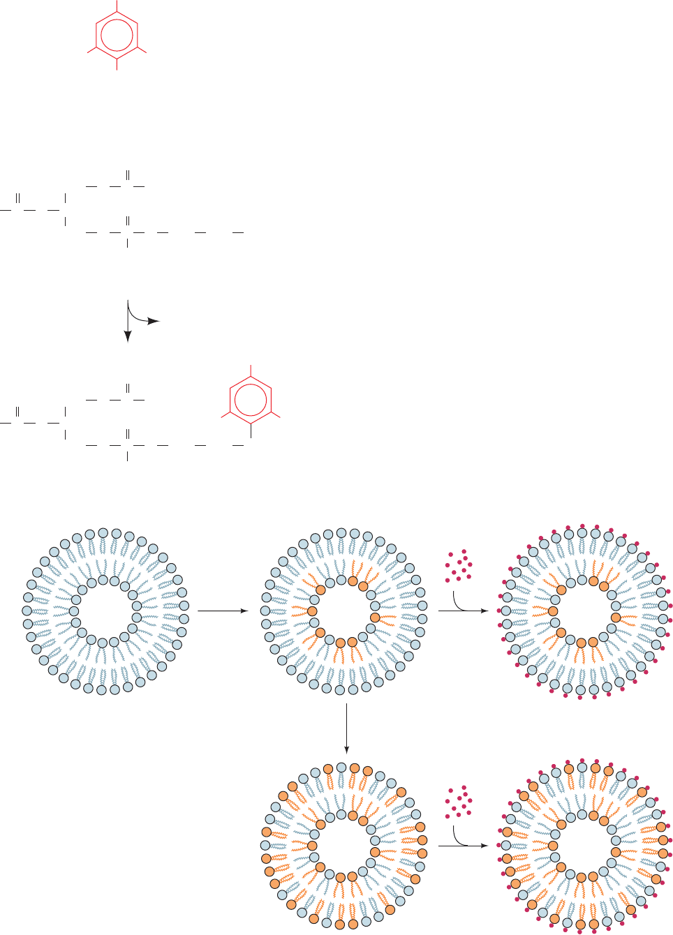
radioactively the phosphoryl groups of only the newly
synthesized phospholipids. Trinitrobenzenesulfonic acid
(TNBS), a membrane-impermeable reagent that combines
with phosphatidylethanolamine (PE; Fig. 12-44), was then
immediately added to the cell suspension. Analysis of the
resulting doubly labeled membrane showed that none of
the TNBS-labeled PE was radioactively labeled. This ob-
servation indicates that newly made PE is synthesized on
the cytoplasmic face of the membrane (Fig. 12-45, top right).
a. Membrane Proteins Catalyze
Phospholipid Flip-Flops
If an interval of only 3 min is allowed to elapse between
the pulse and the TNBS addition, about half of the
32
P-labeled PE is also TNBS labeled (Fig. 12-45, bottom).
This observation indicates that the flip-flop rate of PE in the
bacterial membrane is ⬃100,000-fold greater than it is in bi-
layers consisting of only phospholipids (where, it will be re-
called, the flip-flop rates have half-times of many days).
How do phospholipids synthesized on one side of the
membrane reach its other side so quickly? Phospholipid
flip-flops appear to be facilitated in two ways:
32
PO
3⫺
4
32
PO
3⫺
4
Figure 12-44 Reaction of TNBS with PE.
Figure 12-45 Location of lipid synthesis in a
bacterial membrane. Newly synthesized PE was
labeled by a 1-min pulse of (orange head
groups), and the PE on the cell surface was
independently labeled by treatment with the
membrane-impermeable reagent TNBS.When
TNBS labeling (red dots) occurred immediately
after the
32
P pulse, none of the
32
P-labeled PE
was also TNBS labeled (top right), thereby
indicating that the PE is synthesized on the
cytoplasmic face of the membrane. If, however,
there was even a few minutes’ delay between the
two labeling procedures, much of the TNBS-labeled
PE in the external face of the membrane was also
32
P labeled (bottom).
32
PO
3⫺
4
C
O
CHO
R
2
CH
2
O
C
O
O
R
1
CH
2
O
P
O
O
O
–
CH
2
CH
2
NH
2
+
NO
2
SO
3
–
O
2
N
NO
2
Trinitrobenzenesulfonic acid (TNBS)
Phosphatidylethanolamine (PE)
C
O
CHO
R
2
CH
2
O
C R
1
CH
2
O
P
O
O
O
–
CH
2
CH
2
NH
NO
2
O
2
N
NO
2
H
2
SO
3
Incubation
with radioactive
phosphate
Immediate
incubation
with TNBS
Incubation
with TNBS
3 min
JWCL281_c12_386-466.qxd 6/9/10 12:06 PM Page 419
1. Membranes contain proteins known as flippases that
catalyze the flip-flops of specific phospholipids. These pro-
teins tend to equilibrate the distribution of their corre-
sponding phospholipids across a bilayer; that is, the net
transport of a phospholipid is from the side of the bilayer
with the higher concentration of the phospholipid to the
opposite side. Such a process, as we shall see in Section 20-2,
is a form of facilitated diffusion.
2. Membranes contain proteins known as phospholipid
translocases that transport specific phospholipids across a
bilayer in a process that is driven by ATP hydrolysis.These
proteins can transport certain phospholipids from the side
of a bilayer that has the lower concentration of the phos-
pholipids being translocated to the opposite side, thereby
establishing a nonequilibrium distribution of the phospho-
lipids. Such a process, as we shall see in Section 20-2, is a
form of active transport.
The observed distribution of phospholipids across mem-
branes (e.g., Fig. 12-35) therefore appears to arise from the
membrane orientations of the enzymes that synthesize
phospholipids combined with the countervailing tenden-
cies of ATP-dependent phospholipid translocases to gener-
ate asymmetric phospholipid distributions and those of
flippases to randomize these distributions.
b. A Membrane’s Characteristic Lipid Composition
Can Arise in Several Ways
In eukaryotic cells, lipids are synthesized on the cytoplas-
mic face of the endoplasmic reticulum, from where they are
transported to other membranes. Perhaps the most impor-
tant mechanism of lipid transport is the budding off of
membranous vesicles from the ER and their subsequent fu-
sion with other membranes (Sections 12-4C and 12-4D).
However, this mechanism does not explain the different
lipid compositions of the various membranes in a cell.
Lipids may also be transported between membranes by the
phospholipid exchange proteins present in many cells.
These proteins spontaneously transfer specific phospho-
lipids, one molecule at a time,between two membranes sep-
arated by an aqueous medium.A membrane’s characteristic
lipid composition may also arise through on-site remodel-
ing and/or selective degradation of its component lipids
through the action of specific enzymes (Section 25-8A).
B. The Secretory Pathway
Membrane proteins, as are all proteins, are ribosomally
synthesized under the direction of messenger RNA tem-
plates such that each polypeptide grows from its N-terminus
to its C-terminus by the stepwise addition of amino acid
residues (Section 5-4B). Cytologists have long noted two
classes of eukaryotic ribosomes, those free in the cytosol
and those bound to the endoplasmic reticulum (ER) so
as to form the rough endoplasmic reticulum (RER, so
called because of the knobby appearance its bound
ribosomes give it; Fig. 1-5). Both classes of ribosomes
are nevertheless structurally identical; they differ only in
the nature of the polypeptide they are synthesizing. Free
ribosomes synthesize mostly soluble and mitochondrial
proteins, whereas membrane-bound ribosomes manufac-
ture TM proteins and proteins destined for secretion, opera-
tion within the ER, or incorporation into lysosomes (mem-
branous organelles containing a battery of hydrolytic
enzymes that function to degrade and recycle cell compo-
nents; Section 1-2Ad). These latter proteins initially appear
in the RER.
a. The Secretory Pathway Accounts for the Targeting
of Many Secreted and Membrane Proteins
How are RER-destined proteins differentiated from
other proteins? And how do these large, relatively polar
molecules pass through the RER membrane? These
processes occur via the secretory pathway, which was first
described by Günter Blobel, César Milstein, and David
Sabatini around 1975. Since ⬃25% of the different species
of proteins synthesized by all types of cells are integral pro-
teins and many others are secreted, ⬃40% of the various
types of proteins that a cell synthesizes must be processed via
the secretory pathway or some other protein targeting path-
way (e.g., that which directs proteins to the mitochondrion;
Section 12-4E). In this subsection, we first present an
overview of the secretory pathway and then discuss its var-
ious aspects in detail. The secretory pathway is outlined in
Fig. 12-46:
1. All secreted, ER-resident, and lysosomal proteins, as
well as many TM proteins, are synthesized with leading (N-
terminal) 13- to 36-residue signal peptides. These signal
peptides consist of a 6- to 15-residue hydrophobic core
flanked by several relatively hydrophilic residues that usu-
ally include one or more basic residues near the N-terminus
(Fig. 12-47). Signal peptides otherwise have little sequence
similarity. However, a variety of evidence indicates they
form ␣ helices in nonpolar environments.
2. When the signal peptide first protrudes beyond the
ribosomal surface (when the polypeptide is at least ⬃40
residues long),the signal recognition particle (SRP), a 325-kD
complex of six different polypeptides and a 300-nucleotide
RNA molecule, binds to both the signal peptide and the
ribosome accompanied by replacement of the SRP’s bound
GDP by GTP. The SRP’s resulting conformational change
causes the ribosome to arrest further polypeptide growth,
thereby preventing the RER-destined protein from being
released into the cytosol as well as averting premature
protein folding that would preclude the protein from en-
tering the ER (see below).
3. The SRP–ribosome complex diffuses to the RER
surface, where it is bound by the SRP receptor (SR) in
complex with the translocon, a protein pore in the ER
membrane through which the growing polypeptide will be
extruded. In forming the SR–translocon complex, the SR’s
bound GDP is replaced by GTP.
4. The SRP and SR stimulate each other to hydrolyze
their bound GTP to GDP (which is energetically equiva-
lent to ATP hydrolysis), resulting in conformational
changes that cause them to dissociate from each other and
420 Chapter 12. Lipids and Membranes
JWCL281_c12_386-466.qxd 6/9/10 12:06 PM Page 420
