Voet D., Voet Ju.G. Biochemistry
Подождите немного. Документ загружается.

Section 11-1. Monosaccharides 361
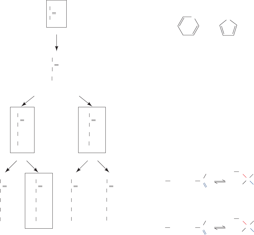
Sugars that differ only by the configuration about one C
atom are known as epimers of one another. Thus
D-glucose
and
D-mannose are epimers with respect to C2, whereas
D-glucose and D-galactose are epimers with respect to C4
(Fig. 11-1). However,
D-mannose and D-galactose are not
epimers of each other because they differ in configuration
about two of their C atoms.
D-Glucose is the only aldose that commonly occurs in na-
ture as a monosaccharide. However, it and several other
monosaccharides including
D-glyceraldehyde, D-ribose,
D-mannose, and D-galactose are important components of
larger biological molecules.
L Sugars are biologically much
less abundant than
D sugars.
The position of their carbonyl group gives ketoses one
less asymmetric center than their isomeric aldoses (e.g.,
compare
D-fructose and D-glucose). n-Carbon ketoses
therefore have 2
n⫺3
stereoisomers. Those with their ketone
function at C2 are the most common form (Fig. 11-2). Note
that some of these ketoses are named by the insertion of
-ul- before the suffix -ose in the name of the corresponding
aldose; thus
D-xylulose is the ketose corresponding to the
aldose
D-xylose. Dihydroxyacetone, D-fructose, D-ribulose,
and
D-xylulose are the biologically most prominent
ketoses.
B. Configurations and Conformations
Alcohols react with the carbonyl groups of aldehydes and
ketones to form hemiacetals and hemiketals, respectively
(Fig. 11-3). The hydroxyl and either the aldehyde or the ke-
tone functions of monosaccharides can likewise react in-
tramolecularly to form cyclic hemiacetals and hemiketals
(Fig. 11-4). The configurations of the substituents to each
carbon atom of these sugar rings are conveniently repre-
sented by their Haworth projection formulas.
A sugar with a six-membered ring is known as a pyra-
nose in analogy with pyran, the simplest compound con-
taining such a ring. Similarly, sugars with five-membered
rings are designated furanoses in analogy with furan.
The cyclic forms of glucose and fructose with six- and five-
membered rings are therefore known as glucopyranose
and fructofuranose, respectively.
a. Cyclic Sugars Have Two Anomeric Forms
The Greek letters preceding the names in Fig. 11-4 still
need to be explained. The cyclization of a monosaccharide
renders the former carbonyl carbon asymmetric. The re-
sulting pair of diastereomers are known as anomers and
the hemiacetal or hemiketal carbon is referred to as the
anomeric carbon. In the ␣ anomer, the OH substituent to
the anomeric carbon is on the opposite side of the sugar
ring from the CH
2
OH group at the chiral center that
O
O
Pyran Furan
Figure 11-2 The stereochemical relationships among the
D-ketoses with three to six carbon atoms. The configuration
about C3 (red) distinguishes the members of each pair.The
biologically most common ketoses are boxed.
Figure 11-3 The reactions of alcohols with (a) aldehydes to
form hemiacetals and (b) ketones to form hemiketals.
Dihydroxyacetone
D-Tagatose
D-Psicose
D-Fructose
D-Sorbose
HCOH
CH
2
OH
HOCH
HOCH
HCOH
CH
2
OH
HOCH
HCOH
HCOH
CH
2
OH
HCOH
HOCH
HCOH
CH
2
OH
HCOH
C
HCOH
D-XyluloseD-Ribulose
CH
2
OH
O
C
CH
2
OH
O
C
CH
2
OH
O
C
CH
2
OH
O
CH
2
OH
HCOH
HOCH
C
CH
2
OH
O
CH
2
OH
HCOH
C
HCOH
CH
2
OH
O
D-Erythrulose
CH
2
OH
HCOH
C
CH
2
OH
O
CH
2
OH
C
CH
2
OH
O
R⬙
R⬙
R⬘ C
Ketone
H
R
⬘
R⬘
C
C
HOR
R OH
+
+
Aldehyde HemiacetalAlcohol
R OH
OH
HemiketalAlcohol
O
R⬘
C
OR
OH
O
(a)
(b)
JWCL281_c11_359-385.qxd 6/3/10 10:35 AM Page 361
362 Chapter 11. Sugars and Polysaccharides
designates the D or L configuration (C5 in hexoses). The
other anomer is known as the  form (Fig. 11-5).
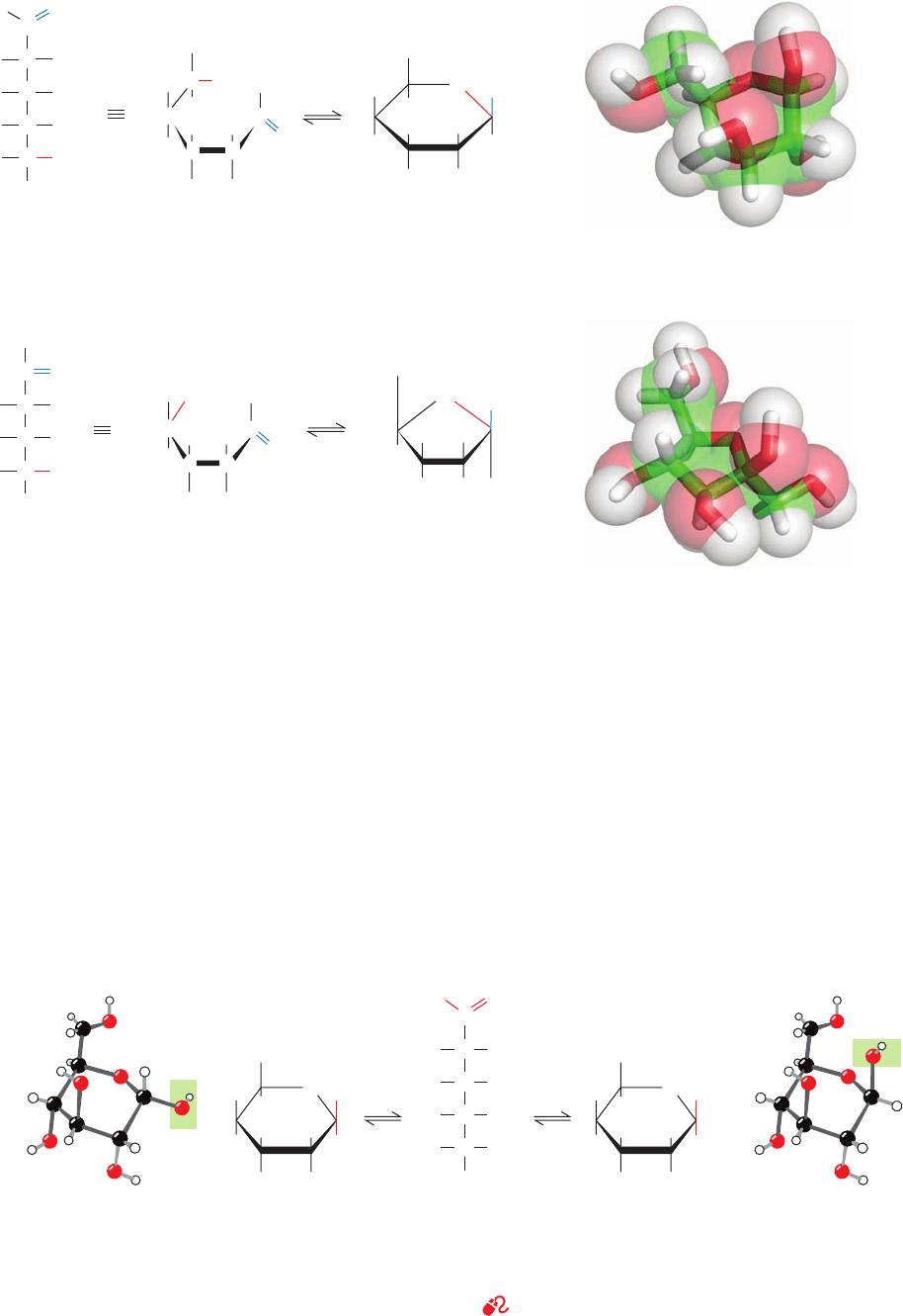
The two anomers of
D-glucose, as any pair of diastere-
omers, have different physical and chemical properties.
For example, the values of the specific optical rotation,
[␣]
20
D
, for ␣-D-glucose and -D-glucose are, respectively,
⫹112.2° and ⫹18.7°. When either of these pure substances
is dissolved in water, however, the specific optical rotation
of the solution slowly changes until it reaches an equilib-
rium value of ⫽⫹52.7°. This phenomenon is known[␣]
20
D
as mutarotation; in glucose, it results from the formation
of an equilibrium mixture consisting of 63.6% of the 
anomer and 36.4% of the ␣ anomer (the optical rotations
of separate molecules in solution are independent of each
other so that the optical rotation of a solution is the
weighted average of the optical rotations of its compo-
nents). The interconversion between these anomers oc-
curs via the linear form of glucose (Fig. 11-5). Yet, since
the linear forms of these monosaccharides are normally
present in only minute amounts, these carbohydrates are
O
OH
CH
2
OH
CH
2
OH
CH
2
OH
CH
2
OH
CH
2
OH
CH
2
OH
C
H
C
OH
OH
C
C
H
HO
H
H
1
2
3
4
5
6
OH H
H
H
H
HO
HOH
C
C
C
C
H
H
OH
H
HO
OH
C
H
O
C
OH
O
OHH
D-Glucose
(linear form)
β-D-Glucopyranose
(Haworth projection)
3
4
5
6
2
1
23
4
5
6
1
H
(a)
O
C
H
C
OH
OH
C
C
HO
H
H
1
2
3
4
5
6
C
C
C
H
HOH
2
C
HOH
2
C
OH
C
O
OH H
D-Fructose
(linear form)
β-D-Fructofuranose
(Haworth projection)
4
5
6
2
(b)
H
HO
3
1
O
H OH
OH
H
OH H
6
2
34
5
CH
2
OH
1
6
CH
2
OH
H
HO
H
O
OH
OH
H
H
HO
OHH
3
5
2
14
6
CH
2
OH
OHH
O
H
OH
H
H
HO
OHH
3
5
2
14
1
C
2
CH
HO
OH
H
3
C
4
CHOH
5
CHOH
6
CH
2
OH
α
-D-Glucopyranose
β
-D-GlucopyranoseD-Glucose
(linear form)
Figure 11-4 Cyclization reactions for hexoses. (a) D-Glucose
in its linear form reacting to yield the cyclic hemiacetal -
D-
glucopyranose and (b)
D-fructose in its linear form reacting to
yield the hemiketal -
D-fructofuranose. The cyclic sugars are
Figure 11-5 The anomeric monosaccharides ␣-
D-
glucopyranose and -
D-glucopyranose, drawn as both Haworth
projections and ball-and-stick models. These pyranose sugars
shown both as Haworth projections and in stick form embedded
in their semitransparent space-filling models with C green,
H white, and O red.
interconvert through the linear form of
D-glucose and differ only
by the configurations about their anomeric carbon atoms, C1.
See Kinemage Exercise 7-1
JWCL281_c11_359-385.qxd 6/7/10 7:14 AM Page 362
Section 11-1. Monosaccharides 363
accurately described as cyclic polyhydroxy hemiacetals or
hemiketals.
b. Sugars Are Conformationally Variable
Hexoses and pentoses may each assume pyranose or fu-
ranose forms. The equilibrium composition of a particular
monosaccharide depends somewhat on conditions but
mostly on the identity of the monosaccharide. For instance,
NMR measurements indicate that whereas glucose almost
exclusively assumes its pyranose form in aqueous solutions,
fructose is 67% pyranose and 33% furanose, and ribose is
75% pyranose and 25% furanose (although in polysaccha-
rides, glucose, fructose, and ribose residues are exclusively
in their respective pyranose, furanose, and furanose forms).
Although, in principle, hexoses and larger sugars can form
rings of seven or more atoms, such rings are rarely observed
because of the greater stabilities of the five- and six-
membered rings that these sugars can also form. The inter-
nal strain of three- and four-membered sugar rings makes
them unstable with respect to linear forms.
The use of Haworth formulas may lead to the erroneous
impression that furanose and pyranose rings are planar.This
cannot be the case, however, because all of the atoms in
these rings are tetrahedrally (sp
3
) hybridized. The pyranose
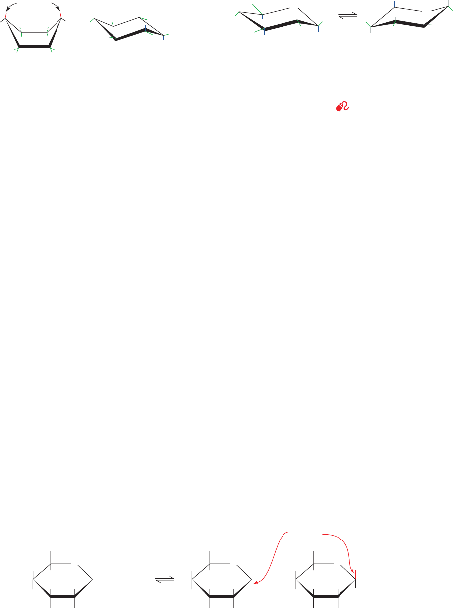
ring, like the cyclohexane ring, may assume a boat or a chair
conformation (Fig. 11-6).The relative stabilities of these var-
ious conformations depend on the stereochemical interac-
tions between the substituents on the ring. The boat con-
former crowds the substituents on its “bow” and “stern” and
eclipses those along its sides, so that in cyclohexane it is ⬃25
kJ ⴢ mol
⫺1
less stable than the chair conformer.The ring sub-
stituents on the chair conformer (Fig. 11-6b) fall into two
geometrical classes: the rather close-fitting axial groups that
extend parallel to the ring’s threefold rotational axis and the
staggered, and therefore minimally encumbered, equatorial
groups. Since the axial and equatorial groups on a cyclo-
hexane ring are conformationally interconvertible, a given
ring has two alternative chair forms (Fig. 11-7); the one that
predominates usually has the lesser crowding among its ax-
ial substituents. The conformational situation of a group di-
rectly affects its chemical reactivity. For example, equatorial
OH groups on pyranoses esterify more readily than do axial
OH groups. Note that -
D-glucose is the only D-aldohexose
that can simultaneously have all five non-H substituents in
the equatorial position (left side of Fig. 11-7). Perhaps this is
why glucose is the most abundant naturally occurring mono-
saccharide. The conformational properties of furanose rings
are discussed in Section 29-2Ab in relation to their effects on
the conformations of nucleic acids.
C. Sugar Derivatives
a. Polysaccharides Are Held Together
by Glycosidic Bonds
The chemistry of monosaccharides is largely that of their
hydroxy and carbonyl groups. For example, in an acid-
catalyzed reaction, the anomeric hydroxyl of a sugar re-
versibly condenses with alcohols to form ␣- and -glycosides
(Greek: glykys, sweet) (Fig. 11-8). The bond connecting the
anomeric carbon to the acetal oxygen is termed a glycosidic
Steric
crowding
(a) Symmetry
axis
(b)
a
a
a
a
a
e
e
e
e
e
e
ChairBoat
a
Figure 11-6 Conformations of the cyclohexane ring. (a) In the
boat conformation, substituents at the “bow” and “stern” (red)
are sterically crowded, whereas those along its sides (green) are
eclipsed. (b) In the chair conformation, the substituents that
extend parallel to the ring’s threefold rotation axis are designated
axial [a] and those that extend roughly outward from this
symmetry axis are designated equatorial [e].The equatorial
substituents about the ring are staggered so that they alternately
extend above and below the mean plane of the ring.
H
CH
2
OH
OH
OH
OH
OH
H
H
H
H
O
O
H
H
H
OH
HO
HO
CH
2
OH
H
H
OH
OCH
3
H
OH
CH
2
OH
H
HO
H
OH
H OH
H
α-D-Glucose
Methyl-α-
D-glucoside
OCH
3
Methyl-β-D-glucoside
+
H
+
+
+
H
2
O
CH
3
OH
Glycosidic
bonds
O
H
OH
CH
2
OH
H
HO
H
H OH
H
O
H
OH
CH
2
OH
H
HO
H
H OH
H
O
Figure 11-7 The two alternative chair conformations of
-
D-glucopyranose. In the conformation on the left, which
predominates, the relatively bulky OH and CH
2
OH substituents
all occupy equatorial positions, whereas in that on the right
(drawn in ball-and-stick form in Fig. 11-5, right) they occupy the
more crowded axial positions.
See Kinemage Exercise 7-1
Figure 11-8 The acid-catalyzed condensation of ␣-D-glucose with methanol to form an
anomeric pair of methyl-
D-glucosides.
JWCL281_c11_359-385.qxd 6/3/10 10:35 AM Page 363

364 Chapter 11. Sugars and Polysaccharides
D-Glucuronic acid
H
H
O
HO HC
C
C
OH
HCOH
HC
COOH
OH
2
1
3
4
5
6
D
-Galacturonic acid
H
H
O
HO HC
C
C
OH
HO C H
HC
COOH
OH
2
1
3
4
5
6
D
-Mannuronic acid
H
HO
O
HO HC
C
C
H
HCOH
HC
COOH
OH
2
1
3
4
5
6
Uronic acids can assume the pyranose, furanose, and linear
forms.
Both aldonic and uronic acids have a strong tendency to
internally esterify so as to form five- and six-membered
lactones (Fig. 11-9). Ascorbic acid (vitamin C, Fig. 11-10) is
a ␥-lactone that is synthesized by plants and almost all ani-
mals except primates and guinea pigs. Its prolonged defi-
ciency in the diet of humans results in the disease known as
scurvy, which is caused by the impairment of collagen for-
mation (Section 8-2B). Scurvy generally results from a lack
of fresh food. This is because, under physiological condi-
tions, ascorbic acid is reversibly oxidized to dehydroascor-
bic acid, which, in turn, is irreversibly hydrolyzed to the
vitamin-inactive diketogulonic acid (Fig. 11-10).
Aldoses and ketoses may be reduced under mild condi-
tions, for example, by treatment with NaBH
4
to yield
acyclic polyhydroxy alcohols known as alditols, which are
named by appending the suffix -itol to the root name of the
parent aldose. Ribitol is a component of flavin coenzymes
(Section 16-2C), and glycerol and the cyclic polyhydroxy
alcohol myo-inositol are important lipid components
bond. Polysaccharides are held together by glycosidic
bonds between neighboring monosaccharide units. The
glycosidic bond is therefore the carbohydrate analog of
the peptide bond in proteins. The bond in a nucleoside
linking its ribose residue to its base is also a glycosidic
bond (Section 5-1A).
The hydrolysis of glycosidic bonds is catalyzed by en-
zymes known as glycosidases that differ in specificity ac-
cording to the identity and anomeric configuration of the
glycoside but are often rather insensitive to the identity of
the alcohol residue. Under basic or neutral conditions and
in the absence of glycosidases, however, the glycosidic
bond is stable, so glycosides do not undergo mutarotation
as do monosaccharides. The methylation of the non-
anomeric OH groups of monosaccharides requires more
drastic conditions than is required for the formation of
methyl glycosides, such as treatment with dimethyl sulfate.
b. Oxidation–Reduction Reactions
Because the cyclic and linear forms of aldoses and ke-
toses interconvert so readily, these sugars undergo reac-
tions typical of aldehydes and ketones. Mild oxidation of
an aldose, either chemically or enzymatically, results in the
conversion of its aldehyde group to a carboxylic acid func-
tion, thereby yielding an aldonic acid such as gluconic acid.
Aldonic acids are named by appending the suffix -onic acid
to the root name of the parent aldose.
Saccharides bearing anomeric carbon atoms that have not
formed glycosides are termed reducing sugars because of
the facility with which the aldehyde group reduces mild ox-
idizing agents. A classic test for the presence of a reducing
sugar is the reduction of Ag
⫹
in an ammonia solution
D-Gluconic acid
1
COOH
2
COHH
3
CHHO
4
COHH
5
COHH
6
CH
2
OH
(Tollens’ reagent) to yield a metallic silver mirror lining on
the inside of the reaction vessel.
The specific oxidation of the primary alcohol group of
aldoses yields uronic acids, which are named by appending
-uronic acid to the root name of the parent aldose.
D-Glucuronic acid, D-galacturonic acid, and D-mannuronic
acid are important components of many polysaccharides.
OH
OH
CH
2
OH
H
H
H
H
OH
O
CO
1
23
4
5
6
␣
␥
␦
HO
OH
H
OH
CHO
H
H
H
C
O
O
1
2
6
3
4
5
␥
␦

␣
D
-Glucono-␦-lactone D-Glucurono-␦-lactone
Figure 11-9 D-Glucono-␦-lactone and D-glucurono-␦-lactone
are, respectively, the lactones of
D-gluconic acid and D-glucuronic
acid. The ␦ indicates that the O atom closing the lactone ring is
also substituent to C
␦
.
CH
2
OH CH
2
OH
H
2
O
CH
2
OH
C
C
H
C
C
HO
H
HO
HO
C
O
C
C
H
C
C
H
HO
CO
O
O
C
C
H
C
C
H
HO
COOH
O
O
OH
–2H
L-Ascorbic
acid
L-Dehydroascorbic
acid
L-Diketogulonic
acid
O O
Figure 11-10 The reversible oxidation of L-ascorbic acid to
L-dehydroascorbic acid. This is followed by the physiologically
irreversible hydrolysis of its lactone ring to form
L-diketogulonic
acid.
JWCL281_c11_359-385.qxd 6/3/10 10:35 AM Page 364
Section 11-2. Polysaccharides 365
HO
HH
H
OH
O
H
OH
H
OH OH
O
HH
HH
OH H
CH
2
OH
CH
3
β-D-2-Deoxyribose
α-
L-Rhamnose
(6-deoxy-
L-mannose)
H
H
H
O
HO
OH
OH
H
OH H
CH
3
α-L-Fucose
(6-deoxy-
L-galactose)
In amino sugars, one or more OH groups are replaced
by an often acetylated amino group.
D-Glucosamine and
D-galactosamine are components of numerous biologically
important polysaccharides.
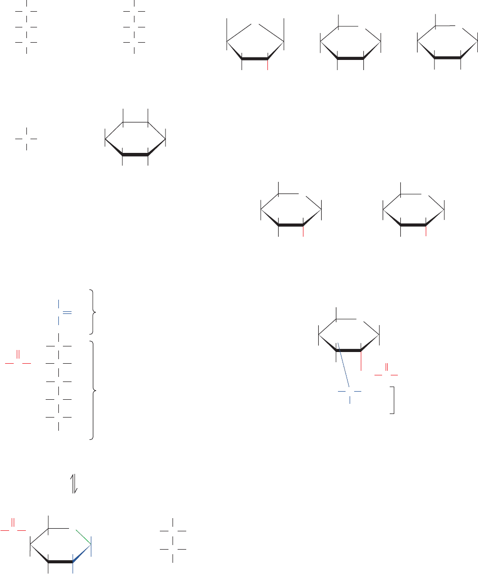
The amino sugar derivative N-acetylmuramic acid, which
consists of N-acetyl-
D-glucosamine in an ether linkage
with
D-lactic acid, is a prominent component of bacterial
cell walls (Section 11-3Ba). N-Acetylneuraminic acid,
which is derived from N-acetylmannosamine and pyruvic
acid (Fig. 11-11), is an important constituent of glycopro-
teins (Section 11-3C) and glycolipids (Section 12-1D).
N-Acetylneuraminic acid and its derivatives are often
referred to as sialic acids.
2 POLYSACCHARIDES
Polysaccharides, which are also known as glycans, con-
sist of monosaccharides linked together by glycosidic
bonds. They are classified as homopolysaccharides or het-
eropolysaccharides if they consist of one type or more than
one type of monosaccharide residue. Homopolysaccha-
rides may be further classified according to the identity of
HO
OH
CH
2
OH
H
H
OH
H
H NH
2
H
H
OH
CH
2
OH
H
HO
H
OH
H NH
2
H
α-D-Glucosamine
(2-amino-2-deoxy-
α-
D-glucopyranose)
α-
D-Galactosamine
(2-amino-2-deoxy-
α-
D-galactopyranose)
O
O
N-Acetylmuramic acid (NAM)
D-Lactic
acid residue
H
O
CH
2
OH
H
HO
H
OH
H
NH CH
3
C
O
H
O
CH
3
COOHC
H
(Section 12-1). Xylitol is a sweetener that is used in “sugar-
less” gum and candies.
c. Other Biologically Important Sugar Derivatives
Monosaccharide units in which an OH group is replaced
by H are known as deoxy sugars. The biologically most im-
portant of these is -
D-2-deoxyribose, the sugar compo-
Ribitol
CH
2
OH
COHH
COHH
COHH
CH
2
OH
Xylitol
CH
2
OH
COHH
CHHO
COHH
CH
2
OH
Glycerol myo-Inositol
CH
2
OH
COHH
CH
2
OH
HO OH
OH
H
H
HO
OH H
H
HOH
H
nent of DNA’s sugar–phosphate backbone (Section 5-1A).
L-Rhamnose and L-fucose are widely occurring polysac-
charide components.
OH
COOH
N-Acetylneuraminic acid
(pyranose form)
COOH
CO
CH
2
HCOH
O
CH
3
CNH
C
H
HO CH
HCOH
HCOH
CH
2
OH
Pyruvic
acid
residue
N-Acetyl-
mannosamine
H
H
N
CCH
3
O
N-Acetylneuraminic acid
(linear form)
HCOH
OHC
H
R =
CH
2
OH
H
H
H
O
OH H
R
Figure 11-11 N-Acetylneuraminic acid in its linear and pyranose
forms. Note that its pyranose ring incorporates the pyruvic acid
residue (blue) and part of the mannose moiety.
JWCL281_c11_359-385.qxd 6/3/10 10:35 AM Page 365
366 Chapter 11. Sugars and Polysaccharides
their monomeric unit. For example, glucans are polymers
of glucose, whereas galactans are polymers of galactose.Al-
though monosaccharide sequences of heteropolysaccha-
rides can, in principle,be as varied as those of proteins, they
are usually composed of only a few types of monosaccha-
rides that alternate in a repetitive sequence.
Polysaccharides, in contrast to proteins and nucleic acids,
form branched as well as linear polymers. This is because
glycosidic linkages can be made to any of the hydroxyls of
a monosaccharide. Fortunately for structural biochemists,
many polysaccharides are linear and those that branch
tend to do so in only a few well-defined ways.
In this section, we discuss the structures of the simplest
polysaccharides, the disaccharides, and then consider the
structures and properties of the most abundant classes of
polysaccharides.We begin by outlining how polysaccharide
structures are elucidated.
A. Carbohydrate Analysis
The purification of carbohydrates can, by and large, be ef-
fected by chromatographic and electrophoretic procedures
similar to those used in protein purification (Sections 6-3
and 6-4), although thin layer chromatography (TLC; Sec-
tion 6-3Dd) is also widely used. Affinity chromatography
(Section 6-3C), using immobilized proteins known as
lectins (Latin: legere, to pick or choose), is a particularly
powerful technique in this regard. Lectins are sugar-binding
proteins that were discovered in plants but are now known
to occur in all organisms, where they participate in a wide
variety of signaling, cell–cell recognition, and adhesion
processes, as well as in targeting newly synthethesized pro-
teins to specific cellular locations (Section 12-4Cg). Lectins
recognize one or more specific monosaccharides with par-
ticular linkages to other sugars in oligosaccharides, usually
with exquisite specificity. Their protein–carbohydrate
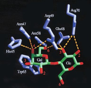
interactions typically include multiple hydrogen bonds,
which often include bridging water molecules, and the
packing of hydrophobic sugar faces against aromatic side
chains (Fig. 11-12). Among the best characterized lectins
are jack bean concanavalin A (Fig. 8-40), which specifi-
cally binds ␣-
D-glucose and ␣-D-mannose residues, and
wheat germ agglutinin (so named because it causes cells
to agglutinate or clump together), which specifically binds
-N-acetylmuramic acid and ␣-N-acetylneuraminic acid.
Characterization of an oligosaccharide requires that the
identities, anomers, linkages, and order of its component
monosaccharides be elucidated.The linkages of the mono-
saccharides may be determined by methylation analysis
(also called permethylation analysis), a technique pio-
neered by Norman Haworth in the 1930s. Methyl ethers not
at the anomeric C atom are resistant to acid hydrolysis but
glycosidic bonds are not. Consequently, if an oligosaccha-
ride is exhaustively methylated and then hydrolyzed,the free
OH groups on the resulting methylated monosaccharides
mark the former positions of the glycosidic bonds. Methyl-
ated monosaccharides are often identified by gas–liquid
chromatography (GLC; a technique in which the station-
ary phase is an inert solid, such as diatomaceous earth,
impregnated with a low-volatility liquid, such as silicone oil,
and the mobile phase is an inert gas, such as He, into which
the sample has been flash evaporated) combined with mass
spectrometry (GLC/MS). HPLC techniques may similarly
be used. Other mass spectrometric techniques for analyz-
ing nonvolatile substances are discussed in Section 7-1I.
Although all aldoses and ketoses with the same number of
C atoms are isomers (Figs. 11-1 and 11-2) and hence have
identical molecular masses, they have characteristic frag-
mentation patterns.
The sequence and anomeric configurations of the
monosaccharides in an oligosaccharide can be determined
through the use of specific exoglycosidases. These enzymes
specifically hydrolyze their corresponding monosaccha-
rides from the nonreducing ends of oligosaccharides (the
ends lacking a free anomeric carbon atom) in a manner
analogous to the actions of exopeptidases on proteins (Sec-
tion 7-1Ab). For example, -galactosidase excises the ter-
minal  anomers of galactose, whereas ␣-mannosidase
does so with the ␣ anomers of mannose. Some of these
exoglycosidases also exhibit specificity for the aglycone, the
sugar chains to which the monosaccharide to be excised
(the glycone) is linked. Through the use of mass spectrom-
etry, the sequence of a polysaccharide may be deduced
from the mass decrements generated by exoglycosidases.
The use of endoglycosidases (hydrolases that cleave glyco-
sidic bonds between nonterminal sugar residues) of vary-
ing specificities can also supply useful sequence informa-
tion. The proton and
13
C NMR spectra of oligosaccharides
can provide the complete sequence of an oligosaccharide if
sufficient material is available. Moreover, two-dimensional
Figure 11-12 Carbohydrate binding by a lectin in the
X-ray structure of human galectin-2 in complex with the
disaccharide lactose. This lectin primarily binds -
D-galactose
residues.The structure is drawn in stick form with the C and O
atoms of lactose’s galactose (Gal) and glucose (Glc) residues
green and red, and the galectin-2 amino acid side chains violet.
Hydrogen bonds between the protein side chains and the sugar
residues are represented by dashed yellow lines. [Courtesy of
Hakon Leffler, Lund University, Sweden. PDBid 1HLC.]
JWCL281_c11_359-385.qxd 6/3/10 10:35 AM Page 366

Section 11-2. Polysaccharides 367
NMR techniques (Section 8-3Ac) can reveal oligosaccha-
ride structures (e.g., see Section 11-2Eb).
B. Disaccharides
We begin our studies of polysaccharides by considering dis-
accharides (Fig. 11-13). Sucrose, the most abundant disac-
charide, occurs throughout the plant kingdom and is famil-
iar to us as common table sugar. Its structure (Fig. 11-13)
was established by methylation analysis as described above
and was later confirmed by its X-ray structure. To name a
polysaccharide systematically, one must specify its compo-
nent monosaccharides, their ring types, their anomeric
forms, and how they are linked together. Sucrose is there-
fore O-␣-
D-glucopyranosyl-(1 S 2)--D-fructofuranoside,
where the symbol (1 S 2) indicates that the glycosidic bond
links C1 of the glucose residue to C2 of the fructose residue.
Note that since these two positions are the anomeric carbon
atoms of their respective monosaccharides, sucrose is not a
reducing sugar (as the suffix -ide implies).
The hydrolysis of sucrose to
D-glucose and D-fructose is
accompanied by a change in optical rotation from dextro to
levo. Consequently, hydrolyzed sucrose is sometimes called
invert sugar and the enzyme that catalyzes this process,
-
D-fructofuranosidase, is archaically named invertase.
Lactose [O--
D-galactopyranosyl-(1 S 4)-D-glucopyra-
nose] or milk sugar (Fig. 11-13) occurs naturally only in
milk, where its concentration ranges from 0 to 7% depend-
ing on the species. The free anomeric carbon of its glucose
residue makes lactose a reducing sugar.
Infants normally express the intestinal enzyme -
D-
galactosidase or lactase that catalyzes the hydrolysis of lac-
tose to its component monosaccharides for absorption into
the bloodstream. Many adults, however, including most
Africans and almost all Asians, have a low level of this en-
zyme (as do most adult mammals, since they normally do
not encounter milk). Consequently, much of the lactose in
any milk they drink moves through their digestive tract to
the colon, where its bacterial fermentation produces large
quantities of CO
2
,H
2
, and irritating organic acids. This re-
sults in an embarrassing and often painful digestive upset
termed lactose intolerance. Perhaps this is why Chinese
cuisine, which is noted for the wide variety of foodstuffs it
employs, is devoid of milk products. However, adult mem-
bers of populations with a tradition of herding cattle,
mainly northern Europeans and certain African groups,
continue expressing the lactase gene and hence can drink
milk without a problem. Modern food technology has
come to the aid of milk lovers who develop lactose intoler-
ance: Milk products in which the lactose has been hy-
drolyzed enzymatically and lactase-containing pills are
now widely available.
There are several common glucosyl–glucose disaccha-
rides.These include maltose [O-␣-
D-glucopyranosyl-(1 S 4)-
D-glucopyranose], an enzymatic hydrolysis product of
starch; isomaltose, its ␣(1 S 6) isomer; and cellobiose, its
(1 S 4) isomer, the repeating disaccharide of cellulose.
Only a few tri- or higher oligosaccharides occur in significant
natural abundance. Not surprisingly, they all occur in plants.
C. Structural Polysaccharides: Cellulose and Chitin
Plants have rigid cell walls (Fig. 1-9) that, in order to main-
tain their shapes, must be able to withstand osmotic pressure
differences between the extracellular and intracellular
spaces of up to 20 atm. In large plants, such as trees, the cell
Figure 11-13 Several common disaccharides. See
Kinemage Exercise 7-2
FructoseGlucose
H
H
OH H
H
H
HO
O
4
HO
OH
H
O
O
Sucrose
2
1
5
4
6
3
5
6
OH
CH
2
OH
H HOCH
2
H
23
1(α)
(
β)
CH
2
OH
Glucose
HOH
HO
HOH
H
O
32 32
H
H
H
H
O
O
OH
Galactose
Lactose
44
H
CH
2
OH CH
2
OH
OH
H
H
6
5
6
1(β)1(β)
OH
5
HOH
OH
H
O
Glucose
H
HOH
H
CH
2
OH
5
6
32
HOH
OH
H
O
O
Glucose
Maltose
H
H
HO
H
CH
2
OH
4
5
4
6
32
1(α)1(β)
HOH
OH
H
O
Glucose
H
H
HO
OH
H
CH
2
5
6
32
HOH
OH
H
O
O
Glucose
Isomaltose
H
H
HO
H
CH
2
OH
4
5
4
6
32
1(α)
1(α)
1(β)1(β)
HOH
OH
H
O
Glucose
H
HOH
H
CH
2
OH
4
5
6
32
HOH
OH
H
O
O
Glucose
Cellobiose
H
H
HO
H
CH
2
OH
4
5
6
32
JWCL281_c11_359-385.qxd 6/7/10 7:14 AM Page 367
368 Chapter 11. Sugars and Polysaccharides
walls also have a load-bearing function. Cellulose, the pri-
mary structural component of plant cell walls (Fig. 11-14),
accounts for over half of the carbon in the biosphere: ⬃10
15
kg of cellulose are estimated to be synthesized and degraded
annually. Although cellulose is predominantly of vegetable
origin, it also occurs in the stiff outer mantles of marine in-
vertebrates known as tunicates (urochordates; Fig. 1-11).
The primary structure of cellulose was determined
through methylation analysis. Cellulose is a linear polymer of
up to 15,000
D-glucose residues (a glucan) linked by (1 S 4)
glycosidic bonds (Fig. 11-15). As is generally true of large
polysaccharides, it has no defined size since, in contrast to
proteins and nucleic acids, there is no genetically deter-
mined template that directs its synthesis.
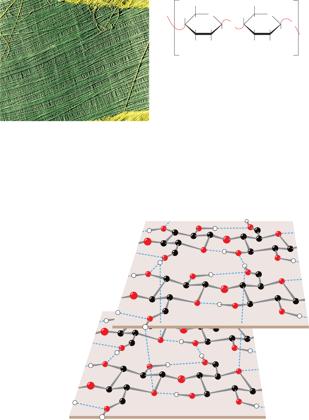
X-ray studies of cellulose fibers led Anatole Sarko to
tentatively propose the model diagrammed in Fig. 11-16.
This highly cohesive, hydrogen bonded structure gives cel-
lulose fibers exceptional strength and makes them water
insoluble despite their hydrophilicity.
Figure 11-14 Electron micrograph of the cellulose fibers in
the cell wall of the alga Chaetomorpha melagonium. Note that
the cell wall consists of layers of parallel fibers. [Biophoto
Associates/Photo Researchers.]
Figure 11-15 The primary structure of cellulose. Here n may
be several thousand.
Glucose
H
OH
HOH
H
O
H
H
H
H
O
O
OH
O
Glucose
Cellulose
H
H
CH
2
OH CH
2
OH
OH
H
n
Figure 11-16 Proposed structural model of cellulose.
Cellulose fibers consist of ⬃40 parallel glucan chains
arranged in an extended fashion. Each of the
(1 S 4)-linked glucose units in a chain is turned
180° with respect to its preceding residue and is
held in this position by intrachain hydrogen bonds
(dashed lines).The glucan chains line up laterally
to form sheets, and these sheets stack vertically
such that they are staggered by half the length
of a glucose unit.The entire assembly is
stabilized by intermolecular hydrogen bonds
between glucose units of neighboring chains.
Hydrogen atoms not participating in
hydrogen bonds have been omitted
for clarity. [Illustration, Irving Geis.
Image from the Irving Geis Collection,
Howard Hughes Medical Institute.
Reprinted with permission.]
JWCL281_c11_359-385.qxd 8/10/10 9:53 AM Page 368
Section 11-2. Polysaccharides 369
In plant cell walls, the cellulose fibers are embedded in
and cross-linked by a matrix of several polysaccharides
that are composed of glucose as well as other monosaccha-
rides. In wood, this cementing matrix also contains a large
proportion of lignin, a plasticlike phenolic polymer. One
has only to watch a tall tree in a high wind to realize the
enormous strength of plant cell walls. In engineering terms,
they are “composite materials,” as is concrete reinforced by
steel rods. Composite materials can withstand large
stresses because the matrix evenly distributes the stresses
among the reinforcing elements.
Although vertebrates themselves do not possess an en-
zyme capable of hydrolyzing the (1 S 4) linkages of cel-
lulose, the digestive tracts of herbivores contain symbiotic
microorganisms that secrete a series of enzymes, collec-
tively known as cellulase, that do so.The same is true of ter-
mites. Nevertheless, the degradation of cellulose is a slow
process because its tightly packed and hydrogen bonded
glucan chains are not easily accessible to cellulase and do
not separate readily even after many of their glycosidic
bonds have been hydrolyzed. The digestion of fibrous
plants such as grass by herbivores is therefore a more com-
plex and time-consuming process than is the digestion of
meat by carnivores (cows, e.g., have multichambered stom-
achs and must chew their cud). Similarly, the decay of dead
plants by fungi, bacteria, and other organisms, and the con-
sumption of wooden houses by termites, often takes years.
Chitin is the principal structural component of the ex-
oskeletons of invertebrates such as crustaceans, insects, and
spiders and is also a major cell wall constituent of most
fungi and many algae. It is estimated that ⬃10
14
kg of chitin
are produced annually, most of it in the oceans, and there-
fore that it is almost as abundant as is cellulose. Chitin is a
homopolymer of (1 S 4)-linked N-acetyl-
D-glucosamine
residue (Fig. 11-17). It differs chemically from cellulose
only in that each C2-OH group is replaced by an acetamido
function. X-ray analysis indicates that chitin and cellulose
have similar structures.
D. Storage Polysaccharides: Starch and Glycogen
a. Starch Is a Food Reserve in Plants and a Major
Nutrient for Animals
Starch is a mixture of glucans that plants synthesize as
their principal food reserve. It is deposited in the cyto-
plasm of plant cells as insoluble granules composed of ␣-
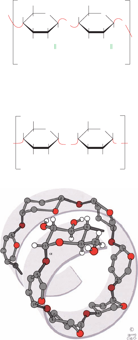
amylose and amylopectin. ␣-Amylose is a linear polymer
of several thousand glucose residues linked by ␣(1 S 4)
bonds (Fig. 11-18a). Note that although ␣-amylose is an
isomer of cellulose, it has very different structural proper-
ties. This is because cellulose’s -glycosidic linkages cause
each successive glucose residue to flip 180° with respect to
the preceding residue, so that the polymer assumes an eas-
ily packed, fully extended conformation (Fig. 11-16). In
contrast, ␣-amylose’s ␣-glycosidic bonds cause it to adopt
an irregularly aggregating helically coiled conformation
(Fig. 11-18b).
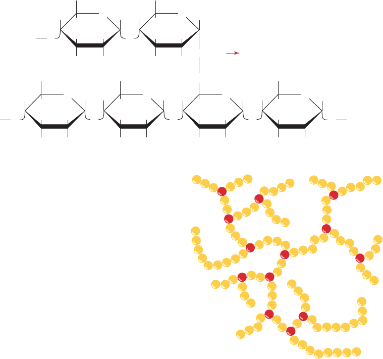
Amylopectin consists mainly of ␣(1 S 4)-linked glucose
residues but is a branched molecule with ␣(1 S 6) branch
Figure 11-17 The primary structure of chitin. Chitin is a
(1 S 4)-linked homopolymer of N-acetyl-
D-glucosamine.
Figure 11-18 ␣-Amylose. (a) The
D-glucose residues of
␣-amylose are linked by ␣(1 S 4) bonds (red). Here n is several
thousand. (b) This regularly repeating polymer forms a left-
handed helix with ⬃6 glucose residues per turn. Note the great
differences in structure and properties that result from changing
␣-amylose’s ␣(1 S 4) linkages to the (1 S 4) linkages of
cellulose (Fig. 11-16). [Illustration, Irving Geis. Image from the
Irving Geis Collection, Howard Hughes Medical Institute.
Reprinted with permission.]
N-Acetylglucosamine
HOH
H NHCCH
3
H
O
H
H
H
H
O
O
O
N-Acetylglucosamine
Chitin
H
H
CH
2
OH CH
2
OH
OH
H
n
O
NHCCH
3
O
O
n
GlucoseGlucose
H
H
OH H
H
H
O
H
OH
H
O
O
O
␣-Amylose
OH
CH
2
OH
HH
OH
H
CH
2
OH
(a)
(b)
JWCL281_c11_359-385.qxd 8/10/10 9:53 AM Page 369
370 Chapter 11. Sugars and Polysaccharides
points every 24 to 30 glucose residues on average (Fig. 11-19).
Amylopectin molecules contain up to 10
6
glucose residues,
which makes them among the largest molecules occurring
in nature. The storage of glucose as starch greatly reduces
the large intracellular osmotic pressures that would result
from its storage in monomeric form because osmotic pres-
sure is proportional to the number of solute molecules in a
given volume.
b. Starch Digestion Occurs in Stages
The digestion of starch, the main carbohydrate source
in the human diet, begins in the mouth. Saliva contains
␣-amylase, which randomly hydrolyzes all the ␣(1 S 4)
glucosidic bonds of starch except its outermost bonds and
those next to branches. By the time thoroughly chewed
food reaches the stomach, where the acidity inactivates
␣-amylase, the average chain length of starch has been
reduced from several thousand to fewer than eight glucose
units. Starch digestion continues in the small intestine
under the influence of pancreatic ␣-amylase, which is simi-
lar to the salivary enzyme. This enzyme degrades starch to
a mixture of the disaccharide maltose, the trisaccharide
maltotriose, which contains three ␣(1 S 4)-linked glucose
residues, and oligosaccharides known as dextrins that
contain the ␣(1 S 6) branches. These oligosaccharides
are hydrolyzed to their component monosaccharides by
specific enzymes contained in the brush border mem-
branes of the intestinal mucosa: an ␣-glucosidase, which
removes one glucose residue at a time from oligosaccha-
rides, an ␣-dextrinase or debranching enzyme, which hy-
drolyzes ␣(1 S 6) and ␣(1 S 4) bonds, a sucrase, and, at
least in infants, a lactase. The resulting monosaccharides
are absorbed by the intestine and transported to the
bloodstream (Section 20-4A).
c. Glycogen Is “Animal Starch”
Glycogen, the storage polysaccharide of animals, is pres-
ent in all cells but is most prevalent in skeletal muscle and
liver, where it occurs as cytoplasmic granules (Fig. 11-20).
The primary structure of glycogen resembles that of amy-
lopectin, but glycogen is more highly branched, with
branch points occurring every 8 to 14 glucose residues.
Glycogen’s degree of polymerization is nevertheless simi-
lar to that of amylopectin. In the cell, glycogen is degraded
for metabolic use by glycogen phosphorylase, which phos-
phorolytically cleaves glycogen’s ␣(1 S 4) bonds sequen-
tially inward from its nonreducing ends to yield glucose-1-
phosphate. Glycogen’s highly branched structure, which
has many nonreducing ends, permits the rapid mobilization
of glucose in times of metabolic need. The ␣(1 S 6)
branches of glycogen are cleaved by a debranching en-
zyme.These enzymes play an important role in glucose me-
tabolism and are discussed further in Section 18-1.
E. Glycosaminoglycans
The extracellular spaces, particularly those of connective
tissues such as cartilage, tendon, skin, and blood vessel
walls, consist of collagen and elastin fibers (Section 8-2B)
Figure 11-19 Amylopectin. (a) Its primary structure near one
of its ␣(1 S 6) branch points (red). (b) Its bushlike structure with
glucose residues at branch points indicated in red.The actual
distance between branch points averages 24 to 30 glucose
residues. Glycogen has a similar structure but is branched every
8 to 14 residues.
CH
2
OH CH
2
OH
CH
2
O
H
HOH HOH
HH
H
OH H OH H
O O
O
CH
2
OH CH
2
OH
HOH HOH
Amylopectin
HH
OH H OH H
O O
HOH
H
OH H
O
CH
2
OH
HOH
H
OH H
O
α(1
6) branch point
Branch
Main
chain
…
…
O
…
(a)
O
HH
O
HH
O
HH
O
HH
H
O
H
(b)
JWCL281_c11_359-385.qxd 6/3/10 10:35 AM Page 370
