Voet D., Voet Ju.G. Biochemistry
Подождите немного. Документ загружается.

Cells contain proteases (enzymes that catalyze the hy-
drolytic cleavage of peptide bonds) and other degradative
enzymes that, on cell lysis, are liberated into solution along
with the protein of interest. Care must be taken that the
protein is not damaged by these enzymes. Degradative en-
zymes may often be rendered inactive at pH’s and temper-
atures that are not harmful to the protein of interest. Alter-
natively, these enzymes can often be specifically inhibited
by chemical agents without affecting the desired protein.
Of course, as the purification of a protein progresses, more
and more of these degradative enzymes are eliminated.
Some proteins are more resistant than others to prote-
olytic degradation.The purification of a protein that is par-
ticularly resistant to proteases may be effected by main-
taining conditions in a crude protein mixture under which
the proteolytic enzymes present are active. This so-called
autolysis technique simplifies the purification of the resist-
ant protein because it is generally far easier to remove se-
lectively the degradation products of contaminating pro-
teins than it is the intact proteins.
Many proteins are denatured by contact with the
air–water interface, and, at low concentrations, a significant
fraction of the protein present may be lost by adsorption to
surfaces. Hence, a protein solution should be handled so as
to minimize frothing and should be kept relatively concen-
trated.There are, of course,other factors to which a protein
may be sensitive, including the oxidation of cysteine
residues to form disulfide bonds; heavy metal contami-
nants, which may irreversibly bind to the protein; and the
salt concentration and polarity of the solution, which must
be kept within the stability range of the protein. Finally,
many microorganisms consider proteins to be delicious, so
protein solutions should be stored under conditions that
inhibit the growth of microorganisms [e.g., in a refrigerator
and/or with small amounts of a toxic substance that does
not react with proteins, such as sodium azide (NaN
3
)].
D. Assay of Proteins
To purify any substance, some means must be found for
quantitatively detecting its presence. A particular protein
rarely comprises more than a few percent by weight of its
tissue of origin and is usually present in much smaller
amounts. Yet much of the material from which it is being
extricated closely resembles the protein of interest. Ac-
cordingly, an assay must be specific for the protein being
purified and highly sensitive to its presence. Furthermore,
the assay must be convenient to use because it may be done
repeatedly, often at every stage of the purification process.
Among the most straightforward of protein assays are
those for enzymes that catalyze reactions with readily de-
tectable products. Perhaps such a product has a character-
istic spectroscopic absorption or fluorescence that can be
monitored. Alternatively, the enzymatic reaction may con-
sume or generate acid so that the enzyme can be assayed
by acid–base titrations. If an enzymatic reaction product is
not easily quantitated, its presence may still be revealed by
further chemical treatment to yield a more readily observ-
able product. Often, this takes the form of a coupled enzy-
matic reaction, in which the product of the enzyme being
assayed is converted, by an added enzyme, to an observ-
able substance.
Proteins that are not enzymes may be assayed through
their ability to bind specific substances or through observa-
tion of their biological effects. For example, receptor pro-
teins are often assayed by incubating them with a radioac-
tive molecule that they specifically bind, passing the
mixture through a protein-retaining filter, and then meas-
uring the amount of radioactivity bound to the filter. The
presence of a hormone may be revealed by its effect on
some standard tissue sample or on a whole organism. The
latter type of assays are usually rather lengthy procedures
because the response elicited by the assay may take days to
develop. In addition, their reproducibility is often less than
satisfactory because of the complex behavior of living sys-
tems. Such assays are therefore used only when no alterna-
tive procedure is available.
a. Immunochemical Techniques Can Readily Detect
Small Quantities of Specific Proteins
Immunochemical procedures provide protein assay
techniques of high sensitivity and discrimination. These
methods employ antibodies, proteins that are produced by
an animal’s immune system in response to the introduction
of a foreign protein and that specifically bind to the foreign
protein (antibodies and the immune system are discussed
in Section 35-2).
Antibodies extracted from the blood serum of an animal
that has been immunized against a particular protein are the
products of many different antibody-producing cells. They
therefore form a heterogeneous mixture of molecules, which
vary in their exact specificities and binding affinities for their
target protein.Antibody-producing cells normally die after a
few cell divisions, so one of them cannot be cloned to pro-
duce a single species of antibody in useful quantities. Such
monoclonal antibodies may be obtained, however, by fusing
a cell producing the desired antibody with a cell of an im-
mune system cancer known as a myeloma (Section 35-2Bd).
The resulting hybridoma cell has an unlimited capacity to di-
vide and, when raised in cell culture, produces large quanti-
ties of the monoclonal antibody.
A protein can be directly detected, or even isolated,
through its precipitation by its corresponding antibodies.
Alternatively, in a so-called radioimmunoassay, a protein
can be indirectly detected by determining the degree with
which it competes with a radioactively labeled standard for
binding to the antibody (Section 19-1Aa). In an enzyme-
linked immunosorbent assay (ELISA; Fig. 6-1):
1. An antibody against the protein of interest is immo-
bilized on an inert solid such as polystyrene.
2. The solution being assayed for the protein is applied
to the antibody-coated surface under conditions in which
the antibody binds the protein. The unbound protein is
then washed away.
3. The resulting protein–antibody complex is further
reacted with a second protein-specific antibody to which
an easily assayed enzyme has been covalently linked.
Section 6-1. Protein Isolation 131
JWCL281_c06_129-162.qxd 2/22/10 2:25 PM Page 131
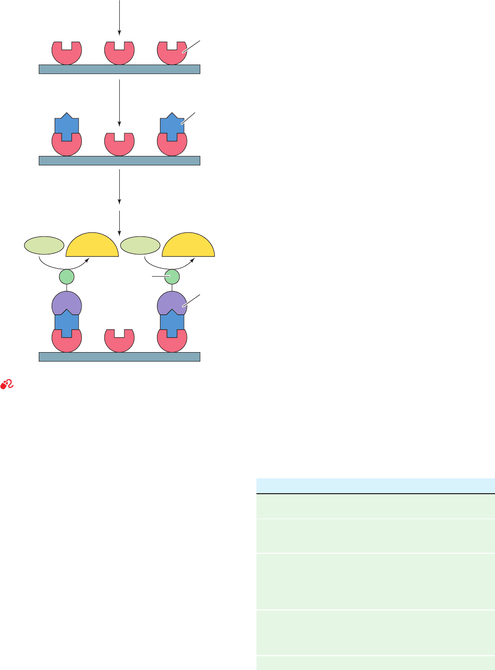
4. After washing away any unbound antibody-linked
enzyme, the enzyme in the immobilized antibody–protein–
antibody–enzyme complex is assayed, thereby indicating
the amount of the protein present.
Both radioimmunoassays and ELISAs are widely used to
detect small amounts of specific proteins and other biolog-
ical substances in both laboratory and clinical applications.
For example, a commonly available pregnancy test, which
is reliably positive within a few days post conception, uses
an ELISA to detect the placental hormone chorionic go-
nadotropin (Section 19-1I) in the mother’s urine.
E. General Strategy of Protein Purification
The fact that proteins are well-defined substances was not
widely accepted until after 1926, when James Sumner first
crystallized an enzyme, jack bean urease. Before that, it
was thought that the high molecular masses of proteins re-
sulted from a colloidal aggregation of rather ill-defined
and mysterious substances of lower molecular mass. Once
it was realized that it was possible, in principle, to purify
proteins, work to do so began in earnest.
In the first half of the twentieth century, the protein pu-
rification methods available were extremely crude by to-
day’s standards. Protein purification was an arduous task
that was as much an art as a science. Usually, the develop-
ment of a satisfactory purification procedure for a given
protein was a matter of years of labor ultimately involving
huge quantities of starting material. Nevertheless, by 1940,
⬃20 enzymes had been obtained in pure form.
Since then, tens of thousands of proteins have been pu-
rified and characterized to varying extents. Modern tech-
niques of separation have such a high degree of discrimina-
tion that one can now obtain, in quantity, a series of
proteins with such similar properties that only a few years
ago their mixture was thought to be a pure substance. Nev-
ertheless, the development of an efficient procedure for the
purification of a given protein may still be an intellectually
challenging and time-consuming task.
Proteins are purified by fractionation procedures.In a se-
ries of independent steps, the various physicochemical
properties of the protein of interest are utilized to separate
it progressively from other substances. The idea here is not
necessarily to minimize the loss of the desired protein, but
to eliminate selectively the other components of the mix-
ture so that only the required substance remains.
It may not be philosophically possible to prove that a
substance is pure. However, the operational criterion for
establishing purity takes the form of the method of exhaus-
tion: the demonstration, by all available methods, that the
sample of interest consists of only one component. There-
fore, as new separation techniques are devised, standards
of purity may have to be revised. Experience has shown
that when a sample of material previously thought to be a
pure substance is subjected to a new separation tech-
nique, it occasionally proves to be a mixture of several
components.
The characteristics of proteins and other biomolecules
that are utilized in the various separation procedures are
solubility, ionic charge,polarity, molecular size,and binding
specificity for other biological molecules. Some of the pro-
cedures we shall discuss and the protein characteristics
they depend on are as follows:
132 Chapter 6. Techniques of Protein and Nucleic Acid Purification
Characteristic Procedure
Solubility 1. Salting in
2. Salting out
Ionic Charge 1. Ion exchange chromatography
2. Electrophoresis
3. Isoelectric focusing
Polarity 1. Adsorption chromatography
2. Paper chromatography
3. Reverse-phase chromatography
4. Hydrophobic interaction
chromatography
Molecular Size 1. Dialysis and ultrafiltration
2. Gel electrophoresis
3. Gel filtration chromatography
4. Ultracentrifugation
Binding Specificity 1. Affinity chromatography
Substrate Substrate
Detectable
product
Detectable
product
First
antibody
Immobilize first
antibody on solid
support
1
Incubate with
protein-containing
sample
2
Add a second antibody
that is covalently linked
to an assayable enzyme
3
Wash and assay
the enzyme
4
Second
antibody
Protein
Enzyme
Solid support
Figure 6-1 An enzyme-linked immunosorbent assay (ELISA).
See the Animated Figures
JWCL281_c06_129-162.qxd 2/22/10 2:25 PM Page 132
Different proteins vary greatly in their solubilities under a
given set of conditions: Certain proteins precipitate from so-
lution under conditions in which others remain quite soluble.
This effect is routinely used as a basis for protein purification.
A. Effects of Salt Concentrations
The solubility of a protein in aqueous solution is a sensitive
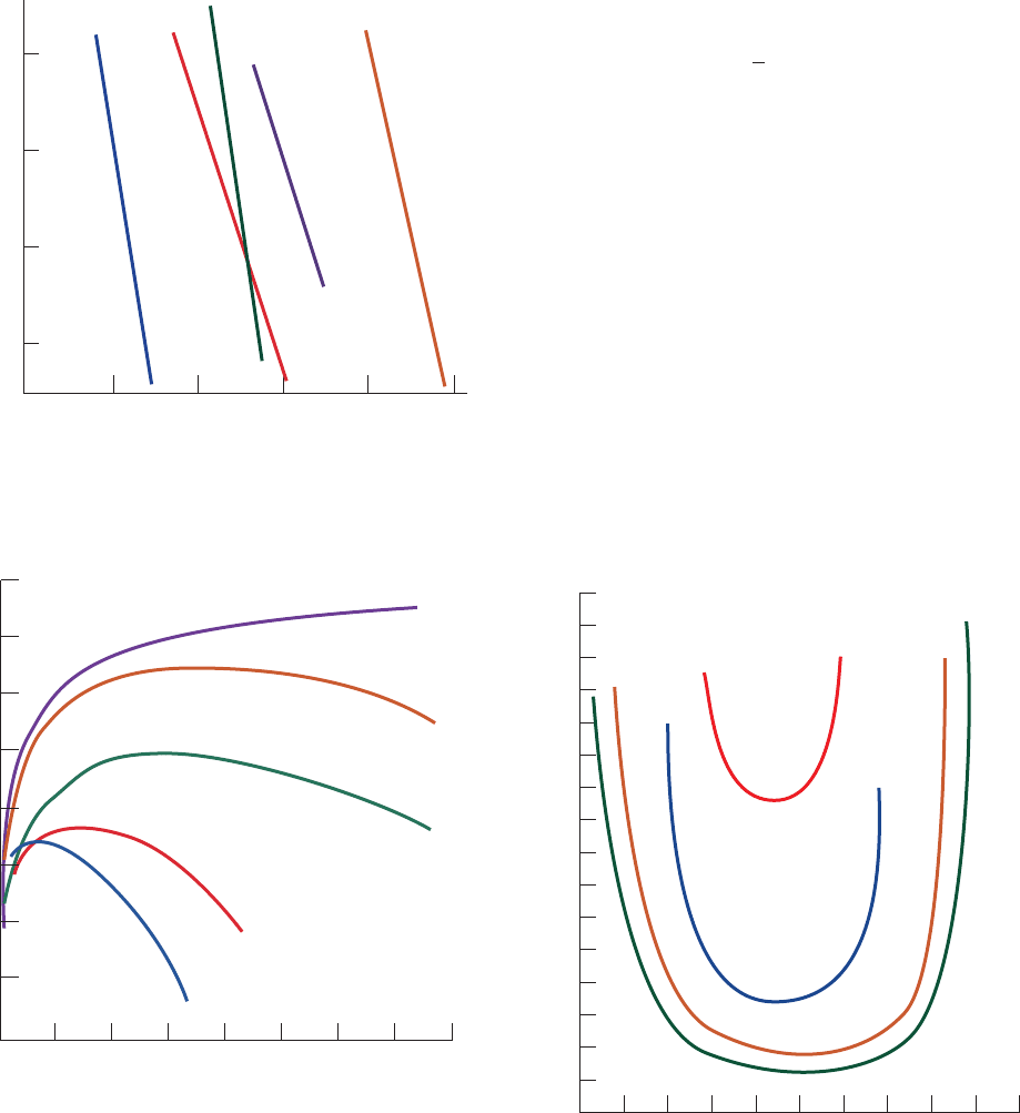
function of the concentrations of dissolved salts (Figs. 6-2
through 6-4). The salt concentration in Figs. 6-2 and 6-3 is
expressed in terms of the ionic strength, I, which is defined
[6.1]
where c
i
is the molar concentration of the ith ionic species
and Z
i
is its ionic charge. The use of this parameter to ac-
count for the effects of ionic charges results from theoreti-
cal considerations of ionic solutions. However, as Fig. 6-3
indicates, a protein’s solubility at a given ionic strength
varies with the types of ions in solution.The order of effec-
tiveness of these various ions in influencing protein solubil-
ity is quite similar for different proteins and is apparently
mainly due to the ions’ size and hydration.
The solubility of a protein at low ionic strength generally
increases with the salt concentration (left side of Fig. 6-3 and
the different curves of Fig. 6-4). The explanation of this
salting in phenomenon is that as the salt concentration of
the protein solution increases, the additional counterions
more effectively shield the protein molecules’ multiple
ionic charges and thereby increase the protein’s solubility.
I ⫽
1
2
a
c
i
Z
2
i
Section 6-2. Solubilities of Proteins 133
In the remainder of this chapter, we discuss these separa-
tion procedures.
2 SOLUBILITIES OF PROTEINS
A protein’s multiple acid–base groups make its solubility
properties dependent on the concentrations of dissolved
salts, the polarity of the solvent, the pH, and the temperature.
Figure 6-2 Solubilities of several proteins in ammonium
sulfate solutions. [After Cohn, E.J. and Edsall, J.T., Proteins,
Amino Acids and Peptides, p. 602, Academic Press (1943).]
Figure 6-3 Solubility of carboxy-hemoglobin at its isoelectric
point as a function of ionic strength and ion type. Here S and S’
are, respectively, the solubilities of the protein in the salt solution
and in pure water.The logarithm of their ratios is plotted so that
the solubility curves can be placed on a common scale. [After
Green,A.A., J. Biol. Chem. 95, 47 (1932).]
Figure 6-4 Solubility of -lactoglobulin as a function of pH at
several NaCl concentrations. [After Fox, S. and Foster, J.S.,
Introduction to Protein Chemistry, p. 242, Wiley (1975).]
0.50
–0.50
0
–1.00
0246810
Log of solubility (g
•
mL )
Ionic strength
Fibrinogen
Pseudoglobulin
Hemoglobin
Serum
albumin C
Myoglobin
–1
0 1.0 2.0 3.0 4.0
⫺0.2
0.2
0.4
0.6
0.8
1.0
1.2
1.4
0
Ionic strength
log(S/S')
NaCl
KCl
MgSO
4
(NH
4
)
2
SO
4
K
2
SO
4
3.2
2.8
2.4
2.0
1.6
Solubility (mg nitrogen
•
mL
–
1
)
1.2
0.4
0
4.8 5.0 5.2
pH
5.4
0.005M
0.010M
0.02M
5.6 5.8
0.8
0.001M
JWCL281_c06_129-162.qxd 2/22/10 2:25 PM Page 133

At high ionic strengths, the solubilities of proteins, as
well as those of most other substances, decrease.This effect,
known as salting out, is primarily a result of the competition
between the added salt ions and the other dissolved solutes
for molecules of solvation. At high salt concentrations, so
many of the added ions are solvated that the amount of bulk
solvent available becomes insufficient to dissolve other
solutes. In thermodynamic terms, the solvent’s activity (effec-
tive concentration; Appendix to Chapter 3) is decreased.
Hence, solute–solute interactions become stronger than
solute–solvent interactions and the solute precipitates.
Salting out is the basis of one of the most commonly
used protein purification procedures. Figure 6-2 shows that
the solubilities of different proteins vary widely as a func-
tion of salt concentration. For example, at an ionic
strength of 3, fibrinogen is much less soluble than the
other proteins in Fig. 6-2. By adjusting the salt concentra-
tion in a solution containing a mixture of proteins to just be-
low the precipitation point of the protein to be purified,
many unwanted proteins can be eliminated from the solu-
tion. Then, after the precipitate is removed by filtration or
centrifugation, the salt concentration of the remaining solu-
tion is increased so as to precipitate the desired protein.In
this manner,a significant purification and concentration of
large quantities of protein can be conveniently effected.
Consequently, salting out is often the initial step in protein
purification procedures. Ammonium sulfate is the most
commonly used reagent for salting out proteins because
its high solubility (3.9M in water at 0°C) permits the
achievement of solutions with high ionic strengths (up to
23.4 in water at 0°C).
Certain ions, notably I
⫺
, , SCN
⫺
,Li
⫹
,Mg
2⫹
,Ca
2⫹
,
and Ba
2⫹
, increase the solubilities of proteins rather than
salting them out.These ions also tend to denature proteins
(Section 8-4E). Conversely, ions that decrease the solubili-
ties of proteins stabilize their native structures, so that pro-
teins which have been salted out are not denatured.
ClO
⫺
4
B. Effects of Organic Solvents
Water-miscible organic solvents, such as acetone and
ethanol, are generally good protein precipitants because
their low dielectric constants reduce the solvating power
of their aqueous solutions for dissolved ions such as pro-
teins. The different solubilities of proteins in these mixed
solvents form the basis of a useful fractionation tech-
nique. This procedure is normally used near 0°C or less
because, at higher temperatures, organic solvents tend to
denature proteins. The lowering of the dielectric constant
by organic solvents also magnifies the differences in the
salting out behavior of proteins, so that these two tech-
niques can be effectively combined. Some water-miscible
organic solvents, however, such as dimethyl sulfoxide
(DMSO) or N,N-dimethylformamide (DMF), are rather
good protein solvents because of their relatively high di-
electric constants.
C. Effects of pH
Proteins generally bear numerous ionizable groups that
have a variety of pK’s. At a pH characteristic for each pro-
tein, the positive charges on the molecule exactly balance
its negative charges. At this pH, the protein’s isoelectric
point, pI (Section 4-1D), the protein molecule carries no
net charge and is therefore immobile in an electric field.
Figure 6-4 indicates that the solubility of the protein
-lactoglobulin is a minimum near its pI of 5.2 in dilute
NaCl solutions and increases more or less symmetrically
about the pI with changes in pH. This solubility behavior,
which is shared by most proteins, is easily explained.
Physicochemical considerations suggest that the solubility
properties of uncharged molecules are insensitive to the
salt concentration. To a first approximation, therefore, a
protein at its isoelectric point should not be subject to salt-
ing in. Conversely, as the pH is varied from a protein’s pI,
that is, as the protein’s net charge increases, it should be
increasingly subject to salting in because the electrostatic
interactions between neighboring molecules that promote
aggregation and precipitation should likewise increase.
Hence, in solutions of moderate salt concentrations, the sol-
ubility of a protein as a function of pH is expected to be at a
minimum at the protein’s pI and to increase about this point
with respect to pH.
Proteins vary in their amino acid compositions and
therefore, as Table 6-1 indicates, in their pI’s.This phenom-
enon is the basis of a protein purification procedure known
as isoelectric precipitation in which the pH of a protein
mixture is adjusted to the pI of the protein to be isolated so
as to selectively minimize its solubility. In practice, this
technique is combined with salting out so that the protein
being purified is usually salted out near its pI.
D. Crystallization
Once a protein has been brought to a reasonable state of
purity, it may be possible to crystallize it. This is usually
134 Chapter 6. Techniques of Protein and Nucleic Acid Purification
Table 6-1 Isoelectric Points of Several Common Proteins
Protein Isoelectric pH
Pepsin ⬍1.0
Ovalbumin (hen) 4.6
Serum albumin (human) 4.9
Tropomyosin 5.1
Insulin (bovine) 5.4
Fibrinogen (human) 5.8
␥-Globulin (human) 6.6
Collagen 6.6
Myoglobin (horse) 7.0
Hemoglobin (human) 7.1
Ribonuclease A (bovine) 7.8
Cytochrome c (horse) 10.6
Histone (bovine) 10.8
Lysozyme (hen) 11.0
Salmine (salmon) 12.1
JWCL281_c06_129-162.qxd 2/22/10 2:25 PM Page 134
done by bringing the protein solution just past its satura-
tion point with the types of precipitating agents discussed
above. On standing for a time (as little as a few minutes, as
much as several months), often while the concentration of
the precipitating agent is being slowly increased, the pro-
tein may precipitate from the solution in crystalline form.
It may be necessary to attempt the crystallization under
different solution conditions and with various precipitat-
ing agents before crystals are obtained. The crystals may
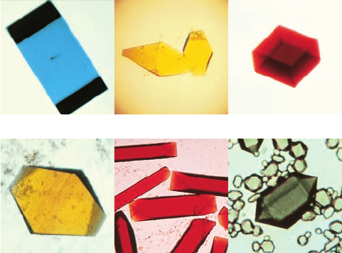
range in size from microscopic to 1 mm or more across.
Crystals of the latter size, which generally require great
care to grow, may be suitable for X-ray crystallographic
analysis (Section 8-3A). Several such crystals are shown
in Fig. 6-5.
3 CHROMATOGRAPHIC SEPARATIONS
In 1903, the Russian botanist Mikhail Tswett described the
separation of plant leaf pigments in solution through the
use of solid adsorbents. He named this process chromato-
graphy (Greek: chroma, color ⫹ graphein, to write), pre-
sumably because of the colored bands that formed in the
adsorbents as the components of the pigment mixtures
separated from one another (and possibly because Tswett
means “color” in Russian).
Modern separation methods rely heavily on chromato-
graphic procedures. In all of them, a mixture of substances
to be fractionated is dissolved in a liquid or gaseous fluid
known as the mobile phase.The resultant solution is perco-
lated through a column consisting of a porous solid matrix
known as the stationary phase, which in certain types of
chromatography may be associated with a bound liquid.
The interactions of the individual solutes with the station-
ary phase act to retard their progress through the matrix in
a manner that varies with the properties of each solute. If
the mixture being fractionated starts its journey through
the column in a narrow band, the different retarding forces
on each component that cause them to migrate at different
rates will eventually cause the mixture to separate into
bands of pure substances.
The power of chromatography derives from the contin-
uous nature of the separation processes. A single purifica-
tion step (or “theoretical plate” as it is often termed in
analogy with distillation processes) may have very little
tendency to separate a mixture into its components. How-
ever,since this process is applied in a continuous fashion so
that it is, in effect, repeated hundreds or even hundreds of
thousands of times, the segregation of the mixture into its
components ultimately occurs. The separated components
can then be collected into separate fractions for analysis
and/or further fractionation.
The various chromatographic methods are classified ac-
cording to their mobile and stationary phases. For example,
in gas–liquid chromatography the mobile and stationary
phases are gaseous and liquid, respectively, whereas in
liquid–liquid chromatography they are immiscible liquids,
one of which is bound to an inert solid support. Chromato-
graphic methods may be further classified according to the
nature of the dominant interaction between the stationary
phase and the substances being separated. For example, if
the retarding force is ionic in character, the separation
technique is referred to as ion exchange chromatography,
whereas if it is a result of the adsorption of the solutes onto
a solid stationary phase, it is known as adsorption chro-
matography.
As has been previously mentioned, a cell contains huge
numbers of different components, many of which closely
resemble one another in their various properties. There-
fore, the isolation procedures for most biological sub-
stances incorporate a number of independent chromato-
graphic steps in order to purify the substance of interest
according to several criteria. In this section, the most
commonly used of these chromatographic procedures are
described.
A. Ion Exchange Chromatography
In the process of ion exchange, ions that are electrostatically
bound to an insoluble and chemically inert matrix are re-
versibly replaced by ions in solution.
Here, R
⫹
A
–
is an anion exchanger in the A
–
form and B
–
represents anions in solution. Cation exchangers similarly
bear negatively charged groups that reversibly bind
R
⫹
A
⫺
⫹ B
⫺
Δ R
⫹
B
⫺
⫹ A
⫺
Section 6-3. Chromatographic Separations 135
(a)
(d) (e)
(b)
(c)
(f)
Figure 6-5 Protein crystals. (a) Azurin from Pseudomonas
aeruginosa,(b) flavodoxin from Desulfovibrio vulgaris,
(c) rubredoxin from Clostridium pasteurianum,(d) azidomet
myohemerythrin from the marine worm Siphonosoma funafuti,
(e) lamprey hemoglobin, and (f ) bacteriochlorophyll a protein
from Prosthecochloris aestuarii.These proteins are colored
because of their associated chromophores (light-absorbing groups);
proteins are colorless in the absence of such bound groups. [Parts
a–c courtesy of Larry Sieker, University of Washington; Parts d
and e courtesy of Wayne Hendrickson, Columbia University; and
Part f courtesy of John Olsen, Brookhaven National Laboratories,
and Brian Matthews, University of Oregon.]
JWCL281_c06_129-162.qxd 2/22/10 2:25 PM Page 135
cations. Polyanions and polycations therefore bind to anion
and cation exchangers, respectively. However, proteins and
other polyelectrolytes (polyionic polymers) that bear both
positive and negative charges can bind to both cation and
anion exchangers depending on their net charge. The affin-
ity with which a particular polyelectrolyte binds to a given
ion exchanger depends on the identities and concentrations
of the other ions in solution because of the competition
among these various ions for the binding sites on the ion ex-
changer. The binding affinities of polyelectrolytes bearing
acid–base groups are also highly pH dependent because of
the variation of their net charges with pH. These principles
are used to great advantage in isolating biological mole-
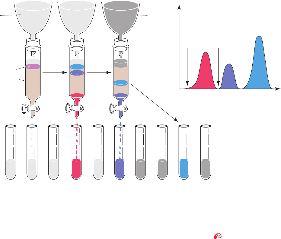
cules by ion exchange chromatography (Fig. 6-6), as de-
scribed below.
In purifying a given protein (or some other polyelec-
trolyte), the pH and the salt concentration of the buffer so-
lution in which the protein is dissolved are chosen so that
the desired protein is strongly bound to the selected ion ex-
changer. A small volume of the impure protein solution is
applied to the top of a column in which the ion exchanger
has been packed, and the column is washed with this buffer
solution.
Various proteins bind to the ion exchanger with differ-
ent affinities. As the column is washed with the buffer, a
process known as elution, those proteins with relatively low
affinities for the ion exchanger move through the column
faster than the proteins that bind to the ion exchanger with
higher affinities.This occurs because the progress of a given
protein through the column is retarded relative to that of
the solvent due to interactions between the protein mole-
cules and the ion exchanger.
The greater the binding affinity of a protein for the ion
exchanger, the more it will be retarded. Thus, proteins
that bind tightly to the ion exchanger can be eluted by
changing the elution buffer to one with a higher salt con-
centration (and/or a different pH), a process called step-
wise elution.
136 Chapter 6. Techniques of Protein and Nucleic Acid Purification
Figure 6-6 Ion exchange chromatography using stepwise
elution. Here the tan region of the column represents the ion
exchanger and the colored bands represent the various proteins.
(a) The protein mixture is bound to the topmost portion of the
ion exchanger in the chromatography column. (b) As the elution
progresses, the various proteins separate into discrete bands as a
consequence of their different affinities for the ion exchanger
Low-salt
elution
buffer
High-salt
elution
buffer
High saltLow salt
Fraction number or volume of effluent
Protein concentration
(a) (b) (c) (d)
Sample mixture
Chromatography
column
Fractions sequentially collected
under the prevailing solution conditions. Here the first band of
protein (red) has passed through the column and is being isolated
as a separate fraction, whereas the other, less mobile, bands
remain near the top of the column. (c) The salt concentration in
the elution buffer is increased to increase the mobility of and
thus elute the remaining bands. (d) The elution diagram of the
protein mixture from the column.
See the Animated Figures
JWCL281_c06_129-162.qxd 2/22/10 2:25 PM Page 136
With the use of a fraction collector,purification of a sub-
stance can be effected by selecting only those fractions of
the column effluent that contain it. Chromatographically
separated materials may be detected in a variety of ways.
The contents of the column effluent may be directly moni-
tored through column-mounted detectors according to its
UV absorbance at a specific wavelength [often 280 nm
for proteins (because the aromatic side chains of Phe, Trp,
and Tyr have strong absorbances at this wavelength; Sec-
tion 9-1Cb) and 260 nm for nucleic acids (their absorption
maximum; Fig. 5-15b)], its fluorescence, its radioactivity, its
refractive index, its pH, or its electrical conductivity. These
properties may also be measured for the individual column
fractions after the chromatographic run has been com-
pleted. In addition, biomolecules may be detected through
their enzymatic and biological activities, as is discussed in
Section 6-1D.
a. Gradient Elution Improves Chromatographic
Separations
The purification process can be further improved by
washing the protein-loaded column using the method of
gradient elution. Here the salt concentration and/or pH is
continuously varied as the column is eluted so as to release
sequentially the various proteins that are bound to the ion
exchanger.This procedure generally leads to a better sepa-
ration of proteins than does elution of the column by a sin-
gle solution or stepwise elution.
Many different types of elution gradients have been suc-
cessfully employed in purifying biological molecules. The
most widely used of these is the linear gradient, in which
the concentration of the eluant solution varies linearly with
the volume of solution passed. A simple device for generat-
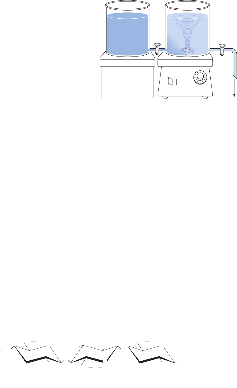
ing such a gradient is illustrated in Fig. 6-7. Here the solute
concentration, c, in the solution being withdrawn from the
mixing chamber, is expressed by
[6.2]
where c
1
is the solution’s initial concentration in the mixing
chamber, c
2
is its concentration in the reservoir chamber,
and f is the remaining fraction of the combined volumes of
the solutions initially present in both reservoirs. Linear gra-
dients of increasing salt concentration are probably more
commonly used than all other means of column elution.
However, gradients of different shapes can be generated
by using two or more chambers of different cross-sectional
areas or programmed mixing devices.
c ⫽ c
2
⫺ (c
2
⫺ c
1
)f
b. Several Types of Ion Exchangers Are Available
Ion exchangers consist of charged groups covalently at-
tached to a support matrix. The chemical nature of the
charged groups determines the types of ions that bind to
the ion exchanger and the strength with which they bind.
The chemical and mechanical properties of the support
matrix govern the flow characteristics, ion accessibility, and
stability of the ion exchanger.
Several classes of materials, colloquially referred to as
resins, are in general use as support matrices for ion ex-
changers in protein purification,including cellulose (Fig.6-8),
polystyrene, agarose gels, and cross-linked dextran gels
(Section 6-3Bb). Table 6-2 contains descriptions of some
commercially available ion exchangers in common use.
Cellulosic ion exchangers are among the materials most
commonly employed to separate biological molecules. The
cellulose, which is derived from wood or cotton, is lightly
derivatized with ionic groups to form the ion exchanger.
The most often used cellulosic anion exchanger is diethyl-
aminoethyl (DEAE)-cellulose, whereas carboxymethyl
Section 6-3. Chromatographic Separations 137
Reservoir
chamber
Solution of
concentration
c
2
Mixing
chamber
Solution of
concentration
c
1
Magnetic stirrer
off on
To
column
Figure 6-7 Device for generating a linear concentration
gradient. Two connected open chambers, which have identical
cross-sectional areas, are initially filled with equal volumes of
solutions of different concentrations.As the solution of
concentration c
1
drains out of the mixing chamber, it is partially
replaced by a solution of concentration c
2
from the reservoir
chamber.The concentration of the solution in the mixing chamber
varies linearly from its initial concentration, c
1
, to the final
concentration, c
2
, as is expressed by Eq. [6.2].
DEAE: R ⫽
CM: R ⫽
CH
2
CH
2
CH
2
NH(CH
2
CH
3
)
2
COO
⫺
⫹
CH
2
O
HO
OH
CH
2
O
OH
HO
R
OH
O
O
CH
2
HO
OH
OH
O
O
O
O
Figure 6-8 Molecular formulas of cellulose-based ion exchangers.
JWCL281_c06_129-162.qxd 2/22/10 2:25 PM Page 137

(CM)-cellulose is the most popular cellulosic cation ex-
changer (Fig. 6-8).
Gel-type ion exchangers can have the same sorts of
charged groups as do cellulosic ion exchangers. The advan-
tage of gel-type ion exchangers is that they combine the
separation properties of gel filtration (Section 6-3B) with
those of ion exchange. Because of their high degree of sub-
stitution of charged groups, which results from their porous
structures, these gels have a higher loading capacity than
do cellulosic ion exchangers.
One disadvantage of cellulosic and gel-type matrices is
that they are easily compressed (usually by the high pres-
sures resulting from attempts to increase the eluant flow
rate), thereby greatly reducing eluant flow. This problem
has been alleviated by the fabrication of noncompressible
matrices such as derivatized silica
or coated glass beads. Such materials allow very high flow
rates and pressures, even when they are very finely pow-
dered, and hence permit more effective chromatographic
separations (see HPLC in Section 6-3Dh).
B. Gel Filtration Chromatography
In gel filtration chromatography, which is also called size
exclusion and molecular sieve chromatography, molecules
are separated according to their size and shape.The station-
ary phase in this technique consists of beads of a hydrated,
spongelike material containing pores that span a relatively
narrow size range of molecular dimensions. If an aqueous
OH
Si
O
O
Si
O
Si O
OH
Si
O
O
Si
Si
Silica
O
OH
Si
O
Si
O
O
O
O
O
solution containing molecules of various sizes is passed
through a column containing such “molecular sieves,” the
molecules that are too large to pass through the pores are
excluded from the solvent volume inside the gel beads.
These larger molecules therefore traverse the column
more rapidly, that is, in a smaller eluant volume, than the
molecules that pass through the pores (Fig. 6-9).
The molecular mass of the smallest molecule unable to
penetrate the pores of a given gel is said to be the gel’s ex-
clusion limit. This quantity is to some extent a function of
molecular shape because elongated molecules, as a conse-
quence of their higher radius of hydration, are less likely to
penetrate a given gel pore than spherical molecules of the
same molecular volume.
The behavior of a molecule on a particular gel column
can be quantitatively characterized. If V
x
is the volume oc-
cupied by the gel beads and V
0
, the void volume, is the vol-
ume of the solvent space surrounding the beads, then V
t
,
the total bed volume of the column, is simply their sum:
[6.3]
V
0
is typically ⬃35% of V
t
.
The elution volume of a given solute, V
e
, is the volume
of solvent required to elute the solute from the column af-
ter it has first contacted the gel. The void volume of a col-
umn is easily measured as the elution volume of a solute
whose molecular mass is larger than the exclusion limit of
the gel.The behavior of a particular solute on a given gel is
therefore characterized by the ratio V
e
/V
0
, the relative elu-
tion volume, a quantity that is independent of the size of
the particular column used.
Molecules with molecular masses ranging below the ex-
clusion limit of a gel will elute from the gel in the order of
their molecular masses, with the largest eluting first. This is
because the pore sizes in any gel vary over a limited range,
so that larger molecules have less of the gel’s interior vol-
ume available to them than do smaller molecules. This ef-
fect is the basis of gel filtration chromatography.
V
t
⫽ V
x
⫹ V
0
138 Chapter 6. Techniques of Protein and Nucleic Acid Purification
Table 6-2 Some Biochemically Useful Ion Exchangers
Name
a
Type Ionizable Group Remarks
DEAE-cellulose Weakly basic Diethylaminoethyl Used to separate acidic
and neutral proteins
CM-cellulose Weakly acidic Carboxymethyl Used to separate basic and
neutral proteins
P-cellulose Strongly and weakly Phosphate Dibasic; binds basic proteins
acidic strongly
Bio-Rex 70 Weakly acidic, Carboxylic acid Used to separate basic
polystyrene-based proteins and amines
DEAE-Sephadex Weakly basic cross- Diethylaminoethyl Combined chromatography
linked dextran gel and gel filtration of acidic
and neutral proteins
SP-Sepharose Strongly acidic cross- Methyl sulfonate Combined chromatography
linked agarose gel and gel filtration of basic
proteins
¬CH
2
SO
3
H
¬CH
2
CH
2
N(C
2
H
5
)
2
¬COOH
¬OPO
3
H
2
¬CH
2
COOH
¬CH
2
CH
2
N(C
2
H
5
)
2
a
Sephadex and Sepharose are products of GE Healthcare; Bio-Rex resins are products of BioRad Laboratories.
JWCL281_c06_129-162.qxd 2/22/10 2:25 PM Page 138
a. Gel Filtration Chromatography Can Be Used
to Estimate Molecular Masses
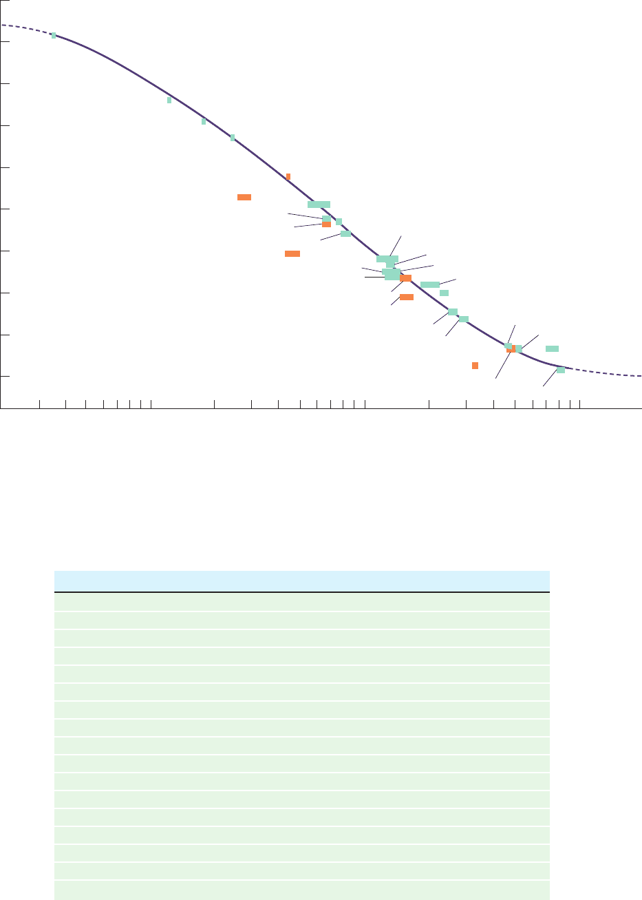
There is a linear relationship between the relative elution
volume of a substance and the logarithm of its molecular
mass over a considerable molecular mass range (Fig. 6-10).
If a plot such as Fig. 6-10 is made for a particular gel filtra-
tion column using macromolecules of known molecular
masses, the molecular mass of an unknown substance can be
estimated from its position on the plot. The precision of this
technique is limited by the accuracy of the underlying as-
sumption that the known and unknown macromolecules
have identical shapes. Nevertheless, gel filtration chro-
matography is often used to estimate molecular masses be-
cause it can be applied to quite impure samples (providing
that the molecule of interest can be identified) and because
it can be rapidly carried out using simple equipment.
b. Most Gels Are Made from Dextran, Agarose,
or Polyacrylamide
The most commonly used materials for making chromato-
graphic gels are dextran (a high molecular mass polymer of
glucose produced by the bacterium Leuconostoc mesen-
teroides), agarose (a linear polymer of alternating
D-galac-
tose and 3,6-anhydro-
L-galactose from red algae), and poly-
acrylamide (Section 6-4B).The properties of several gels that
are commonly employed in separating biological molecules
are listed in Table 6-3. The porosity of dextran-based gels,
sold under the trade name Sephadex,is controlled by the mo-
lecular mass of the dextran used and the introduction of glyc-
eryl ether units that cross-link the hydroxyl groups of the
polyglucose chains. The several classes of Sephadex that
are available have exclusion limits between 0.7 and 600 kD.
The pore size in polyacrylamide gels is similarly controlled
by the extent of cross-linking of neighboring polyacrylamide
molecules (Section 6-4B). They are commercially available
under the trade name of Bio-Gel P and have exclusion limits
between 0.2 and 400 kD.Very large molecules and supramol-
ecular assemblies can be separated using agarose gels, sold
under the trade names Sepharose and Bio-Gel A, which have
exclusion limits ranging up to 150,000 kD.
Gel filtration is often used to “desalt” a protein solution.
For example, an ammonium sulfate–precipitated protein
Section 6-3. Chromatographic Separations 139
Solvent
Volume of effluent
Amount of solute
(a) (b) (c) (d)
Gel
beads
(e)
Gel
matrix
Gel
bead
Large
molecules
Small
molecules
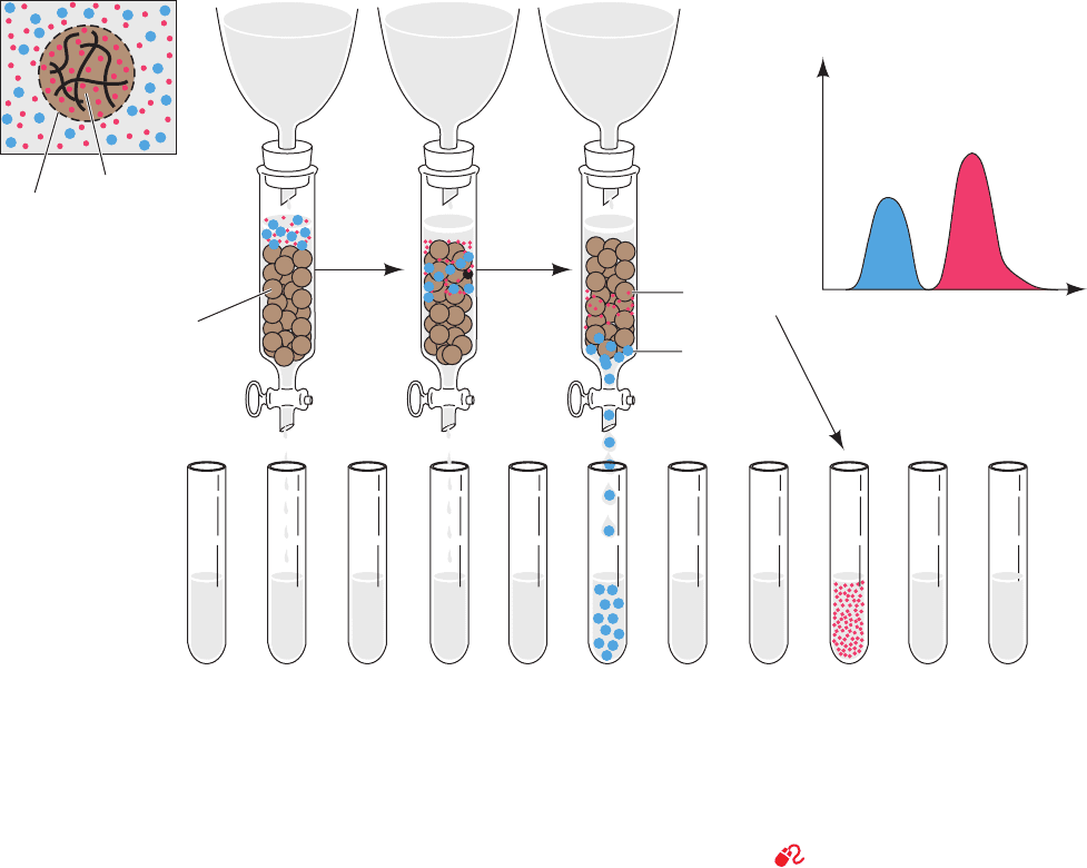
Figure 6-9 Gel filtration chromatography. (a) A gel bead,
whose periphery is represented by a dashed line, consists of a gel
matrix (wavy solid lines) that encloses an internal solvent space.
Smaller molecules (red dots) can freely enter the internal solvent
space of the gel bead from the external solvent space. However,
larger molecules (blue dots) are too large to penetrate the gel
pores. (b) The sample solution begins to enter the gel column (in
which the gel beads are now represented by brown spheres).
(c) The smaller molecules can penetrate the gel and consequently
migrate through the column more slowly than the larger molecules
that are excluded from the gel. (d) The larger molecules emerge
from the column to be collected separately from the smaller
molecules, which require additional solvent for elution from the
column. (e) The elution diagram of the chromatogram indicating
the complete separation of the two components, with the larger
component eluting first.
See the Animated Figures
JWCL281_c06_129-162.qxd 2/22/10 2:25 PM Page 139
140 Chapter 6. Techniques of Protein and Nucleic Acid Purification
Table 6-3 Some Commonly Used Gel Filtration Materials
Name
a
Type Fractionation Range (kD)
Sephadex G-10 Dextran 0.05–0.7
Sephadex G-25 Dextran 1–5
Sephadex G-50 Dextran 1–30
Sephadex G-100 Dextran 4–150
Sephadex G-200 Dextran 5–600
Sephacryl S-100 Dextran, cross-linked 1–100
Sephacryl S-200 Dextran, cross-linked 5–250
Sephacryl S-300 Dextran, cross-linked 4–150
Sephacryl S-400 Dextran, cross-linked 20–8000
Bio-Gel P-2 Polyacrylamide 0.1–1.8
Bio-Gel P-6 Polyacrylamide 1–6
Bio-Gel P-10 Polyacrylamide 1.5–20
Bio-Gel P-30 Polyacrylamide 2.5–40
Bio-Gel P-100 Polyacrylamide 5–100
Sepharose 6B Agarose 10–4,000
Sepharose 4B Agarose 60–20,000
Sepharose 2B Agarose 70–40,000
a
Sephadex, Sephacryl, and Sepharose are products of GE Healthcare; Bio-Gel gels are products of BioRad
Laboratories.
Figure 6-10 Molecular mass determination by gel filtration
chromatography. The graph shows the relative elution volume
versus the logarithm of molecular mass for a variety of proteins
on a cross-linked dextran column (Sephadex G-200) at pH 7.5.
3.0
e
1.0
2.5
2.0
1.5
10 100 1000
Molecular mass (kD)
Glucagon
Sucrose
Myoglobin
Chymotrypsinogen
Ovalbumin
Malate dehydrogenase
Ovomucoid
Bovine serum albumin
Transferrin
Fetuin
Lactoperoxidase
Glyceraldehyde-3-phosphate dehydrogenase
Lactate dehydrogenase
Aldolase
Fumarase
Catalase
Apoferritin
Ferritin
Blue dextran
Fibrinogen
Urease
β-Galactosidase
α−Crystallin
α−Conarachin
R–Phycoerythrin
γ−Globulins
Ceruloplasmin
Yeast alcohol dehydrogenase
E. coli phosphatase
Cytochrome c
V
/
V
0
Serum albumin dimer
Orange bars represent glycoproteins (proteins with attached
carbohydrate groups). [After Andrews, P., Biochem. J. 96, 597
(1965).]
JWCL281_c06_129-162.qxd 2/22/10 2:25 PM Page 140
