Voet D., Voet Ju.G. Biochemistry
Подождите немного. Документ загружается.

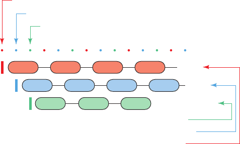
tain the proper register between the mRNA and the incom-
ing tRNAs, that is, that the ribosome maintain the correct
reading frame. As is illustrated in Fig. 5-30, a shift of even
one nucleotide along an mRNA will lead to the synthesis of
an entirely different polypeptide from the point of the shift
onward. Thus, the AUG codon that initiates polypeptide
synthesis also sets the polypeptide’s reading frame. Yet
AUG also specifies a polypeptide’s internal Met residues,
and an mRNA is likely to contain numerous AUGs in differ-
ent reading frames. How then does the ribosome select the
initiation codon from among the many AUGs in an mRNA?
In prokaryotes, the answer is that each mRNA contains a se-
quence on the upstream (5¿) side of the initiating codon (a
region that does not encode polypeptide chain) through
which the ribosome identifies this codon. In eukaryotes, the
answer is simpler; the initiating codon is usually the first
AUG that is downstream of the mRNA’s 5¿ cap.
e. Prokaryotic mRNAs Have Short Lifetimes
In prokaryotes, transcription and translation both take
place in the same cellular compartment, the cytosol (Figs.
1-2 and 1-13). Consequently ribosomes often attach to the
5¿ end of an mRNA before its synthesis is complete and
commence synthesizing the corresponding polypeptide.
This is essential because, since the mRNAs in prokaryotes
have average lifetimes of only 1 to 3 minutes before being
hydrolytically degraded by enzymes known as nucleases,
the 5¿ end of an mRNA may be degraded before its 3¿ end
is synthesized.This rapid turnover of its mRNAs permits a
prokaryote to respond quickly to changes in its environ-
ment by synthesizing the proteins appropriate for its new
situation within minutes of the change (recall that prokary-
otes are adapted to live in environments in which there are
rapid fluctuations in the available nutrients; Section 1-2).
Eukaryotic cells, in contrast, mostly lead a more seden-
tary existence. Their RNAs are transcribed and post-tran-
scriptionally modified in the nucleus, whereas ribosomes
occupy the cytosol where translation takes place (Fig. 1-5).
Hence, mature mRNAs must be transported from the nu-
cleus to the cytosol in order to participate in translation.
Eukaryotic mRNAs therefore tend to have lifetimes on the
order of several days.
f. Proteins Are Subject to Post-Translational
Modifications and Degradation
Newly synthesized polypeptides often require post-
translational modifications to become functional. In many
proteins, the leading (N-terminal) Met residue that was
specified by its mRNA’s initiating codon is excised by a
specific protease (an enzyme that hydrolytically cleaves
peptide bonds). Proteins are then subject to numerous
other chemical modifications at specific residues, including
specific proteolytic cleavages, acylation, hydroxylation,
methylation, and phosphorylation (Section 4-3A). In addi-
tion, eukaryotic proteins, but not prokaryotic proteins, are
subject to glycosylation (the addition of polysaccharides)
at specific sites (Sections 11-3C and 23-3B). Indeed, glyco-
proteins (proteins that have been glycosylated) are the
most common type of eukaryotic protein and can consist of
up to 90% or more by mass of polysaccharide groups.
All cells have several mechanisms for degrading pro-
teins to their component amino acids. This enables cells to
eliminate damaged or abnormal proteins, destroy proteins
that are no longer needed, and utilize proteins as nutrients.
The lifetime of a protein in a cell can be surprisingly short,
as little as a fraction of a minute, although many proteins in
eukaryotes have lifetimes of days or weeks. Thus cells are
dynamic entities that are constantly turning over most of
their components, in particular their RNA and proteins.
C. DNA Replication
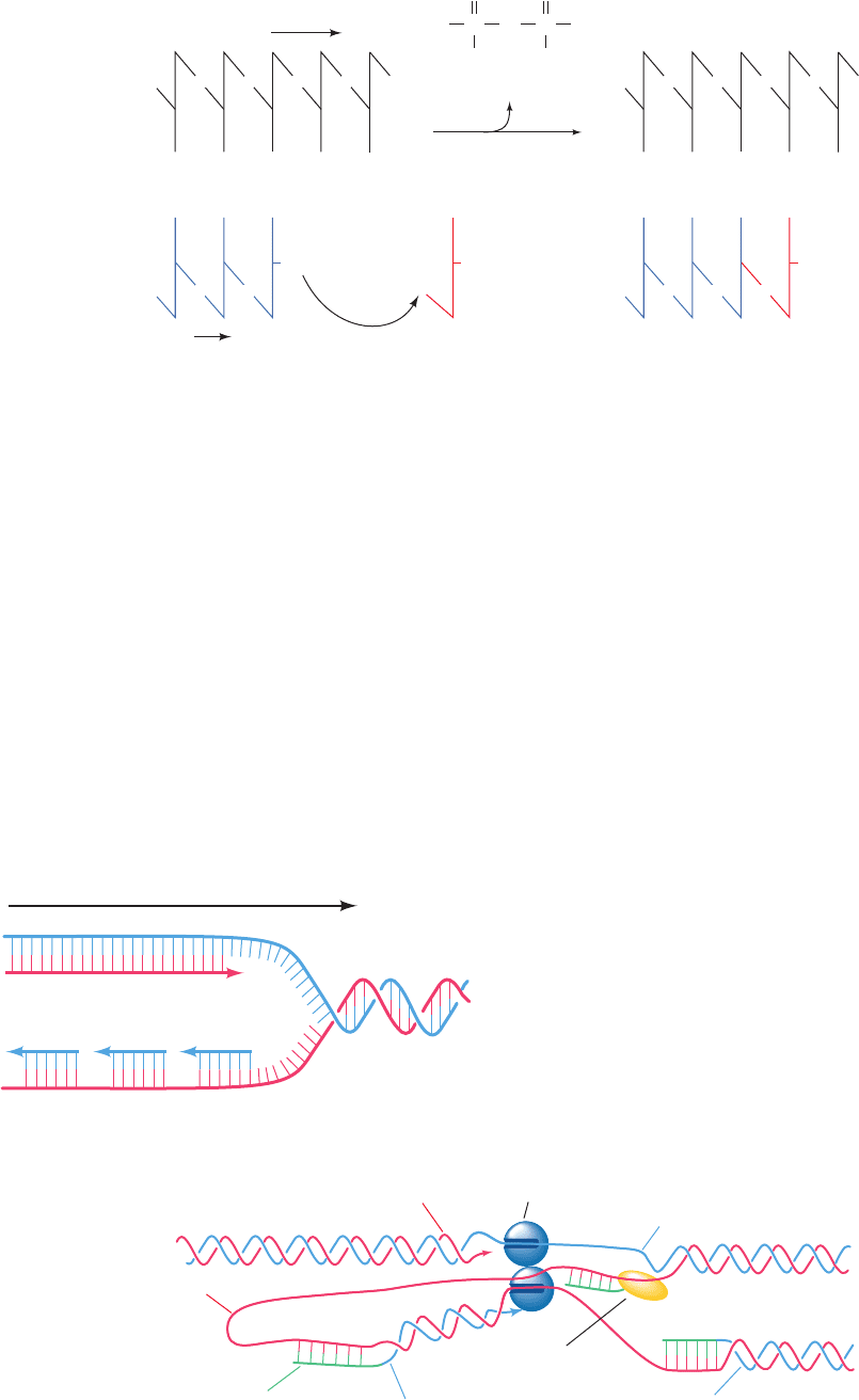
The chemical reaction by which DNA is replicated (Fig.
5-31) is nearly identical to that synthesizing RNA (Fig. 5-23),
but with two major differences: (1) deoxynucleoside
triphosphates (dNTPs) rather than nucleoside triphos-
phates are the reactants and (2) the enzyme that catalyzes
the reaction is DNA polymerase rather than RNA poly-
merase. The properties of DNA polymerase result in a
third major difference between RNA and DNA synthesis:
Whereas RNA polymerase can link together two nu-
cleotides on a DNA template, DNA polymerase can only
extend (in the 5¿ to 3¿ direction) an existing polynucleotide
that is base paired to the DNA’s template strand. Thus,
whereas RNA polymerase can initiate RNA synthesis de
novo (from the beginning), DNA polymerase requires an
oligonucleotide primer, which it lengthens.
a. Primers Are RNA
If DNA polymerase cannot synthesize DNA de novo,
where do primers come from? It turns out that they are not
DNA, as might be expected, but rather RNA.In E. coli, these
RNA primers are synthesized by both RNA polymerase (the
same enzyme that synthesizes all other RNAs) and by a spe-
cial RNA polymerase known as primase. DNA polymerase
then extends this RNA primer, which is eventually excised
and replaced by DNA, as is explained below.This extra com-
plexity in DNA synthesis increases the fidelity of DNA repli-
cation. Whereas a cell makes many copies of an RNA and
Section 5-4. Gene Expression and Replication: An Overview 101
Third reading frame start
Second reading frame start
First reading frame start
Third reading frame
Second reading frame
First reading frame
. . .
GUUCA GCCUA AAG
. . .
. . .
Val
Phe
Ser
Gln
Ser
Ala
Pro
Leu
Stop
Lys
Arg
Figure 5-30 Nucleotide reading frames. An mRNA might be
read in any of three different reading frames, each of which
yields a different polypeptide.
JWCL281_c05_082-128.qxd 2/19/10 4:46 PM Page 101
hence can tolerate an occasional mistake in its synthesis, a
mistake (mutation) in the synthesis of DNA, the archive of
genetic information, may be passed on to all of the cell’s de-
scendants. Since a Watson–Crick base pair is partially stabi-
lized by its neighboring base pairs (a cooperative interac-
tion), the first few base pairs that are formed in a newly
synthesized polynucleotide will initially be less stable than
the base pairs that are formed later. Consequently, these first
few bases are more likely to be erroneously incorporated due
to mispairing than those at the end of a longer chain. If a
primer were DNA, there would be no way to differentiate it
from other DNA so as to selectively replace it with more ac-
curately synthesized DNA. Since the primer is RNA, how-
ever, it is readily identified and replaced.
b. DNA’s Two Strands Are Replicated in
Different Ways
A fourth major difference between RNA and DNA syn-
thesis is that, whereas only one DNA strand at a time is
transcribed, in most cases both of its strands are simultan-
eously replicated. This takes place at a replication fork, the
junction where the two strands of the parental DNA are
pried apart and where the two daughter strands are synthe-
sized (Fig. 1-17), each by a different molecule of DNA poly-
merase. One of these DNA polymerase molecules continu-
ously copies the parental strand that extends in its 3¿ to 5¿
direction from the replication fork, thereby synthesizing the
resulting daughter strand, which is known as the leading
strand, in its 5¿ to 3¿ direction. However, since the second
102 Chapter 5. Nucleic Acids, Gene Expression, and Recombinant DNA Technology
Figure 5-31 Action of DNA polymerases. DNA polymerases
assemble incoming deoxynucleoside triphosphates on single-
DNA polymerase
...
5′ Replicated DNA
Pyrophosphate ion
3′
B
1
′ B
2
′ B
4
′
B
4
B
3
′
B
1
B
2
OH
B
3
B
5
′
OH
+
ppp
p
pp
...
...
p
p
p p pp
.
.
.
.
.
.
.
.
.
3′
Template DNA
5′
...
B
1
′ B
2
′ B
4
′
B
4
B
3
′
B
1
B
2
B
3
B
5
′
OH
p
pp
...
...
p
p p
p p pp
.
.
.
.
.
.
.
.
.
.
.
.
O
P
–
O
O
–
O
PO
O
–
O
–
Figure 5-32 Replication of duplex DNA in E. coli. (a) Since
the two DNA polymerase molecules at the replication fork are
linked together and DNA polymerase can only synthesize DNA
in its 5¿ to 3¿ direction, the leading strand can be synthesized con-
tinuously but the lagging strand must be synthesized discontinu-
ously, that is, in segments. (b) This is because the lagging strand
template can only be copied if it loops around so as to feed through
the DNA polymerase in its 3¿ to 5¿ direction. Consequently, when
the DNA polymerase that is synthesizing the lagging strand en-
counters the previously synthesized lagging strand segment, it
releases the lagging strand template and rebinds to it farther up-
stream so as to extend the next RNA primer to be synthesized.
3′
5′
3′
5′
3′
5′
5′
3′
Leading strand
Lagging strand
Parental strands
Motion of
replication
fork
(a)
5′
3′
Lagging
strand
template
Leading
strand
Leading
strand
template
DNA
polymerases
RNA primer
5′
3′
5′
3′
Newly initiated lagging
strand segment
Previously synthesized
lagging strand segment
Primase synthesizing
new RNA primer
Parental strands
(b)
stranded DNA templates such that the growing strand is elon-
gated in the 5¿ to 3¿ direction.
JWCL281_c05_082-128.qxd 5/31/10 2:02 PM Page 102
DNA polymerase at the replication fork also synthesizes
DNA in the 5¿ to 3¿ direction and yet must travel with the
replication fork, how does it copy the parental strand that
extends from the replication fork in its 5¿ to 3¿ direction?
The answer is that it synthesizes the so-called lagging strand
discontinuously, that is, in pieces (Fig. 5-32a, opposite). It
does so by binding the looped-around lagging strand tem-
plate so as to extend its newly synthesized RNA primer in
its 5¿ to 3¿ direction (Fig. 5-32b; in effect, reversing its direc-
tion of travel) until it encounters the previously synthesized
primer.The DNA polymerase then disengages from the lag-
ging strand template and rebinds to it upstream of its previ-
ous position, where it then extends the next RNA primer to
be synthesized. Thus the lagging strand is synthesized dis-
continuously, whereas the leading strand is synthesized con-
tinuously. The synthesis of lagging strand primers in E. coli
is catalyzed by primase, which accompanies the replication
fork (Fig. 5-32b), whereas the synthesis of leading strand
primers, a much rarer event, occurs most efficiently when
both primase and RNA polymerase are present.
c. Lagging Strand Synthesis Requires
Several Enzymes
Escherichia coli contains two species of DNA polymerase
that are essential for its survival. Of these, DNA polymerase
III (Pol III) is the DNA replicase, that is, it synthesizes the
leading strand and most of the lagging strand. DNA poly-
merase I (Pol I) has a different function, that of removing
the RNA primers and replacing them with DNA. Pol I can
do so because it has a second enzymatic activity besides that
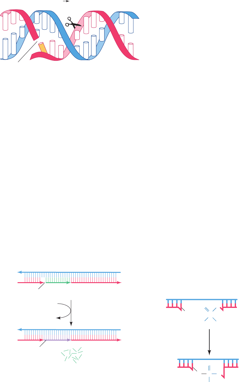
of a DNA polymerase; it is also a 5ⴕ S 3ⴕ exonuclease (an
exonuclease hydrolytically removes one or more nu-
cleotides from the end of a polynucleotide rather than cleav-
ing it at an internal position). The 5¿S3¿ exonuclease func-
tion binds to single-strand nicks (places where successive
nucleotides are not covalently linked such as on the 5¿ side of
an RNA primer after the succeeding lagging strand segment
has been synthesized). It then excises a 1- to 10-nucleotide
segment of the nicked strand in the 5¿ to 3¿ (5¿S3¿) direc-
tion past the nick (Fig. 5-33). Pol I’s 5¿S3¿ exonuclease and
DNA polymerase activities work in concert, so as Pol I’s
5¿S3¿ exonuclease removes the primer, its DNA polymerase
activity replaces this RNA with DNA (Fig. 5-34).
The synthesis of the leading strand is completed by the
replacement of its single RNA primer with DNA. How-
ever, the completion of lagging strand synthesis requires
that the nicks between its multiple discontinuously synthe-
sized segments be sealed.This is the job of an independent
enzyme named DNA ligase that covalently links adjacent
3¿-OH and 5¿-phosphate groups (Fig. 5-35).
d. Errors in DNA Sequences Are Subject
to Correction
In E. coli, RNA polymerase has an error rate of ⬃1
wrong base for every 10
4
nucleotides it transcribes. In con-
trast, newly replicated DNA contains only ⬃1 error per 10
8
to 10
10
base pairs. We have already seen that the use of
RNA primers increases the fidelity of lagging strand syn-
thesis. However, the main reason for the enormous fidelity
of DNA replication is that both Pol I and Pol III have
3ⴕ S 5ⴕ exonuclease activities. The 3¿S5¿ exonuclease
Section 5-4. Gene Expression and Replication: An Overview 103
3′
5′
5′
3′
Nick
RNA
primer
DNA
Template DNA
Ribonucleotides
3′
5′
5′
3′
Newly synthesized
DNA
+
DNA polymerase I
dNTPs
pyrophosphate ion
Figure 5-34 Replacement of RNA primers by DNA in lagging
strand synthesis. In E. coli, the RNA primer on the 5¿ end of a
newly synthesized DNA segment is excised through the action of
DNA polymerase I’s 5¿S3¿ exonuclease activity and is simulta-
neously replaced by DNA as catalyzed by the enzyme’s DNA
polymerase activity.
O
–
OH
P
–
O
OO
PO
O
O
O
–
ATP
+
AMP
++
DNA ligase
P
2
O
7
4–
Figure 5-35 Function of DNA ligase. DNA ligase seals single-
strand nicks in duplex DNA. It does so in a reaction that is pow-
ered by the hydrolysis of ATP or a similar compound.
C
G
T
...
A
5′ 3′ Exonuclease
hydrolysis site
Sin
g
le-strand nick
G
A
T
C
A
A
T
C
G
A
T
C
T
C
A
G
G
C
5′
3′ 3′
5′
p
...
...
...
...
...
...
...
...
...
X
...
...
G
A
T
Figure 5-33 The 5ⴕ S 3ⴕ exonuclease function of DNA
polymerase I. This enzymatic activity excises up to 10 nucleotides
from the 5¿ end of a single-strand nick.The nucleotide
immediately past the nick (X) may or may not be base paired.
JWCL281_c05_082-128.qxd 2/19/10 4:46 PM Page 103
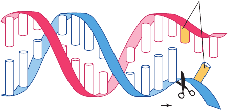
degrades the newly synthesized 3¿ end of a daughter strand
one nucleotide at a time (Fig. 5-36), thereby annulling the
polymerase reaction. This enzymatic function is activated
by non-Watson–Crick base pairing and consequently acts
to edit out the occasional mistakes made by the poly-
merase function, thereby greatly increasing the fidelity of
replication. However, in addition to this proofreading func-
tion on both Pol I and Pol III, all cells contain batteries of
enzymes that detect and correct residual errors in replica-
tion as well as damage which DNA incurs through the ac-
tion of such agents as UV radiation and mutagens (sub-
stances that damage DNA by chemically reacting with it)
as well as by spontaneous hydrolysis (Section 30-5). In E.
coli, Pol I also functions to replace the damaged DNA seg-
ments that these enzymes have excised.
5 MOLECULAR CLONING
A major problem in almost every area of biochemical re-
search is obtaining sufficient quantities of the substance of
interest. For example, a 10-L culture of E. coli grown to its
maximum titer of ⬃10
10
cells ⴢ mL
⫺1
contains, at most, 7 mg
of DNA polymerase I, and many of its proteins are present
in far lesser amounts. Yet it is rare that even as much as half
of any protein originally present in an organism can be re-
covered in pure form (Chapter 6). Eukaryotic proteins may
be even more difficult to obtain because many eukaryotic
tissues, whether acquired from an intact organism or grown
in tissue culture, are available only in small quantities.As far
as the amount of DNA is concerned, our 10-L E. coli culture
would contain ⬃0.1 mg of any 1000-bp length of chromoso-
mal DNA (a length sufficient to contain most prokaryotic
genes), but its purification in the presence of the rest of the
chromosomal DNA (which consists of 4.6 million bp) would
be an all but impossible task. These difficulties have been
largely eliminated through the development of molecular
cloning techniques (a clone is a collection of identical organ-
isms that are derived from a single ancestor). These meth-
ods, which are also referred to as genetic engineering and re-
combinant DNA technology, deserve much of the credit for
the enormous progress in biochemistry and the dramatic rise
of the biotechnology industry since the late 1970s.
The main idea of molecular cloning is to insert a DNA
segment of interest into an autonomously replicating DNA
molecule, a so-called cloning vector or vehicle, so that the
DNA segment is replicated with the vector. Cloning such a
chimeric vector (chimera: a monster in Greek mythology
that has a lion’s head, a goat’s body, and a serpent’s tail) in
a suitable host organism such as E. coli or yeast results in
the production of large amounts of the inserted DNA seg-
ment.If a cloned gene is flanked by the properly positioned
control sequences for transcription and translation, the
host may also produce large quantities of the RNA and
protein specified by that gene. The techniques of genetic
engineering, whose understanding is prerequisite to under-
standing many of the experiments discussed in this text-
book, are outlined in this section.
A. Restriction Endonucleases
In order to effectively carry out molecular cloning,it is nec-
essary to be able to manipulate precisely sequence-defined
DNA fragments. This is done through the use of enzymes
known as restriction endonucleases.
Bacteriophages that propagate efficiently on one bacter-
ial strain, such as E. coli K12, have a very low rate of infec-
tion (⬃0.001%) in a related bacterial strain such as E. coli
B. However, the few viral progeny of this latter infection
propagate efficiently in the new host but only poorly in the
original host. Evidently, the new host modifies these bacte-
riophages in some way. What is the molecular basis of this
host-specific modification? Werner Arber showed that it re-
sults from a restriction–modification system in the bacterial
host, which consists of a restriction endonuclease (alterna-
tively, restriction enzyme; endonucleases are enzymes that
hydrolytically cleave polynucleotides at internal sites) and a
matched DNA methyltransferase. Restriction endonucle-
ases recognize a specific base sequence of four to eight bases
in double-stranded DNA and cleave both strands of the du-
plex. DNA methyltransferases methylate a specific base (at
the amino group of an adenine or either the 5 position or
the amino group of a cytosine) in the same base sequence
recognized by the matched restriction enzyme.
A restriction enzyme does not cleave its corresponding
methylated DNA. A newly replicated strand of bacterial
DNA, which is protected from degradation by the methy-
lated parent strand with which it forms a duplex, is
methylated before the next cycle of replication. A restric-
tion–modification system therefore protects the bacterium
against invasion by foreign (usually viral) DNAs which,
once they have been cleaved by a restriction endonuclease,
are further degraded by bacterial exonucleases. Invading
DNAs are only rarely methylated before being attacked by
restriction enzymes. Yet if a viral DNA does become
methylated, it is able to reproduce in its new host. Its prog-
eny, however,are no longer methylated in the way that per-
mits them to propagate in the original host (which has dif-
ferent restriction–modification systems).
There are four types of restriction endonucleases, Types
I, II, III, and IV. Type I and Type III restriction enzymes
104 Chapter 5. Nucleic Acids, Gene Expression, and Recombinant DNA Technology
G
A
T
G
A
...
G
3′ 5′
Exonuclease
hydrolysis site
Mismatched
bases
p
G
C
T
T
C
A
A
C
T
A
G
G
OH
C
T
C
T
T
G
C
5′
3′
3′
5′
...
...
...
...
...
...
...
...
...
...
C
Figure 5-36 The 3ⴕ S 5ⴕ exonuclease function of DNA poly-
merase I and DNA polymerase III. In E. coli, this enzymatic ac-
tivity excises mispaired nucleotides from the 3¿ end of a growing
DNA strand.
JWCL281_c05_082-128.qxd 2/19/10 4:46 PM Page 104

each carry both the endonuclease and the DNA methyl-
transferase activity on a single protein molecule. Type I re-
striction enzymes cleave the DNA at a possibly random
site located at least 1000 bp from the recognition sequence,
Type III enzymes do so 24 to 26 bp distant from the recog-
nition sequence, and Type IV enzymes cleave methylated
DNA. Type II restriction enzymes, which were discovered
and characterized by Hamilton Smith and Daniel Nathans
in the late 1960s, are separate entities from their correspon-
ding DNA methyltransferases. They cleave DNAs at spe-
cific sites within or near the recognition sequence,a property
that makes Type II restriction enzymes indispensable bio-
chemical tools for DNA manipulation. In what follows, we
discuss only Type II restriction enzymes.
Nearly 4000 species of Type II restriction enzymes from
a variety of bacteria that have over 270 different sequence
specificities have been characterized. Several of the more
widely used species are listed in Table 5-4.A restriction en-
donuclease is named by the first letter of the genus of the
bacterium that produced it and the first two letters of its
species, followed by its serotype or strain designation, if
any, and a roman numeral if the bacterium expresses more
than one type of restriction enzyme. For example, EcoRI is
produced by E. coli strain RY13.
a. Most Restriction Endonucleases Recognize
Palindromic DNA Sequences
Most Type II restriction enzyme recognition sites pos-
sess exact twofold rotational symmetry, as is diagrammed
in Fig. 5-37. Such sequences are known as palindromes (a
palindrome is a word, verse, or sentence that reads the
same backward and forward; two examples are “Madam,
I’m Adam” and “Sex at noon taxes”). Many restriction en-
zymes, such as EcoRI (Fig. 5-37a), catalyze cleavage of the
two DNA strands at positions that are symmetrically stag-
gered about the center of the palindromic recognition se-
quence. This yields restriction fragments with complemen-
tary single-stranded ends that are from one to four
nucleotides in length. Restriction fragments with such co-
hesive or sticky ends can associate by complementary base
pairing with other restriction fragments generated by the
same restriction enzyme. Some restriction cuts, such as that
of EcoRV (Fig. 5-37b), pass through the twofold axis of the
Section 5-5. Molecular Cloning 105
Figure 5-37 Restriction sites. The recognition sequences of the
restriction endonucleases (a) EcoRI and (b) EcoRV have
twofold (palindromic) symmetry (red symbol).The cleavage sites
are indicated (arrows). Note that EcoRI generates DNA
fragments with sticky ends, whereas EcoRV generates
blunt-ended fragments.
(a)
A
.
.
.
T
A
.
.
.
T
G
.
.
.
C
C
.
.
.
G
T
.
.
.
A
T
.
.
.
A
3′
5′
(b)
Cleavage site Twofold symmetry axis
EcoRI EcoRV
5′
3′
3′
5′
G
.
.
.
C
A
.
.
.
T
A
.
.
.
T
T
.
.
.
A
T
.
.
.
A
C
.
.
.
G
5′
3′
Table 5-4 Recognition and Cleavage Sites of Some Type II Restriction Enzymes
Enzyme Recognition Sequence
a
Microorganism
AluI AGTC*T Arthrobacter luteus
BamHI GTGATC*C Bacillus amyloliquefaciens H
BglI GCCNNNNTNGCC Bacillus globigii
BglII ATGATCT Bacillus globigii
EcoRI GTAA*TTC Escherichia coli RY13
EcoRII TCC*(
A
T
)GG Escherichia coli R245
EcoRV GA*TTATC Escherichia coli J62 pLG74
HaeII RGCGCTY Haemophilus aegyptius
HaeIII GGTC*C Haemophilus aegyptius
HindIII A*TAGCTT Haemophilus influenzae R
d
HpaII CTC*GG Haemophilus parainfluenzae
MspI C*TCGG Moraxella species
PstI CTGCA*TG Providencia stuartii 164
PvuII CAGTC*TG Proteus vulgaris
SalI GTTCGAC Streptomyces albus G
TaqI TTCGA* Thermus aquaticus
XhoI CTTCGAG Xanthomonas holcicola
a
The recognition sequence is abbreviated so that only one strand, reading 5¿ to 3¿, is given.The cleavage site is
represented by an arrow (
T) and the modified base, where it is known, is indicated by a following asterisk (A*
is N
6
-methyladenine and C* is 5-methylcytosine). R, Y, and N represent purine nucleotide, pyrimidine
nucleotide, and any nucleotide, respectively.
Source: REBASE. The restriction enzyme database (http://rebase.neb.com).
JWCL281_c05_082-128.qxd 2/19/10 4:46 PM Page 105
palindrome to yield restriction fragments with fully base
paired blunt ends. Since a given base has a one-fourth
probability of occurring at any nucleotide position (assum-
ing the DNA has equal proportions of all bases), a restric-
tion enzyme with an n-base pair recognition site produces
restriction fragments that are, on average, 4
n
base pairs
long. Thus AluI (4-bp recognition sequence) and EcoRI
(6-bp recognition sequence) restriction fragments should
average 4
4
⫽ 256 and 4
6
⫽ 4096 bp in length, respectively.
b. Restriction-Fragment Length Polymorphisms
Provide Markers for Characterizing Genes
The treatment of a DNA molecule with a restriction en-
donuclease produces a series of precisely defined fragments
that can be separated according to size by gel electrophore-
sis (Fig. 5-38). (In gel electrophoresis, charged molecules are
applied to one end of a thin slab of polyacrylamide or
agarose gel and are separated through the application of an
electric field. Under the conditions used to separate DNA
fragments, the molecules move according to size, with the
smallest fragments moving fastest. Gel electrophoresis is
further discussed in Section 6-4B.) Complementary single
strands can be separated either by melting the DNA and
subjecting it to gel electrophoresis, or by using density gra-
dient ultracentrifugation in alkaline CsCl solution (recall
that DNA is denatured under alkaline conditions).
Individuality in humans and other species derives from
their genetic polymorphism; homologous human chromo-
somes differ in sequence, on average, every ⬃1250 bp.
These genetic differences create and eliminate restriction
sites (Fig. 5-39). Restriction enzyme digests of the corre-
sponding segments from homologous chromosomes there-
fore contain fragments with different lengths; that is, these
DNAs have restriction-fragment length polymorphisms
(RFLPs; Fig. 5-40). Since, with the exception of identical
twins, each individual has a unique set of RFLPs (its haplo-
type), RFLPs can be used for purposes of identification.
B. Cloning Vectors
Plasmids, viruses, and artificial chromosomes are used as
cloning vectors in genetic engineering.
a. Plasmid-Based Cloning Vectors
Plasmids are circular DNA duplexes of 1 to 200 kb that
contain the requisite genetic machinery, such as a replica-
tion origin (a site at which DNA replication is initiated;
Section 30-3Ca), to permit their autonomous propagation
in a bacterial host or in yeast. Plasmids may be considered
molecular parasites but in many instances they benefit
106 Chapter 5. Nucleic Acids, Gene Expression, and Recombinant DNA Technology
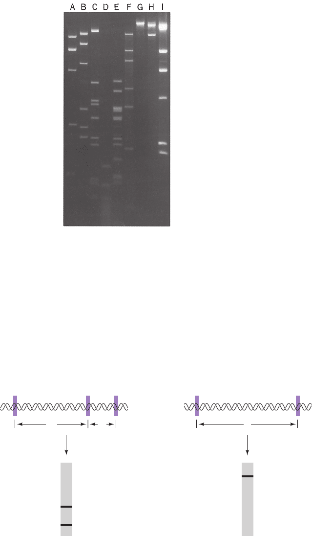
Figure 5-38 Agarose gel electrophoretogram of restriction
digests. The Agrobacterium radiobacter plasmid pAgK84 was
digested with (A) BamHI, (B) PstI, (C) BglII, (D) HaeIII, (E)
HincII, (F) SacI, (G) XbaI, and (H) HpaI. Lane I contains
phage DNA digested with HindIII as standards since these frag-
ments have known sizes.The DNA fragments in the elec-
trophoretogram are made visible by fluorescence against a black
background. [From Slota, J.E. and Farrand, S.F., Plasmid 8, 180
(1982). Copyright © 1982 by Academic Press.]
Figure 5-39 Restriction-fragment length
polymorphisms. A mutational change that
affects a restriction site in a DNA segment
alters the number and sizes of its restriction
fragments.
Cleave with
restriction enzyme
and electrophorese
Chromosome I
DNA has
3 target sites
Chromosome II
DNA has only
2 of the target sites
Fragment C is
the size of
A + B combined
A
B
CB
A
C
123 1 3
JWCL281_c05_082-128.qxd 2/19/10 4:46 PM Page 106
their host by providing functions, such as resistance to an-
tibiotics, that the host lacks. Indeed, the widespread and
alarming appearance, since antibiotics came into use,of an-
tibiotic-resistant pathogens is partially the result of the
rapid proliferation among these organisms of plasmids
containing genes that confer resistance to antibiotics.
Some types of plasmids, which are present in one or a
few copies per cell, replicate once per cell division as does
the bacterial chromosome; their replication is said to be un-
der stringent control. Most plasmids used in molecular
cloning, however, are under relaxed control; they are nor-
mally present in 10 to as many as 700 copies per cell. More-
over, if protein synthesis in the bacterial host is inhibited,
for example, by the antibiotic chloramphenicol (Section 32-
3Gb), thereby preventing cell division, these plasmids con-
tinue to replicate until 2 or 3 thousand copies have accumu-
lated per cell (which represents about half of the cell’s total
DNA).The plasmids that have been constructed (by genetic
engineering techniques; Section 5-5C) for use in molecular
cloning are relatively small, replicate under relaxed control,
carry genes specifying resistance to one or more antibiotics,
and contain a number of conveniently located restriction
endonuclease sites into which the DNA to be cloned may
be inserted. Indeed, many plasmid vectors contain a strate-
gically located short (⬍100 bp) segment of DNA known as
a polylinker that has been synthesized to contain a variety
of restriction sites that are not present elsewhere in the
plasmid.The E.coli plasmid designated pUC18 (Fig. 5-41) is
representative of the cloning vectors presently in use
(“pUC” stands for “plasmid-Universal Cloning”).
The expression of a chimeric plasmid in a bacterial host
was first demonstrated in 1973 by Herbert Boyer and Stan-
ley Cohen.The host bacterium takes up a plasmid when the
two are mixed together in a process that is greatly en-
hanced by the presence of divalent cations such as Ca
2⫹
Section 5-5. Molecular Cloning 107
pUC18
(2.69 kb)
XmnI
ScaI
PvuI
A
vaII
AvaII
HgiEII
Af lIII
PvuII
PvuII
PvuI
MstI
MstI
BglI
BglI
NarI
NdeI, HgiEII
EcoO109
AatII
SspI
1000
2000
Polylinker
lacI
lacZ'
amp
R
0
Figure 5-41 The pUC18 cloning vector. A
restriction map of the plasmid pUC18 indicates
the positions of its amp
R
, lacZⴕ, and lacI genes.
The amp
R
gene confers resistance to the antibi-
otic ampicillin (a penicillin derivative; Section
11-3Bb); lacZ¿ is a modified form of the lacZ
gene, which encodes the enzyme -galactosi-
dase (Section 11-2B); and lacI encodes the lac
repressor, a protein that controls the transcrip-
tion of lacZ (Section 5-4Aa).The polylinker,
which encodes an 18-residue polypeptide
segment inserted near the N-terminus of
-galactosidase, incorporates 13 different
restriction sites that do not occur elsewhere
in the plasmid.
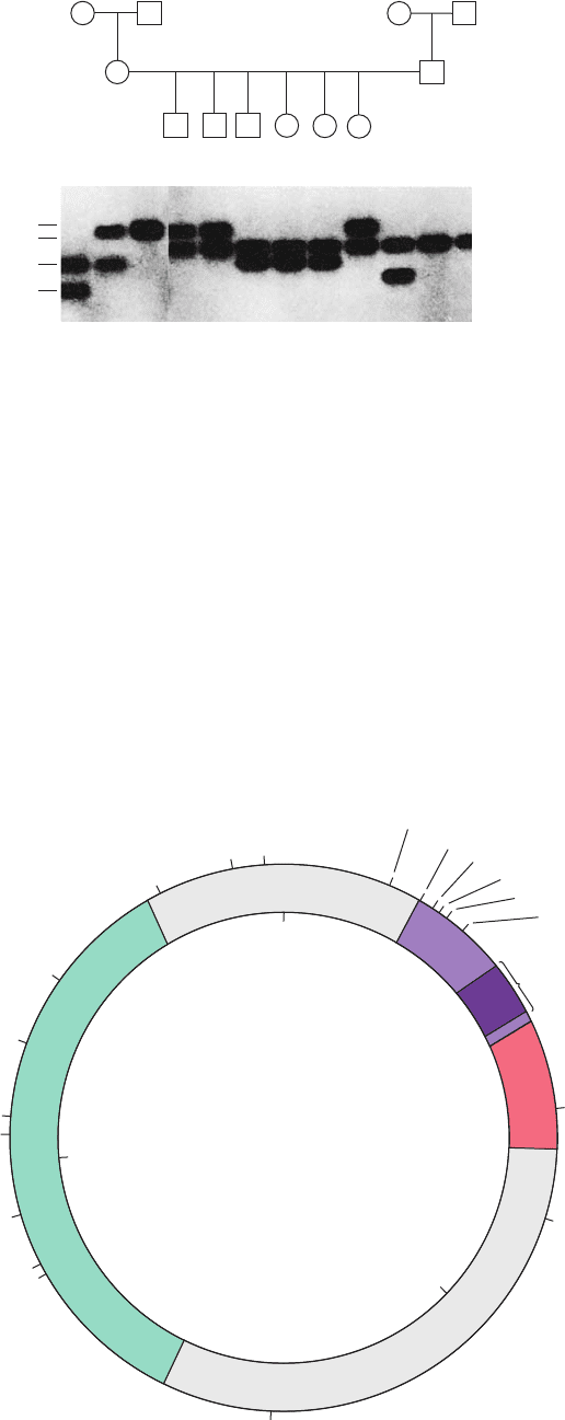
Figure 5-40 Inheritance of RFLPs according to the rules of
Mendelian genetics. Four alleles of a particular gene, each char-
acterized by different restriction markers, can occur in all possi-
ble pairwise combinations and segregate independently in each
generation (circles in the upper panel represent females and
squares represent males). In the P (parental) generation, two in-
dividuals have heterozygous haplotypes (CD and BD) and the
other two have homozygous haplotypes (AA and BB).Their
children, the F
1
generation, have the haplotypes AC or BB. Con-
sequently, every individual in the F
2
generation (grandchildren)
inherited either an A or a C from their mother and a B from
their father.The lower panel shows a gel electrophoretogram of
these restriction fragments run in parallel lanes. [Courtesy of Ray
White, University of Utah Medical School.]
Pedigree and genotypes
P
CD AA
Alleles
F
1
AC
F
2
BB
BD
BB
AB AB BC BC
BC
AB
A
B
C
D
JWCL281_c05_082-128.qxd 2/19/10 4:46 PM Page 107
and brief heating to ⬃42°C (which increases cell mem-
brane permeability to DNA; such cells are said to be trans-
formation competent). Nevertheless, an absorbed plasmid
vector becomes permanently established in its bacterial
host (transformation) with an efficiency of only ⬃0.1%.
Plasmid vectors cannot be used to clone DNAs of more
than ⬃10 kb. This is because the time required for plasmid
replication increases with plasmid size. Hence intact plas-
mids with large, unessential (to them) inserts are lost
through the faster proliferation of plasmids that have elim-
inated these inserts by random deletions.
b. Virus-Based Cloning Vectors
Bacteriophage (Fig. 5-42) is an alternative cloning vehi-
cle that can be used to clone DNAs of up to 16 kb.The cen-
tral third of this virus’s 48.5-kb genome is not required for
phage infection (Section 33-3Aa) and can therefore be re-
placed by foreign DNAs of up to slightly greater size using
techniques discussed in Section 5-5C. The chimeric phage
DNA can then be introduced into the host cells by infecting
them with phages formed from the DNA by an in vitro pack-
aging system (Section 33-3Bc).The use of phages as cloning
vectors has the additional advantage that the chimeric DNA
is produced in large amounts and in easily purified form.
l Phages can be used to clone even longer DNA inserts.
The viral apparatus that packages DNA into phage heads
requires only that the DNA have a specific 16-bp sequence
known as a cos site located at each end and that these ends
be 36 to 51 kb apart (Section 33-3Bc). Placing two cos sites
the proper distance apart on a plasmid vector yields, via an
in vitro packaging system, a so-called cosmid vector, which
can contain foreign DNA of up to ⬃49 kb. Cosmids have
no phage genes and hence, on introduction into a host cell
via phage infection, reproduce as plasmids.
The filamentous bacteriophage M13 (Fig. 5-43) is also a
useful cloning vector.It has a single-stranded circular DNA
that is contained in a protein tube composed of ⬃2700 he-
lically arranged identical protein subunits. This number is
controlled, however, by the length of the phage DNA being
coated; insertion of foreign DNA in a nonessential region
of the M13 chromosome results in the production of longer
phage particles. Although M13 cloning vectors cannot sta-
bly maintain DNA inserts of ⬎1 kb,they are widely used in
the production of DNA for sequence analysis (Section 7-
2Ba) because these phages directly produce the single-
stranded DNA that the technique requires.
Baculoviruses are a large and diverse group of pathogenic
viruses that infect mainly insects (but not vertebrates, so that
they are safe for laboratory use) and hence can be grown in
cultures of insect cells. A segment of the double-stranded
DNA that forms the genome of some of these viruses is un-
necessary for viral replication in tissue cultures of insect cells
and hence can be replaced by a foreign DNA of up to 15 kb.
c. YAC and BAC Vectors
DNA segments larger than those that can be carried by cos-
mids may be cloned in yeast artificial chromosomes (YACs)
and in bacterial artificial chromosomes (BACs). YACs are
linear DNA segments that contain all the molecular para-
phernalia required for replication in yeast: a replication
108 Chapter 5. Nucleic Acids, Gene Expression, and Recombinant DNA Technology
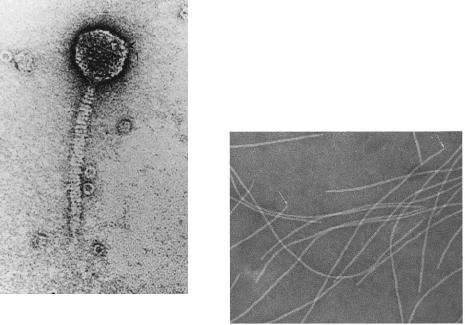
Figure 5-42 Electron micrograph of bacteriophage . Bac-
teriophage reproduces in certain strains of E. coli. On binding
to a susceptible E. coli, the DNA contained in the “head” of the
phage particle is injected, through its “tail,” into the bacterial
cell, where it is replicated ⬃100 times and packaged to form
progeny phage (Section 33-3). [Courtesy of A.F. Howatson. From
Lewin, B., Gene Expression,Vol. 3, Fig. 5.23, Wiley (1977).]
Figure 5-43 Electron micrograph of the filamentous bacterio-
phage M13. Note that some filaments appear to be pointed at
one end (arrows). [Courtesy of Robley Williams, Stanford
University, and Harold Fisher, University of Rhode Island.]
JWCL281_c05_082-128.qxd 2/19/10 4:46 PM Page 108
origin [known as an autonomously replicating sequence
(ARS)], a centromere (the chromosomal segment that at-
taches to the spindle during mitosis and meiosis), and telom-
eres (the ends of linear chromosomes that permit their repli-
cation; Section 30-4D). BACs, which replicate in E. coli, are
derived from circular plasmids that normally replicate long
regions of DNA and are maintained at a level of approxi-
mately one copy per cell (properties similar to those of ac-
tual chromosomes). These vectors contain the minimal se-
quences required for autonomous replication, copy-number
control, and proper partitioning of the plasmid during cell
division. YACs and BACs containing inserts of several hun-
dred kilobase pairs have been successfully cloned.
C. Gene Manipulation
A DNA to be cloned is, in many cases, obtained as a se-
quence-defined fragment through the application of restric-
tion endonucleases (for M13 vectors, the restriction en-
zymes’ requirement of duplex DNA necessitates
converting this phage DNA to its double-stranded form
through the use of DNA polymerase I). Recall that most
restriction endonucleases cleave duplex DNA at specific
palindromic sites so as to yield single-stranded ends that
are complementary to each other (cohesive or sticky ends;
Section 5-5Aa). Therefore, as Janet Mertz and Ron Davis
first demonstrated in 1972, a restriction fragment may be in-
serted into a cut made in a cloning vector by the same restric-
tion enzyme (Fig. 5-44). The complementary (cohesive)
ends of the two DNAs specifically associate under annealing
conditions and are covalently joined (spliced) through the
action of DNA ligase (Fig. 5-35; the DNA ligase produced
by bacteriophage T4 must be used for blunt-ended restric-
tion cuts such as those generated by AluI, EcoRV, and
HaeIII; Table 5-4). A great advantage of using a restriction
enzyme to construct a chimeric vector is that the DNA insert
can be precisely excised from the cloned vector by cleaving
it with the same restriction enzyme.
If the foreign DNA and cloning vector have no common
restriction sites at innocuous positions, they may still be
spliced, using a procedure pioneered by Dale Kaiser and Paul
Berg, through the use of terminal deoxynucleotidyl trans-
ferase (terminal transferase). This mammalian enzyme adds
nucleotides to the 3¿-terminal OH group of a DNA chain; it is
the only known DNA polymerase that does not require a
template. Terminal transferase and dTTP, for example, can
build up poly(dT) tails of ⬃100 residues on the 3¿ ends of the
DNA segment to be cloned (Fig. 5-45). The cloning vector is
enzymatically cleaved at a specific site and the 3¿ ends of the
cleavage site are similarly extended with poly(dA) tails. The
complementary homopolymer tails are annealed,any gaps re-
sulting from differences in their lengths filled in by DNA poly-
merase I, and the strands joined by DNA ligase.
A disadvantage of the above technique is that it elimi-
nates the restriction sites that were used to generate the
foreign DNA insert and to cleave the vector. It may there-
fore be difficult to recover the insert from the cloned vec-
tor.This difficulty is circumvented by a technique in which
a chemically synthesized palindromic “linker” having a re-
striction site matching that of the cloning vector is ap-
pended to both ends of the foreign DNA (the chemical
Section 5-5. Molecular Cloning 109
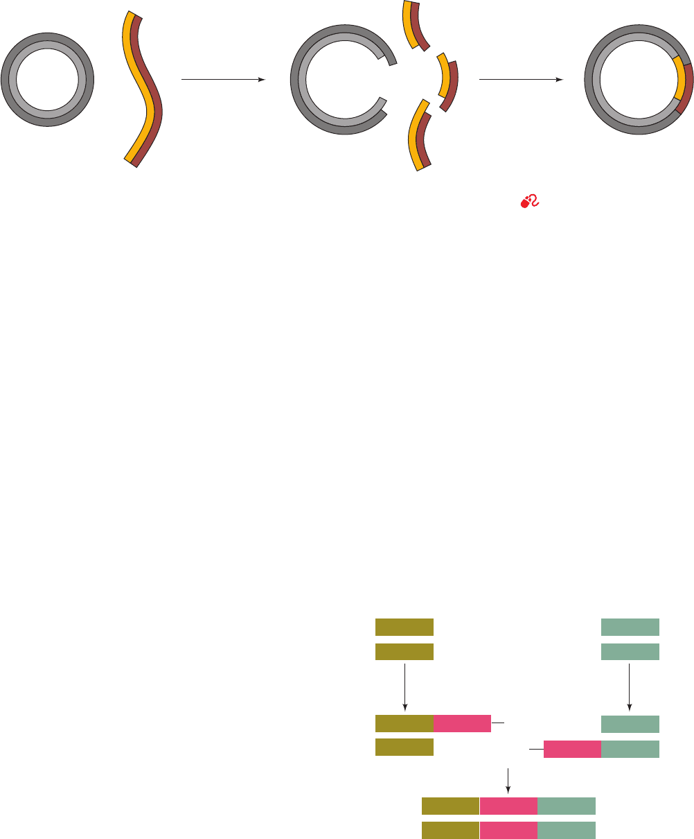
Figure 5-44 Construction of a recombinant DNA molecule. A
restriction fragment is inserted in a cloning vector’s correspon-
ding restriction cut.The sticky ends of the vector and the foreign
Cloning
vector
Chimeric
DNA
Anneal foreign
DNA fragment
to cloning vector
and ligate
restriction
endonuclease
Foreign DNA
++
DNA to be cloned Cloning vector
dATP dTTP
terminal
transferase
terminal
transferase
OH
OH
Anneal and ligate
Recombinant DNA
5' 3'
5'
5'
3'
5'
5'
3'
3'
3'
5'
3'
AAAAAA
TTTTTT
AAAAAA
TTTTTT
+
+
Figure 5-45 Splicing DNA using terminal transferase. Tw o
DNA fragments may be joined through the generation of
complementary homopolymer tails via the action of the enzyme
terminal transferase. The poly(dA) and poly(dT) tails shown
in this example may be replaced by poly(dC) and poly(dG) tails.
DNA anneal and are subsequently covalently joined by DNA
ligase to yield a chimeric DNA.
See the Animated Figures
JWCL281_c05_082-128.qxd 2/19/10 4:46 PM Page 109
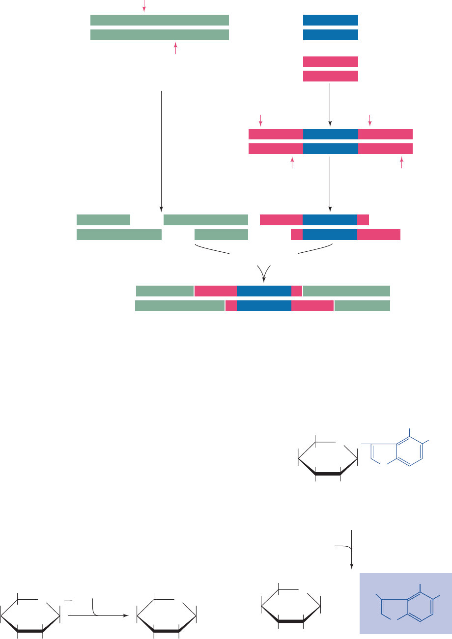
synthesis of oligonucleotides is discussed in Section 7-6A).
The linker is attached to the foreign DNA by blunt end lig-
ation with T4 DNA ligase and then cleaved with the appro-
priate restriction enzyme to yield the correct cohesive ends
for ligation to the vector (Fig. 5-46).
a. Properly Transformed Cells Must Be Selected
Both transformation and the proper construction of
chimeric vectors occur with low efficiency. How can one se-
lect only those host organisms that have been transformed
by the properly constructed vector? In the case of plasmid
transformation, this is usually done through a double screen
using antibiotics and/or chromogenic (color-producing)
substrates. For example, the pUC18 plasmid contains the
lacZⴕ gene (Fig. 5-41; a modified form of the lac operon’s
Z gene; Fig. 5-25). The lacZ¿ gene encodes the enzyme
-galactosidase, which catalyzes the hydrolysis of the bond
from O1 of the sugar -
D-galactose to a substituent.
O
HOCH
2
H
OH H
HOH
HH
O
HO
H
2
O
β-galactosidase
R
4
1
3
5
2
-D-Galactose
OH
HOCH
2
H
OH H
HOH
HH
O
HO
+ ROH
6
Thus, when grown in the presence of 5-bromo-4-chloro-3-
indolyl--
D-galactoside (commonly known as X-gal), a col-
orless substance which when hydrolyzed by -galactosi-
dase yields a blue product,
5-Bromo-4-chloro-3-indolyl--D-galactoside (X-gal)
(colorless)
O
HOCH
2
H
OH H
HOH
HH
O
HO
N
H
Cl
Br
-D-Galactose 5-Bromo-4-chloro-3-hydroxyindole
(blue)
OH
HOCH
2
H
OH H
HOH
HH
O
HO
N
H
ClCl
BrBr
HOHO
⫹
H
2
O
β-galactosidase
110 Chapter 5. Nucleic Acids, Gene Expression, and Recombinant DNA Technology
Figure 5-46 Construction of a recombinant DNA molecule
through the use of synthetic oligonucleotide adaptors. In this ex-
Foreign DNA
to be cloned
Cloning
vector
Recombinant
DNA
Synthetic
adaptor
T4 DNA ligase
EcoRI
EcoRI
Restriction
sites
Anneal and ligate
CTTAAG
GAATTC
CTTAA G
G AATTC
CTTAA
G
CTTAAG
GAATTC
CTTAAGCTTAAG
GAATTC
CTTAAG
GAATTC
GAATTC
CTTAAG
GAATTC
G
AATTC
+
ample, the adaptor and the cloning vector have EcoRI restriction
sites (red arrows).
JWCL281_c05_082-128.qxd 2/19/10 4:46 PM Page 110
