Voet D., Voet Ju.G. Biochemistry
Подождите немного. Документ загружается.


aromatic side chains, which are characterized by bulk as
well as nonpolarity.
b. Uncharged Polar Side Chains Have Hydroxyl,
Amide, or Thiol Groups
Six amino acids are commonly classified as having un-
charged polar side chains. Serine and threonine bear hy-
droxylic R groups of different sizes. Asparagine and gluta-
mine (Fig. 4-4) have amide-bearing side chains of different
sizes. Tyrosine has a phenolic group, which, together with
the aromatic groups of phenylalanine and tryptophan, ac-
counts for most of the UV absorbance and fluorescence ex-
hibited by proteins (Section 9-1Cb). Cysteine has a thiol
group that is unique among the 20 amino acids in that it of-
ten forms a disulfide bond to another cysteine residue
through the oxidation of their thiol groups (Fig. 4-5). This
disulfide bond has great importance in protein structure: It
can join separate polypeptide chains or cross-link two cys-
teines in the same chain. Two disulfide-linked cysteines are
referred to in the older biochemical literature as the amino
acid cystine because they were originally thought to form a
unique amino acid. However, the discovery that cystine
residues arise through the cross-linking of two cysteine
residues after polypeptide biosynthesis has occurred has
caused the name cystine to become less commonly used.
c. Charged Polar Side Chains May Be Positively or
Negatively Charged
Five amino acids have charged side chains. The basic
amino acids are positively charged at physiological pH val-
ues; they are lysine, which has a butylammonium side
chain, arginine, which bears a guanidino group, and histi-
dine, which carries an imidazolium moiety. Of the 20
␣-amino acids, only histidine, with pK
R
⫽ 6.0, ionizes within
the physiological pH range. At pH 6.0, its imidazole side
group is only 50% charged so that histidine is neutral at the
basic end of the physiological pH range.As a consequence,
histidine side chains often participate in the catalytic reac-
tions of enzymes. The acidic amino acids, aspartic acid and
glutamic acid, are negatively charged above pH 3; in their
ionized state, they are often referred to as aspartate and
glutamate. Asparagine and glutamine are, respectively, the
amides of aspartic acid and glutamic acid.
The allocation of the 20 amino acids among the three
different groups is, of course, somewhat arbitrary. For ex-
ample, glycine and alanine, the smallest of the amino acids,
and tryptophan, with its heterocyclic ring,might just as well
be classified as uncharged polar amino acids. Similarly, ty-
rosine and cysteine, with their ionizable side chains, might
also be thought of as charged polar amino acids, particu-
larly at higher pH’s, whereas asparagine and glutamine are
nearly as polar as their corresponding carboxylates, aspar-
tate and glutamate.
The 20 amino acids vary considerably in their physico-
chemical properties such as polarity, acidity, basicity, aro-
maticity, bulk, conformational flexibility, ability to cross-link,
ability to hydrogen bond, and chemical reactivity. These sev-
eral characteristics, many of which are interrelated, are
largely responsible for proteins’ great range of properties.
Section 4-1. The Amino Acids of Proteins 71
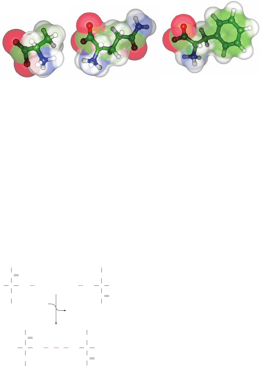
Figure 4-4 Structures of the ␣-amino acids alanine, glutamine,
and phenylalanine. The amino acids are shown as ball-and-stick
models embedded in their transparent space-filling models.The
Figure 4-5 The reaction linking two cysteine residues by a
disulfide bond.
Alanine PhenylalanineGlutamine
C
C
NH
H
O
CH
2
SH
Cysteine
residue
+
C
C
NH
H
O
CH
2
HS
Cysteine
residue
[O]
H
2
O
C
C
NH
H
O
CH
2
S
C
C
NH
H
O
CH
2
S
atoms are colored according to type with C green, H white, N
blue, and O red.
JWCL281_c04_065-081.qxd 5/31/10 1:37 PM Page 71

72 Chapter 4. Amino Acids
D. Acid–Base Properties
Amino acids and proteins have conspicuous acid–base
properties. The ␣-amino acids have two or, for those with
ionizable side groups, three acid–base groups. The titration
curve of glycine, the simplest amino acid, is shown in Fig. 4-6.
At low pH values, both acid–base groups of glycine are
fully protonated so that it assumes the cationic form
⫹
H
3
NCH
2
COOH. In the course of the titration with a
strong base, such as NaOH, glycine loses two protons in the
stepwise fashion characteristic of a polyprotic acid.
The pK values of glycine’s two ionizable groups are suf-
ficiently different so that the Henderson–Hasselbalch
equation:
[2.6]
closely approximates each leg of its titration curve. Conse-
quently, the pK for each ionization step is that of the mid-
point of its corresponding leg of the titration curve (Sec-
tions 2-2A & 2-2C): At pH 2.35 the concentrations of the
cationic form,
⫹
H
3
NCH
2
COOH, and the zwitterionic form,
⫹
H
3
NCH
2
COO
⫺
, are equal; similarly, at pH 9.78 the con-
centrations of the zwitterionic form and the anionic form,
H
2
NCH
2
COO
⫺
, are equal. Note that amino acids never as-
sume the neutral form in aqueous solution.
The pH at which a molecule carries no net electric
charge is known as its isoelectric point, pI. For the ␣-amino
acids, the application of the Henderson–Hasselbalch equa-
tion indicates that, to a high degree of precision,
[4.1]pI ⫽
1
2
(pK
i
⫹ pK
j
)
pH ⫽ pK ⫹ log
a
[A
⫺
]
[HA]
b
where K
i
and K
j
are the dissociation constants of the two
ionizations involving the neutral species. For monoamino,
monocarboxylic acids such as glycine, K
i
and K
j
represent
K
1
and K
2
. However, for aspartic and glutamic acids, K
i
and
K
j
are K
1
and K
R
, whereas for arginine, histidine, and ly-
sine, these quantities are K
R
and K
2
.
Acetic acid’s pK (4.76), which is typical of aliphatic
monocarboxylic acids, is ⬃2.4 pH units higher than the pK
1
of its ␣-amino derivative glycine.This large difference in pK
values of the same functional group is caused, as is dis-
cussed in Section 2-2C, by the electrostatic influence of
glycine’s positively charged ammonium group; that is, its
group helps repel the proton from its COOH group.
Conversely, glycine’s carboxylate group increases the basic-
ity of its amino group (pK
2
⫽ 9.78) with respect to that of
glycine methyl ester (pK ⫽ 7.75). However, the ¬NH
⫹
3
groups of glycine and its esters are significantly more acidic
than are aliphatic amines (pK ⬇ 10.7) because of the electron-
withdrawing character of the carboxyl group.
The electronic influence of one functional group on an-
other is rapidly attenuated as the distance between the
groups increases. Hence, the pK values of the ␣-carboxy-
late groups of amino acids and the side chain carboxylates
of aspartic and glutamic acids form a series that is progres-
sively closer in value to the pK of an aliphatic monocar-
boxylic acid. Likewise, the ionization constant of lysine’s
side chain amino group is indistinguishable from that of an
aliphatic amine.
a. Proteins Have Complex Titration Curves
The titration curves of the ␣-amino acids with ionizable
side chains, such as that of glutamic acid, exhibit the ex-
pected three pK values. However, the titration curves of
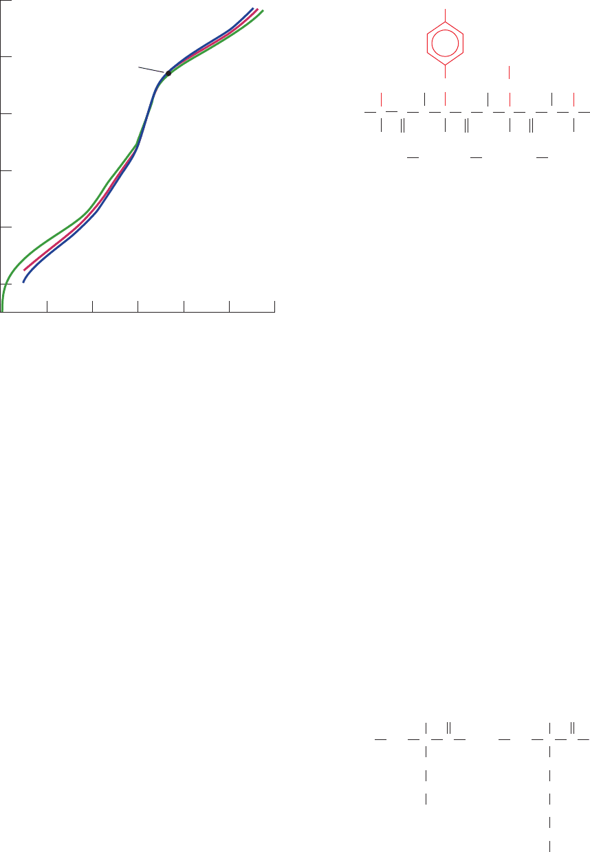
polypeptides and proteins, an example of which is shown in
Fig. 4-7, rarely provide any indication of individual pK val-
ues because of the large numbers of ionizable groups they
represent (typically 30% of a protein’s amino acid side
chains are ionizable; Table 4-1). Furthermore, the covalent
and three-dimensional structure of a protein may cause the
pK of each ionizable group to shift by as much as several
pH units from its value in the free ␣-amino acid as a result
of the electrostatic influence of nearby charged groups,
medium effects arising from the proximity of groups of low
dielectric constant, and the effects of hydrogen bonding as-
sociations.The titration curve of a protein is also a function
of the salt concentration, as is shown in Fig. 4-7,because the
salt ions act electrostatically to shield the side chain
charges from one another, thereby attenuating these
charge–charge interactions.
E. A Few Words on Nomenclature
The three-letter abbreviations for the 20 amino acid
residues are given in Table 4-1. It is worthwhile memorizing
these symbols because they are widely used throughout the
biochemical literature, including this text. These abbrevia-
tions are, in most cases, taken from the first three letters of
the corresponding amino acid’s name; they are conversa-
tionally pronounced as read.
¬NH
⫹
3
Figure 4-6 Titration curve of glycine. Other monoamino,
monocarboxylic acids ionize in a similar fashion. [After Meister,
A., Biochemistry of the Amino Acids (2nd ed.), Vol. 1, p. 30,
Academic Press (1965).]
See the Animated Figures
2.01.51.00.50
pH
12
10
8
6
4
2
H
+
ions dissociated/molecule
pK
2
pI
pK
1
H
3
NCH
2
COO
–
+ H
+
+
+
H
3
NCH
2
COOH
H
3
NCH
2
COO
–
+
H
2
NCH
2
COO
–
+ H
+
JWCL281_c04_065-081.qxd 5/31/10 1:37 PM Page 72

The symbol Glx means Glu or Gln and, similarly, Asx
means Asp or Asn. These ambiguous symbols stem from
laboratory experience: Asn and Gln are easily hydrolyzed
to aspartic acid and glutamic acid, respectively, under the
acidic or basic conditions that are usually used to excise
them from proteins.Therefore, without special precautions,
we cannot determine whether a detected Glu was origi-
nally Glu or Gln, and likewise for Asp and Asn.
The one-letter symbols for the amino acids are also
given in Table 4-1. This more compact code is often used
when comparing the amino acid sequences of several simi-
lar proteins and hence should also be memorized. Note
that the one-letter symbols are usually the first letter of the
amino acid residue’s name. However, for those sets of
residues that have the same first letter, this is only true of
the most abundant residue of the set.
Amino acid residues in polypeptides are named by
dropping the suffix -ine in the name of the amino acid and
replacing it by -yl. Polypeptide chains are described by
starting at the amino terminus (known as the N-terminus)
and sequentially naming each residue until the carboxyl
terminus (the C-terminus) is reached. The amino acid at
the C-terminus is given the name of its parent amino acid.
Thus the compound shown in Fig. 4-8 is alanyltyrosylas-
partylglycine. Of course such names for polypeptide chains
of more than a few residues are extremely cumbersome.
The use of abbreviations for amino acid residues partially
relieves this problem. Thus the foregoing tetrapeptide is
Ala-Tyr-Asp-Gly using the three-letter abbreviations and
AYDG using the one-letter symbols. Note that these ab-
breviations are always written so that the N-terminus of
the polypeptide chain is to the left and the C-terminus is to
the right.
The various nonhydrogen atoms of the amino acid side
chains are often named in sequence with the Greek alpha-
bet (␣, , ␥, ␦, ε, , , …) starting at the carbon atom adja-
cent to the peptide carbonyl group (the C
␣
atom). There-
fore, as Fig. 4-9 indicates, Glu has a ␥-carboxyl group and
Lys has a -amino group (alternatively known as an ε-
amino group because the N atom is substituent to C
ε
). Un-
fortunately, this labeling system is ambiguous for several
amino acids. Consequently, standard numbering schemes
for organic molecules are also employed. These are indi-
cated in Table 4-1 for the heterocyclic side chains.
2 OPTICAL ACTIVITY
The amino acids as isolated by the mild hydrolysis of pro-
teins are, with the exception of glycine, all optically active;
that is, they rotate the plane of plane-polarized light (see
below).
Optically active molecules have an asymmetry such that
they are not superimposable on their mirror image in the
same way that a left hand is not superimposable on its mir-
ror image, a right hand. This situation is characteristic of
substances that contain tetrahedral carbon atoms that
have four different substituents. The two such molecules
Section 4-2. Optical Activity 73
Figure 4-7 Titration curves of the enzyme ribonuclease A at
25ⴗC. The concentration of KCl is 0.01M for the blue curve, 0.03M
for the red curve, and 0.15M for the green curve. [After Tanford,
C. and Hauenstein, J.D., J. Am. Chem. Soc. 78, 5287 (1956).]
Figure 4-8 The tetrapeptide Ala-Tyr-Asp-Gly.
Figure 4-9 Greek lettering scheme used to identify the atoms
in the glutamyl and lysyl R groups.
0
12
10
8
6
4
2
pH
H
+
ions dissociated/molecule
302520151050
Isoelectric
point
C
H
C
C
CH
2
OH
O
H
N
CH
2
COO
–
C
C
O
N
COO
–
H
H
HH
H
C
H
3
N
CH
3
C
O
N
H
Ala Tyr Asp Gly
+
H
2
C
β
H
2
C
γ
H
2
C
δ
H
2
C
ε
Glu Lys
H
NH
H
+
N
ζ
C
α
C
O
3
H
2
C
β
H
2
C
γ
COO
–
H
NH
C
α
C
O
JWCL281_c04_065-081.qxd 5/31/10 1:37 PM Page 73

74 Chapter 4. Amino Acids
depicted in Fig. 4-10 are not superimposable since they are
mirror images. The central atoms in such atomic constella-
tions are known as asymmetric centers or chiral centers
and are said to have the property of chirality (Greek: cheir,
hand).The C
␣
atoms of all the amino acids, with the excep-
tion of glycine, are asymmetric centers. Glycine, which has
two H atoms substituent to its C
␣
atom, is superimposable
on its mirror image and is therefore not optically active.
Molecules that are nonsuperimposable mirror images
are known as enantiomers of one another. Enantiomeric
molecules are physically and chemically indistinguishable
by most techniques. Only when probed asymmetrically, for
example, by plane-polarized light or by reactants that also
contain chiral centers, can they be distinguished and/or dif-
ferentially manipulated.
There are three commonly used systems of nomencla-
ture whereby a particular stereoisomer of an optically ac-
tive molecule can be classified. These are explained in the
following sections.
A. An Operational Classification
Molecules are classified as dextrorotatory (Greek: dexter,
right) or levorotatory (Greek: laevus, left) depending on
whether they rotate the plane of plane-polarized light
clockwise or counterclockwise from the point of view of
the observer. This can be determined by an instrument
known as a polarimeter (Fig. 4-11).A quantitative measure
of the optical activity of the molecule is known as its spe-
cific rotation:
[4.2]
where the superscript 25 refers to the temperature at which
polarimeter measurements are customarily made (25⬚C)
and the subscript D indicates the monochromatic light that
is traditionally employed in polarimetry, the so-called D-
line in the spectrum of sodium (589.3 nm). Dextrorotatory
and levorotatory molecules are assigned positive and neg-
ative values of [␣]
25
D
. Dextrorotatory molecules are there-
fore designated by the prefix (⫹) and their levorotatory
enantiomers have the prefix (⫺). In an equivalent but ar-
chaic nomenclature, the lowercase letters d (dextro) and l
(levo) are used.
The sign and magnitude of a molecule’s specific rotation
depend on the structure of the molecule in a complicated
and poorly understood manner.It is not yet possible to pre-
dict reliably the magnitude or even the sign of a given mol-
ecule’s specific rotation. For example, proline, leucine, and
arginine, which are isolated from proteins, have specific ro-
tations in pure aqueous solutions of ⫺86.2⬚, ⫺10.4⬚, and
⫹12.5⬚, respectively. Their enantiomers exhibit values of
of the same magnitude but of opposite signs. As
might be expected from the acid–base nature of the amino
acids, these quantities vary with the solution pH.
A problem with this operational classification system
for optical isomers is that it provides no presently inter-
pretable indication of the absolute configuration (spatial
arrangement) of the chemical groups about a chiral center.
Furthermore, a molecule with more than one asymmetric
center may have an optical rotation that is not obviously
[␣]
25
D
[␣]
25
D
⫽
observed rotation (degrees)
optical path
length (dm)
⫻
concentration
(g ⴢ cm
⫺3
)
Figure 4-10 The two enantiomers of fluorochlorobromo-
methane. The four substituents are tetrahedrally arranged
about the central atom with the dotted lines indicating that a
substituent lies behind the plane of the paper, a triangular line
indicating that it lies above the plane of the paper, and a thin line
indicating that it lies in the plane of the paper.The mirror plane
relating the enantiomers is represented by a vertical dashed line.
Figure 4-11 Schematic diagram of a polarimeter. This device is used to measure optical rotation.
HH
Cl
C
F
Br
F
C
Cl
Br
Mirror plane
Analyzer
(can be rotated)
Degree scale
(fixed)
Polarimeter
tube
Fixed
polarizer
Light
source
Plane of polarization
of the emerging light
is not the same as
that of the entering
polarized light
Optically active
substance in solution
in the tube causes
the plane of the polarized
light to rotate
+
+90°
–90°
180°
0°
–
JWCL281_c04_065-081.qxd 5/31/10 1:37 PM Page 74

related to the rotatory powers of the individual asymmetric
centers. For this reason, the following relative classification
scheme is more useful.
B. The Fischer Convention
In this system, the configuration of the groups about an
asymmetric center is related to that of glyceraldehyde, a
molecule with one asymmetric center. By a convention in-
troduced by Fischer in 1891, the (⫹) and (⫺) stereoisomers
of glyceraldehyde are designated
D-glyceraldehyde and
L-glyceraldehyde, respectively (note the use of small
uppercase letters).With the realization that there was only
a 50% chance that he was correct, Fischer assumed that
the configurations of these molecules were those shown in
Fig. 4-12. Fischer also proposed a convenient shorthand
notation for these molecules, known as Fischer projec-
tions, which are also given in Fig. 4-12. In the Fischer con-
vention, horizontal bonds extend above the plane of the
paper and vertical bonds extend below the plane of the
paper as is explicitly indicated by the accompanying
geometrical formulas.
The configuration of groups about a chiral center can
be related to that of glyceraldehyde by chemically con-
verting these groups to those of glyceraldehyde using re-
actions of known stereochemistry. For ␣-amino acids, the
arrangement of the amino, carboxyl, R, and H groups
about the C
␣
atom is related to that of the hydroxyl, alde-
hyde, CH
2
OH, and H groups, respectively, of glyceralde-
hyde. In this way,
L-glyceraldehyde and L-␣-amino acids
are said to have the same relative configurations (Fig. 4-13).
Through the use of this method, the configurations of the
␣-amino acids can be described without reference to their
specific rotations.
All a-amino acids derived from proteins have the
L stereo-
chemical configuration; that is, they all have the same rela-
tive configuration about their C
␣
atoms. In 1949, it was
demonstrated by a then new technique in X-ray crystallog-
raphy that Fischer’s arbitrary choice was correct: The des-
ignation of the relative configuration of chiral centers is the
same as their absolute configuration.The absolute configu-
ration of
L-␣-amino acid residues may be easily remem-
bered through the use of the “CORN crib” mnemonic that
is diagrammed in Fig. 4-14.
a. Diastereomers Are Chemically and Physically
Distinguishable
A molecule may have multiple asymmetric centers. For
such molecules, the terms stereoisomers and optical iso-
mers refer to molecules with different configurations about
at least one of their chiral centers, but that are otherwise
identical. The term enantiomer still refers to a molecule
that is the mirror image of the one under consideration,
that is, different in all its chiral centers. Since each asym-
metric center in a chiral molecule can have two possible
configurations, a molecule with n chiral centers has 2
n
dif-
ferent possible stereoisomers and 2
n⫺1
enantiomeric pairs.
Threonine and isoleucine each have two chiral centers and
hence 2
2
⫽ 4 possible stereoisomers. The forms of threo-
nine and isoleucine that are isolated from proteins, which
are by convention called the
L forms, are indicated in Table
4-1.The mirror images of the
L forms are the D forms.Their
other two optical isomers are said to be diastereomers (or
allo forms) of the enantiomeric
D and L forms.The relative
Section 4-2. Optical Activity 75
HO
CHO
C
H
C
Mirror plane
CH
2
OH
OHH
CHO
CH
2
OH
Fischer projection
HO
CHO
C
H
C
CH
2
OH
OHH
CHO
CH
2
OH
Geometric formulas
L-Glyceraldehyde D-Glyceraldehyde
C
CH
2
OH
CHO
HHO
L-Glyceraldehyde
C
+
R
COO
–
HH
3
N
L-α-Amino acid
R
N
CO
H
C
α
Figure 4-12 Fischer convention configurations for naming the
enantiomers of glyceraldehyde. Glyceraldehyde enantiomers are
represented by geometric formulas (top) and their corresponding
Fischer projection formulas (bottom). Note that in Fischer
projections, all horizontal bonds point above the page and all
vertical bonds point below the page. The mirror planes relating the
enantiomers are represented by a vertical dashed line. (Fischer
projection formulas, as traditionally presented, omit the central
C symbolizing the chiral carbon atom.The Fischer projection
formulas in this text, however, will generally have a central C.)
Figure 4-13 Configurations of
L-glyceraldehyde and
L-␣-amino acids.
Figure 4-14 “CORN crib” mnemonic for the hand of
L-amino
acids. Looking at the C
␣
atom from its H atom substituent, its
other substituents should read in the clockwise
direction as shown. Here CO, R, and N, respectively, represent
the carbonyl group, side chain, and main chain nitrogen atom.
[After Richardson, J.S., Adv. Protein Chem. 34, 171 (1981).]
CO¬R¬N
JWCL281_c04_065-081.qxd 5/31/10 1:37 PM Page 75

configurations of all four stereoisomers of threonine are
given in Fig. 4-15. Note the following points:
1. The
D-allo and L-allo forms are mirror images of each
other, as are the
D and L forms. Neither allo form is sym-
metrically related to either of the
D or L forms.
2. In contrast to the case for enantiomeric pairs, di-
astereomers are physically and chemically distinguishable
from one another by ordinary means such as melting
points, spectra, and chemical reactivity; that is, they are re-
ally different compounds in the usual sense.
A special case of diastereoisomerism occurs when the
two asymmetric centers are chemically identical. Two of
the four Fischer projections of the sort shown in Fig. 4-15
then represent the same molecule. This is because the two
asymmetric centers in this molecule are mirror images of
each other.Such a molecule is superimposible on its mirror
image and is therefore optically inactive. This so-called
meso form is said to be internally compensated. The three
optical isomers of cystine are shown in Fig. 4-16, where it
can be seen that the
D and L isomers are mirror images of
each other as before. Only
L-cystine occurs in proteins.
C. The Cahn–Ingold–Prelog System
Despite its usefulness, the Fischer scheme is awkward and
often ambiguous for molecules with more than one asym-
metric center. For this reason, the following absolute
nomenclature scheme was formulated in 1956 by Robert
Cahn, Christopher Ingold, and Vladimir Prelog. In this sys-
tem, the four groups surrounding a chiral center are ranked
according to a specific although arbitrary priority scheme:
Atoms of higher atomic number bonded to a chiral center
are ranked above those of lower atomic number. For exam-
ple, the oxygen atom of an OH group takes precedence
over the carbon atom of a CH
3
group that is bonded to the
same chiral C atom. If any of the first substituent atoms are
of the same element, the priority of these groups is estab-
lished from the atomic numbers of the second, third, etc.,
atoms outward from the asymmetric center. Hence a
CH
2
OH group takes precedence over a CH
3
group. There
are other rules (given in the references and in many or-
ganic chemistry textbooks) for assigning priority ratings to
substituents with multiple bonds or differing isotopes. The
order of priority of some common functional groups is
Note that each of the groups substituent to a chiral center
must have a different priority rating; otherwise the center
could not be asymmetric.
The prioritized groups are assigned the letters W, X,Y, Z
such that their order of priority rating is W ⬎ X ⬎ Y ⬎ Z.
To establish the configuration of the chiral center, it is
viewed from the asymmetric center toward the Z group
(lowest priority). If the order of the groups W S X S Y as
seen from this direction is clockwise, then the configuration
of the asymmetric center is designated (R) (Latin: rectus,
right). If the order of W S X S Y is counterclockwise, the
asymmetric center is designated (S) (Latin: sinister, left).
L-Glyceraldehyde is therefore designated (S)-glyceraldehyde
(Fig.4-17) and, similarly,
L-alanine is (S)-alanine (Fig.4-18).
In fact, all the
L-amino acids from proteins are (S)-amino
acids, with the exception of
L-cysteine, which is (R)-cysteine.
A major advantage of this so-called Cahn–Ingold– Prelog
or (RS) system is that the chiralities of compounds with mul-
tiple asymmetric centers can be unambiguously described.
Thus, in the (RS) system,
L-threonine is (2S,3R)-threonine,
whereas
L-isoleucine is (2S,3S)-isoleucine (Fig. 4-19).
⬎ CH
2
OH ⬎ C
6
H
5
⬎ CH
3
⬎
2
H ⬎
1
H
SH ⬎ OH ⬎ NH
2
⬎ COOH ⬎ CHO
76 Chapter 4. Amino Acids
Figure 4-15 Fischer projections of threonine’s four stereoisomers.
The
D and L forms are mirror images as are the D-allo and L-allo
forms.
D- and L-threonine are each diastereomers of both D-allo-
and
L-allo-threonine.
Figure 4-16 The three stereoisomers of cystine. The
D and L forms are related by mirror symmetry,
whereas the meso form has internal mirror symmetry and therefore lacks optical activity.
COO
⫺
⫹
CHH
3
N
C OHH
CH
3
L
-Threonine
Mirror
plane
COO
⫺
⫹
CNH
3
H
C HHO
CH
3
D
-Threonine
COO
⫺
⫹
CHH
3
N
C HHO
CH
3
L
-allo-Threonine
COO
⫺
⫹
CNH
3
H
C OHH
CH
3
D
-allo-Threonine
COO
⫺
⫹
CHH
3
N
CH
2
H
S
COO
⫺
⫹
CH
3
N
CH
2
L
-Cystine
Mirror plane
S
COO
⫺
⫹
CHH
3
N
CH
2
NH
3
S
COO
⫺
⫹
NH
3
⫹
NH
3
⫹
CH
CH
2
meso-Cystine
Mirror plane
S
COO
⫺
CH
CH
2
S
COO
⫺
CH
CH
2
D
-Cystine
S
JWCL281_c04_065-081.qxd 5/31/10 1:37 PM Page 76

a. Prochiral Centers Have Distinguishable
Substituents
Two chemically identical substituents to an otherwise chi-
ral tetrahedral center are geometrically distinct; that is, the
center has no rotational symmetry so that it can be unam-
biguously assigned left and right sides. Consider, for exam-
ple, the substituents to the C1 atom of ethanol (the CH
2
group; Fig. 4-20a). If one of the H atoms were converted to
another group (not CH
3
or OH), C1 would be a chiral cen-
ter. The two H atoms are therefore said to be prochiral. If
we arbitrarily assign the H atoms the subscripts a and b
(Fig. 4-20), then H
b
is said to be pro-R because in sighting
from C1 toward H
a
(as if it were the Z group of a chiral
center), the order of priority of the other substituents de-
creases in a clockwise direction (Fig. 4-20b). Similarly, H
a
is
said to be pro-S (Fig. 4-20c).
Planar objects with no rotational symmetry also have the
property of prochirality. For example, in many enzymatic
reactions, stereospecific addition to a trigonal carbon atom
occurs from a particular side of that carbon atom to yield a
chiral center (Section 13-2A). If a trigonal carbon is facing
the viewer such that the order of priority of its substituents
decreases in a clockwise manner (Fig. 4-21a), that face is
designated as the re face (after rectus).The opposite face is
designated as the si face (after sinister) since the priorities
of its substituents decrease in the counterclockwise direc-
tion (Fig. 4-21b). Comparison of Figs. 4-20b and 4-21a indi-
cates that an H atom adding to the re side of acetaldehyde
atom C1 occupies the pro-R position of the resulting tetra-
hedral center. Conversely, a pro-S H atom is generated by
si side addition to this trigonal center (Figs. 4-20c and
4-21b).
Closely related compounds that have the same configu-
rational representation under the Fischer
DL convention
may have different representations under the (RS) system.
Consequently, we shall use the Fischer convention in most
cases. The (RS) system, however, is indispensable for de-
scribing prochirality and stereospecific reactions, so we
shall find it invaluable for describing enzymatic reactions.
Section 4-2. Optical Activity 77
Figure 4-17 The structural formula of L-glyceraldehyde. Its
equivalent (RS) system representation indicates that it is
(S)-glyceraldehyde. In the latter drawing, the chiral C atom is
represented by the large circle, and the H atom, which is located
behind the plane of the paper, is represented by the smaller
concentric dashed circle.
Figure 4-18 The structural formula of
L-alanine. Its equivalent
(RS) system representation indicates that it is (S)-alanine.
C
CH
2
OH
CHO
HHO
L-Glyceraldehyde (S)-Glyceraldehyde
OH
CHO
CH
2
OH
H
(Z)
(W)
(X)
(Y)
COO
⫺
OOC
H
3
C
H
(Z)
⫹
CHH
3
N
CH
3
L
-Alanine (S)-Alanine
NH
3
⫺
(W)
(Y)
(X)
H
3
C
OH
H
H
⫺
OOC
(2S,3R)-Threonine
H
3
C
CH
2
CH
3
H
H
⫺
OOC NH
3
⫹
NH
3
⫹
(2S,3S)-Isoleucine
OH
OH
C
H
b
H
b
( pro-R)
H
a
CH
3
(a)
(b)
(c)
H
3
C
H
a
OH
H
a
(pro-S)
H
3
C
H
b
C
H
C
O
O
H
3
C
H
3
C
(a)
(b)
H
Figure 4-21 Views of acetaldehyde. (a) Its re face and (b) its si
face.
Figure 4-19 Newman projection diagrams of the stereoisomers
of threonine and isoleucine derived from proteins. Here the
bond is viewed end on.The nearer atom, C
␣
, is represented
by the confluence of the three bonds to its substituents, whereas
the more distant atom, C

, is represented by a circle from which
its three substituents project.
C
␣
¬C

Figure 4-20 Views of ethanol. (a) Note that H
a
and H
b
, although
chemically identical, are distinguishable: Rotating the molecule
by 180⬚ about the vertical axis so as to interchange these two
hydrogen atoms does not yield an indistinguishable view of the
molecule because the rotation also interchanges the chemically
different OH and CH
3
groups. (b) Looking from C1 to H
a
, the
pro-S hydrogen atom (the dotted circle). (c) Looking from C1 to
H
b
, the pro-R hydrogen atom.
JWCL281_c04_065-081.qxd 5/31/10 1:37 PM Page 77

D. Chirality and Biochemistry
The ordinary chemical synthesis of chiral molecules pro-
duces racemic mixtures of these molecules (equal amounts
of each member of an enantiomeric pair) because ordinary
chemical and physical processes have no stereochemical
bias. Consequently, there are equal probabilities for an
asymmetric center of either hand to be produced in any
such process. In order to obtain a product with net optical
activity, a chiral process must be employed. This usually
takes the form of using chiral reagents, although, at least in
principle, the use of any asymmetric influence such as light
that is plane polarized in one direction can produce a net
asymmetry in a reaction product.
One of the most striking characteristics of life is its pro-
duction of optically active molecules. The biosynthesis of a
substance possessing asymmetric centers almost invariably
produces a pure stereoisomer. The fact that the amino acid
residues of proteins all have the
L configuration is just one
example of this phenomenon. This observation has
prompted the suggestion that a simple diagnostic test for
the past or present existence of extraterrestrial life, be it on
moon rocks or in meteorites that have fallen to Earth,
would be the detection of net optical activity in these mate-
rials. Any such finding would suggest that the asymmetric
molecules thereby detected had been biosynthetically pro-
duced. Thus, even though ␣-amino acids have been ex-
tracted from carbonaceous meteorites, the observation
that they come in racemic mixtures suggests that they are
of chemical rather than biological origin.
One of the enigmas of the origin of life is why terrestrial
life is based on certain chiral molecules rather than their
enantiomers, that is, on
L-amino acids, for example, rather
than
D-amino acids.Arguments that physical effects such as
polarized light might have promoted significant net asym-
metry in prebiotically synthesized molecules (Section
1-5B) have not been convincing. Perhaps
L-amino
acid–based life-forms arose at random and simply “ate”
any
D-amino acid–based life-forms.
The importance of stereochemistry in living systems is
also a concern of the pharmaceutical industry. Many drugs
are chemically synthesized as racemic mixtures, although
only one enantiomer has biological activity. In most cases,
the opposite enantiomer is biologically inert and is there-
fore packaged along with its active counterpart. This is
true, for example, of the widely used anti-inflammatory
agent ibuprofen, only one enantiomer of which is physio-
logically active (Fig. 4-22). Occasionally, the inactive enan-
tiomer of a useful drug produces harmful effects and must
therefore be eliminated from the racemic mixture. The
most striking example of this is the drug thalidomide (Fig.
4-23), a mild sedative whose “inactive” enantiomer causes
severe birth defects. Partly because of the unanticipated
problems caused by “inactive” drug enantiomers, chiral or-
ganic synthesis has become an active area of medicinal
chemistry.
3 “NONSTANDARD” AMINO ACIDS
The 20 common amino acids are by no means the only
amino acids that occur in biological systems. “Nonstan-
dard” amino acid residues are often important constituents
of proteins and biologically active polypeptides. Many
amino acids, however, are not constituents of proteins. To-
gether with their derivatives, they play a variety of biologi-
cally important roles.
A. Amino Acid Derivatives in Proteins
The “universal” genetic code, which is nearly identical in all
known life-forms (Section 5-4Bb), specifies only the 20
“standard” amino acids of Table 4-1. Nevertheless, many
other amino acids, a selection of which is given in Fig. 4-24,
are components of certain proteins. In all known cases but
two (Section 32-2De), however, these unusual amino acids
result from the specific modification of an amino acid
residue after the polypeptide chain has been synthesized.
Among the most prominent of these modified amino acid
residues are 4-hydroxyproline and 5-hydroxylysine. Both
of these amino acid residues are important structural con-
stituents of the fibrous protein collagen, the most abundant
protein in mammals (Section 8-2B). Amino acids of pro-
teins that form complexes with nucleic acids are often
modified. For example, the chromosomal proteins known
as histones may be specifically methylated, acetylated,
and/or phosphorylated at specific Lys, Arg, and Ser
residues (Section 34-3Baa). Several of these derivatized
78 Chapter 4. Amino Acids
Figure 4-22 Ibuprofen. Only the enantiomer shown has
anti-inflammatory action.The chiral carbon is red.
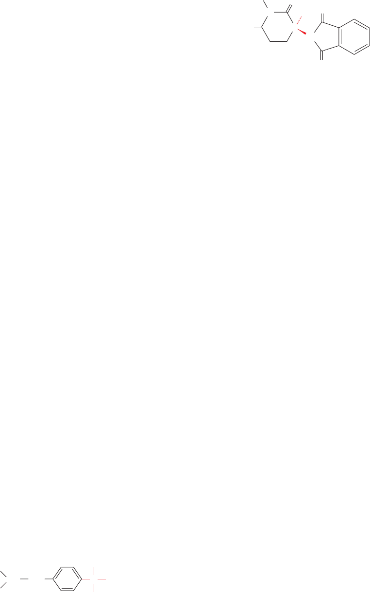
Figure 4-23 Thalidomide. This drug was widely used in Europe
as a mild sedative in the early 1960s. Its inactive enantiomer (not
shown), which was present in equal amounts in the formulations
used, causes severe birth defects in humans when taken during
the first trimester of pregnancy.Thalidomide was often prescribed
to alleviate the nausea (morning sickness) that is common during
this period.
CH
2
H
3
C
H
3
C
CH C
H
CH
3
COOH
Ibuprofen
C
N
O
O
H
Thalidomide
H
O
O
N
JWCL281_c04_065-081.qxd 5/31/10 1:37 PM Page 78

amino acid residues are presented in Fig. 4-24. N-Formyl-
methionine is initially the N-terminal residue of all
prokaryotic proteins, but is usually removed as part of the
protein maturation process (Section 32-3Ca). ␥-Carboxy-
glutamic acid is a constituent of several proteins involved
in blood clotting (Section 35-1Ba). Note that in most cases,
these modifications are important, if not essential, for the
function of the protein.
D-Amino acid residues are components of many of the
relatively short (⬍20 residues) bacterial polypeptides that
are enzymatically rather than ribosomally synthesized.
These polypeptides are perhaps most widely distributed as
constituents of bacterial cell walls (Section 11-3Ba), which
D-amino acids render less susceptible to attack by the pep-
tidases (enzymes that hydrolyze peptide bonds) that many
organisms employ to digest bacterial cell walls. Likewise,
D-amino acids are components of many bacterially produced
peptide antibiotics including valinomycin, gramicidin A
(Section 20-2C), and actinomycin D (Section 31-2Cc).
D-Amino acid residues are also functionally essential com-
ponents of several ribosomally synthesized polypeptides of
eukaryotic as well as prokaryotic origin. These
D-amino
acid residues are posttranslationally formed, most proba-
bly through the enzymatically mediated inversion of the
preexisting
L-amino acid residues.
B. Specialized Roles of Amino Acids
Besides their role in proteins, amino acids and their deriva-
tives have many biologically important functions.A few ex-
amples of these substances are shown in Fig. 4-25. This al-
ternative use of amino acids is an example of the biological
opportunism that we shall repeatedly encounter: Nature
tends to adapt materials and processes that are already pres-
ent to new functions.
Amino acids and their derivatives often function as chem-
ical messengers in the communications between cells. For
example, glycine, ␥-aminobutyric acid (GABA; a glutamate
Section 4-3. “Nonstandard” Amino Acids 79
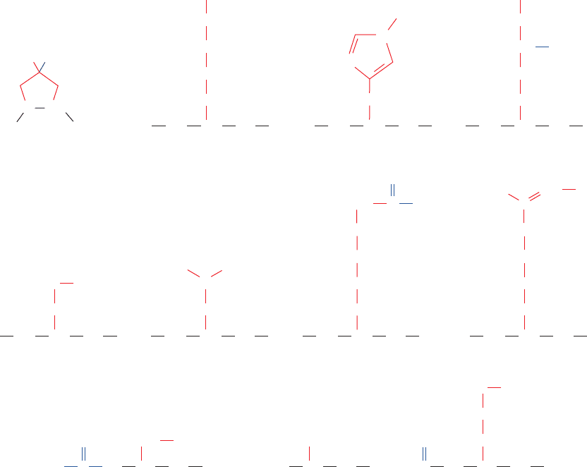
Figure 4-24 Some uncommon amino acid residues that are
components of certain proteins. All of these residues are modified
from one of the 20 “standard” amino acids after polypeptide chain
COO
–
4-Hydroxyproline
NH
CO
CH
NH
CO
CH
α
ε-N-Acetyllysine
NH
CO
3-Methylhistidine 5-Hydroxylysine
CH
OH
NH
CO
CH
α
γ-CarboxyglutamateO-Phosphoserine
N-FormylmethionineN,N,N-TrimethylalanineN-Acetylserine
CH
2
O
NH
CH
2
CH
2
CH
2
CH
2
ε
δ
γ
β
CH
2
CH
γ
β
COO
–
CH
2
N
N
4
3
2
1
5
PO
3
2
–
C
O
NH CH CO
–
OOC
OH
CH
3
CH
3
CH
3
CH
3
H
4
3
5
NH
CO
CH
ω-N-Methylarginine
NH
CH
2
CH
2
CH
2
NH
CO
CH
α
ε-N,N,N-Trimethyllysine
N(CH
3
)
3
CH
2
CH
2
CH
2
CH
2
ε
δ
γ
β
CH
2
OH
HC NH CH CO
O
C
O
CH
2
CH
2
SCH
3
(CH
3
)
3
NCHCO
CH
3
NCH
⫹
NH
CO
CH
21
NH
⫹
3
CH
2
CH
CH
2
CH
2
6
5
4
3
NH
⫹
C
H
2
N
⫹
12
biosynthesis.Those amino acid residues that are derivatized at
their N
␣
position occur at the N-termini of proteins.
JWCL281_c04_065-081.qxd 5/31/10 1:37 PM Page 79

decarboxylation product), and dopamine (a tyrosine deriv-
ative) are neurotransmitters (substances released by nerve
cells to alter the behavior of their neighbors; Section 20-5C);
histamine (the decarboxylation product of histidine) is a
potent local mediator of allergic reactions; and thyroxine
(a tyrosine derivative) is an iodine-containing thyroid hor-
mone that generally stimulates vertebrate metabolism
(Section 19-1D).
Certain amino acids are important intermediates in vari-
ous metabolic processes.Among them are citrulline and or-
nithine, intermediates in urea biosynthesis (Section 26-2B);
homocysteine, an intermediate in amino acid metabolism
(Section 26-3Ea); and S-adenosylmethionine, a biological
methylating reagent (Section 26-3Ea).
Nature’s diversity is remarkable. Over 700 different
amino acids have been found in various plants, fungi, and
bacteria, most of which are ␣-amino acids. For the most
part, their biological roles are obscure although the fact
that many are toxic suggests that they have a protective
function. Indeed, some of them, such as azaserine, are med-
ically useful antibiotics. Many of these amino acids are sim-
ple derivatives of the 20 “standard” amino acids although
some of them, including azaserine and -cyanoalanine
(Fig. 4-25), have unusual structures.
80 Chapter 4. Amino Acids
1 The Amino Acids of Proteins Proteins are linear poly-
mers that are synthesized from the same 20 “standard”
␣-amino acids through their condensation to form peptide
bonds. These amino acids all have a carboxyl group with a pK
near 2.2 and an amino substituent with a pK near 9.4 attached
to the same carbon atom, the C
␣
atom. The ␣-amino acids are
zwitterionic compounds, , in the physio-
logical pH range. The various amino acids are usually classi-
fied according to the polarities of their side chains, R, which
are substituent to the C
␣
atom. Glycine, alanine, valine, leucine,
isoleucine, methionine, proline (which is really a secondary
amino acid), phenylalanine, and tryptophan are nonpolar
amino acids; serine, threonine, asparagine, glutamine, tyrosine,
and cysteine are uncharged and polar; and lysine,arginine, his-
tidine, aspartic acid, and glutamic acid are charged and polar.
The side chains of many of these amino acids bear acid–base
groups, and hence the properties of the proteins containing
them are pH dependent.
2 Optical Activity The C
␣
atoms of all ␣-amino acids ex-
⫹
H
3
N¬CHR¬COO
⫺
cept glycine each bear four different substituents and are
therefore chiral centers. According to the Fischer convention,
which relates the configuration of
D- or L-glyceraldehyde to
that of the asymmetric center of interest, all the amino acids of
proteins have the
L configuration; that is, they all have the
same absolute configuration about their C
␣
atom. According
to the Cahn–Ingold–Prelog (RS) system of chirality nomen-
clature, they are, with the exception of cysteine, all (S)-amino
acids.The side chains of threonine and isoleucine also contain
chiral centers. A prochiral center has no rotational symmetry,
and hence its substituents, in the case of a central atom, or its
faces, in the case of a planar molecule, are distinguishable.
3 “Nonstandard” Amino Acids Amino acid residues
other than the 20 from which proteins are synthesized also
have important biological functions. These “nonstandard”
residues result from the specific chemical modifications of
amino acid residues in preexisting proteins. Amino acids and
their derivatives also have independent biological roles such
as neurotransmitters, metabolic intermediates, and poisons.
CHAPTER SUMMARY
Figure 4-25 Some biologically produced derivatives of “standard” amino acids and amino
acids that are not components of proteins.
+
CH
2
H
3
N
CH
2
H
3
N
CH
2
CH
2
–
OOC
α
β
γ
γ-Aminobutyric acid (GABA)
+
CH
2
H
3
N
+
H
3
N
+
Histamine
CH
2
N
N
H
CH
Thyroxine
COO
–
H
3
N
+
COO
–
CH
2
O
CH
2
HO
HO
Dopamine
I
I
I
HO
I
H
3
N
CO
O
⫺
CH
Citrulline
HN
CH
2
CH
2
CH
2
C
O
NH
2
C
O
CH N
+
N
–
Azaserine S-Adenosylmethionine
+
CH
Ornithine
CH
2
CH
2
CH
2
NH
3
⫹
-Cyanoalanine
N
C
H
3
N
+
COO
–
CH
CH
2
Homocysteine
H
3
N
+
COO
–
CH
CH
2
CH
2
SH
CH
2
O
H
3
N
+
COO
–
CH
N
N
N
N
HH
H
OH
H
OH
O
NH
2
H
3
N
CO
O
⫺
CH
S
CH
2
CH
2
CH
2
CH
3
+
+
JWCL281_c04_065-081.qxd 5/31/10 1:37 PM Page 80
