Voet D., Voet Ju.G. Biochemistry
Подождите немного. Документ загружается.


Problems 81
1. Name the 20 standard amino acids without looking them
up. Give their three-letter and one-letter symbols.Identify the two
standard amino acids that are isomers and the two others that, al-
though not isomeric, have essentially the same molecular mass for
the neutral molecules.
2. Draw the following oligopeptides in their predominant
ionic forms at pH 7: (a) Phe-Met-Arg, (b) tryptophanyllysylaspar-
tic acid, and (c) Gln-Ile-His-Thr.
3. How many different pentapeptides are there that contain
one residue each of Gly, Asp,Tyr, Cys, and Leu?
4. Draw the structures of the following two oligopeptides with
their cysteine residues cross-linked by a disulfide bond: Val-Cys,
Ser-Cys-Pro.
*5. What are the concentrations of the various ionic species in
a 0.1M solution of lysine at pH 4, 7, and 10?
6. Derive Eq. [4.1] for a monoamino, monocarboxylic acid
(use the Henderson–Hasselbalch equation).
*7. The isoionic point of a compound is defined as the pH of a
pure water solution of the compound.What is the isoionic point of
a 0.1M solution of glycine?
8. Normal human hemoglobin has an isoelectric point of 6.87.
A mutant variety of hemoglobin, known as sickle-cell hemoglo-
bin, has an isoelectric point of 7.09. The titration curve of
hemoglobin indicates that, in this pH range, 13 groups change
ionization states per unit change in pH. Calculate the difference in
ionic charge between molecules of normal and sickle-cell hemo-
globin.
9. Indicate whether the following familiar objects are chiral,
prochiral, or nonchiral.
(a) A glove (g) A snowflake
(b) A tennis ball (h) A spiral staircase
(c) A good pair of scissors (i) A flight of normal stairs
(d) A screw (j) A paper clip
(e) This page (k) A shoe
(f) A toilet paper roll (l) A pair of glasses
10. Draw four equivalent Fischer projection formulas for
L-alanine (see Figs. 4-12 and 4-13).
*11. (a) Draw the structural formula and the Fischer projec-
tion formula of (S)-3-methylhexane. (b) Draw all the stereoiso-
mers of 2,3-dichlorobutane. Name them according to the (RS) sys-
tem and indicate which of them has the meso form.
12. Identify and name the prochiral centers or faces of the fol-
lowing molecules:
(a) Acetone (d) Alanine
(b) Propene (e) Lysine
(c) Glycine (f) 3-Methylpyridine
13. Write out the dominant structural formula, at pH 12.0, of
the pentapeptide Thr-Tyr-His-Cys-Lys. Indicate the positions of its
chiral centers and its prochiral centers.Assume that the pK’s of its
ionizable groups are the same as those in the corresponding free
amino acid.
PROBLEMS
History
Vickery, H.B. and Schmidt, C.L.A.,The history of the discovery of
amino acids, Chem. Rev. 9, 169–318 (1931).
Vickery, H.B., The history of the discovery of the amino acids. A
review of amino acids discovered since 1931 as components of
native proteins, Adv. Protein Chem. 26, 81–171 (1972).
Properties of Amino Acids
Barrett, G.C. and Elmore, D.T., Amino Acids and Peptides, Chap-
ters 1–4, Cambridge University Press (1998).
Cohn, E.J. and Edsall, J.T., Proteins, Amino Acids and Peptides as
Ions and Dipolar Ions, Academic Press (1943). [A classic work
in its field.]
Meister, A., Biochemistry of the Amino Acids (2nd ed.), Vol. 1,
Academic Press (1965). [A compendium of information on
amino acid properties.]
Optical Activity
Cahn, R.S., An introduction to the sequence rule, J. Chem. Ed. 41,
116–125 (1964). [A presentation of the Cahn–Ingold–Prelog
system of nomenclature.]
Huheey, J.E., A novel method for assigning R,S labels to enan-
tiomers, J. Chem. Ed. 63, 598–600 (1986).
Lamzin, V.S., Dauter, Z., and Wilson, K.S., How nature deals with
stereoisomers, Curr. Opin. Struct. Biol. 5, 830–836 (1995). [Dis-
cusses proteins synthesized from
D-amino acids.]
Mislow, K., Introduction to Stereochemistry, Benjamin (1966).
Solomons, T.W.G. and Fryhle, C.B., Organic Chemistry (9th ed.),
Chapter 5, Wiley (2008). [A discussion of chirality. Most other
organic chemistry textbooks contain similar material.]
“Nonstandard” Amino Acids
Fowden, L., Lea, P.J., and Bell, E.A., The non-protein amino acids
of plants, Adv. Enzymol. 50, 117–175 (1979).
Fowden, L., Lewis, D., and Tristram, H., Toxic amino acids: their
action as antimetabolites, Adv. Enzymol. 29, 89–163 (1968).
Kleinkauf, H. and Döhren, H., Nonribosomal polypeptide forma-
tion on multifunctional proteins, Trends Biochem. Sci. 8,
281–283 (1993).
Mor,A., Amiche, M., and Nicholas, P., Enter a new post-transcrip-
tional modification:
D-amino acids in gene-encoded peptides,
Trends Biochem. Sci. 17, 481–485 (1992).
REFERENCES
JWCL281_c04_065-081.qxd 5/31/10 1:37 PM Page 81

82
CHAPTER 5
Nucleic Acids,
Gene Expression,
and Recombinant
DNA Technology
1 Nucleotides and Nucleic Acids
A. Nucleotides, Nucleosides, and Bases
B. The Chemical Structures of DNA and RNA
2 DNA Is the Carrier of Genetic Information
A. Transforming Principle Is DNA
B. The Hereditary Molecule of Many Bacteriophages Is DNA
3 Double Helical DNA
A. The Watson–Crick Structure: B-DNA
B. DNA Is Semiconservatively Replicated
C. Denaturation and Renaturation
D. The Size of DNA
4 Gene Expression and Replication: An Overview
A. RNA Synthesis: Transcription
B. Protein Synthesis: Translation
C. DNA Replication
5 Molecular Cloning
A. Restriction Endonucleases
B. Cloning Vectors
C. Gene Manipulation
D. The Identification of Specific DNA Sequences:
Southern Blotting
E. Genomic Libraries
F. The Polymerase Chain Reaction
G. Production of Proteins
H. Transgenic Organisms and Gene Therapy
I. Social, Ethical, and Legal Considerations
Knowledge of how genes are expressed and how they can
be manipulated is becoming increasingly important for un-
derstanding nearly every aspect of biochemistry. Conse-
quently, although we do not undertake a detailed discus-
sion of these processes until Part V of this textbook, we
outline their general principles in this chapter.We do so by
describing the chemical structures of nucleic acids, how we
have come to know that DNA is the carrier of genetic in-
formation, the structure of the major form of DNA,and the
general principles of how the information in genes directs
the synthesis of RNA and proteins (how genes are ex-
pressed) and how DNA is replicated. The chapter ends
with a discussion of how DNA is experimentally manipu-
lated and expressed, processes that are collectively re-
ferred to as genetic engineering.These processes have rev-
olutionized the practice of biochemistry.
1 NUCLEOTIDES AND NUCLEIC ACIDS
Nucleotides and their derivatives are biologically ubiqui-
tous substances that participate in nearly all biochemical
processes:
1. They form the monomeric units of nucleic acids and
thereby play central roles in both the storage and the ex-
pression of genetic information.
2. Nucleoside triphosphates, most conspicuously ATP
(Section 1-3B), are the “energy-rich” end products of the
majority of energy-releasing pathways and the substances
whose utilization drives most energy-requiring processes.
3. Most metabolic pathways are regulated, at least in
part, by the levels of nucleotides such as ATP and ADP.
Moreover, certain nucleotides, as we shall see, function as
intracellular signals that regulate the activities of numer-
ous metabolic processes.
4. Nucleotide derivatives, such as nicotinamide adenine
dinucleotide (Section 13-2A), flavin adenine dinucleotide
(Section 16-2C), and coenzyme A (Section 21-2), are re-
quired participants in many enzymatic reactions.
5. As components of the enzymelike nucleic acids
known as ribozymes, nucleotides have important catalytic
activities themselves.
A. Nucleotides, Nucleosides, and Bases
Nucleotides are phosphate esters of a five-carbon sugar
(which is therefore known as a pentose; Section 11-1A)
in which a nitrogenous base is covalently linked to C1¿
of the sugar residue. In ribonucleotides (Fig. 5-1a), the
monomeric units of RNA, the pentose is
D-ribose, whereas
in deoxyribonucleotides (or just deoxynucleotides; Fig. 5-1b),
O
HH
H
H
OH
Ribonucleotides
OH
CH
2
–2
O
3
PO
4⬘
5⬘
3⬘ 2⬘
1⬘
Base
O
HH
H
H
OH H
CH
2
–2
O
3
PO
4⬘
5⬘
3⬘ 2⬘
1⬘
Base
Deoxyribonucleotides
(a) (b)
Figure 5-1 Chemical structures of (a) ribonucleotides and
(b) deoxyribonucleotides.
JWCL281_c05_082-128.qxd 2/19/10 4:46 PM Page 82

a
The presence of a 2¿-deoxyribose unit in place of ribose, as occurs in DNA, is implied by the prefixes “deoxy” or
“d.” For example, the deoxynucleoside of adenine is deoxyadenosine or dA. However, for thymine-containing
residues, which rarely occur in RNA, the prefix is redundant and may be dropped.The presence of a ribose unit
may be explicitly implied by the prefixes “ribo” or “r.”Thus the ribonucleotide of thymine is ribothymidine or rT.
b
The position of the phosphate group in a nucleotide may be explicitly specified as in, for example, 3¿-AMP
and 5¿-GMP.
the monomeric units of DNA, the pentose is 2ⴕ-deoxy-
D-ribose (note that the “primed” numbers refer to the
atoms of the ribose residue;“unprimed” numbers refer to
atoms of the nitrogenous base).The phosphate group may
be bonded to C5¿ of the pentose to form a 5ⴕ-nucleotide
(Fig. 5-1) or to its C3¿ to form a 3ⴕ-nucleotide. If the phos-
phate group is absent, the compound is known as a nucle-
oside. A 5¿-nucleotide, for example, may therefore be re-
ferred to as a nucleoside-5ⴕ-phosphate. In all naturally
occurring nucleotides and nucleosides, the bond linking
the nitrogenous base to the pentose C1¿ atom (which is
called a glycosidic bond; Section 11-1Ca) extends from
the same side of the ribose ring as does the C4¿¬C5¿ bond
(the so-called  configuration; Section 11-1Ba) rather
than from the opposite side (the ␣ configuration). Note
that nucleotide phosphate groups are doubly ionized at
physiological pH’s; that is, nucleotides are moderately
strong acids.
The nitrogenous bases are planar, aromatic, heterocyclic
molecules which, for the most part, are derivatives of either
purine or pyrimidine.
The structures, names, and abbreviations of the common
bases, nucleosides, and nucleotides are given in Table 5-1.
The major purine components of nucleic acids are adenine
and guanine residues; the major pyrimidine residues are
those of cytosine, uracil (which occurs mainly in RNA),
and thymine (5-methyluracil, which occurs mainly in
DNA). The purines form glycosidic bonds to ribose via
N
NN
H
N
6
7
8
9
51
4
3
2
N
4
53
6
1
2
N
Purine Pyrimidine
Section 5-1. Nucleotides and Nucleic Acids 83
Table 5-1 Names and Abbreviations of Nucleic Acid Bases, Nucleosides, and Nucleotides
Base Base Nucleoside Nucleotide
b
Formula (X ⫽ H) (X ⫽ ribose
a
) (X ⫽ ribose phosphate
a
)
Adenine Adenosine Adenylic acid
Ade Ado Adenosine monophosphate
A A AMP
Guanine Guanosine Guanylic acid
Gua Guo Guanosine monophosphate
G G GMP
Cytosine Cytidine Cytidylic acid
Cyt Cyd Cytidine monophosphate
C C CMP
Uracil Uridine Uridylic acid
Ura Urd Uridine monophosphate
U U UMP
Thymine Deoxythymidine Deoxythymidylic acid
Thy dThd Deoxythymidine monophosphate
T dT dTMP
N
N
NH
2
N
X
N
N
H
2
N
H
O
N
N
X
N
N
N
NH
2
X
O
O
H
N
X
N
O
dX
CH
3
O
N
N
H
N
O
JWCL281_c05_082-128.qxd 2/19/10 4:46 PM Page 83
their N9 atoms, whereas pyrimidines do so through their
N1 atoms (note that purines and pyrimidines have dissimi-
lar atom numbering schemes).
B. The Chemical Structures of DNA and RNA
The chemical structures of the nucleic acids were eluci-
dated by the early 1950s largely through the efforts of
Phoebus Levene, followed by the work of Alexander Todd.
Nucleic acids are, with few exceptions, linear polymers of
nucleotides whose phosphate groups bridge the 3¿ and 5¿ po-
sitions of successive sugar residues (e.g., Fig. 5-2). The phos-
phates of these polynucleotides, the phosphodiester
groups, are acidic, so that, at physiological pH’s, nucleic
acids are polyanions. Polynucleotides have directionality,
that is, each has a 3ⴕ end (the end whose C3¿ atom is not
linked to a neighboring nucleotide) and a 5ⴕ end (the end
whose C5¿ atom is not linked to a neighboring nucleotide).
a. DNA’s Base Composition Is Governed
by Chargaff’s Rules
DNA has equal numbers of adenine and thymine
residues (A ⫽ T) and equal numbers of guanine and cyto-
sine residues (G ⫽ C).These relationships, known as Char-
gaff’s rules, were discovered in the late 1940s by Erwin
Chargaff, who first devised reliable quantitative methods
for the separation and analysis of DNA hydrolysates. Char-
gaff also found that the base composition of DNA from a
given organism is characteristic of that organism; that is, it
is independent of the tissue from which the DNA is taken
as well as the organism’s age, its nutritional state, or any
other environmental factor. The structural basis for Char-
gaff’s rules is that in double-stranded DNA,G is always hy-
drogen bonded (forms a base pair) with C, whereas A al-
ways forms a base pair with T (Fig. 1-16).
DNA’s base composition varies widely among different
organisms. It ranges from ⬃25% to 75% G ⫹ C in different
84 Chapter 5. Nucleic Acids, Gene Expression, and Recombinant DNA Technology
HOCH
2
N
N
N
N
HH
HH
OH
O
NH
2
1
2
3
4
5
6
7
8
9
A
1⬘
2⬘
3⬘
4⬘
5⬘
CH
2
HN
(CH
3
)
N
HH
HH
OH
O
P
–
O
O
O
1
2
3
4
5
6
1⬘
2⬘
3⬘
4⬘
5⬘
5⬘ end
O
O
O
U (T)
CH
2
N
N
HH
HH
OH
O
P
–
O
O
O
1
2
3
4
5
6
1⬘
2⬘
3⬘
4⬘
5⬘
O
O
C
NH
2
CH
2
HH
HH
OH
O
PO
O
O
–
1⬘
2⬘
3⬘
4⬘
5⬘
O
HN
N
N
N
H
2
N
1
2
3
4
5
6
7
8
9
G
O
OPO
3
2–
3⬘ end
5⬘
3⬘
(a)
5⬘
OH
HO
2⬘
3⬘
A
5⬘
OH2⬘
3⬘
U
5⬘
OH2⬘
3⬘
C
5⬘
OH2⬘
3⬘
G
p p p p
(b)
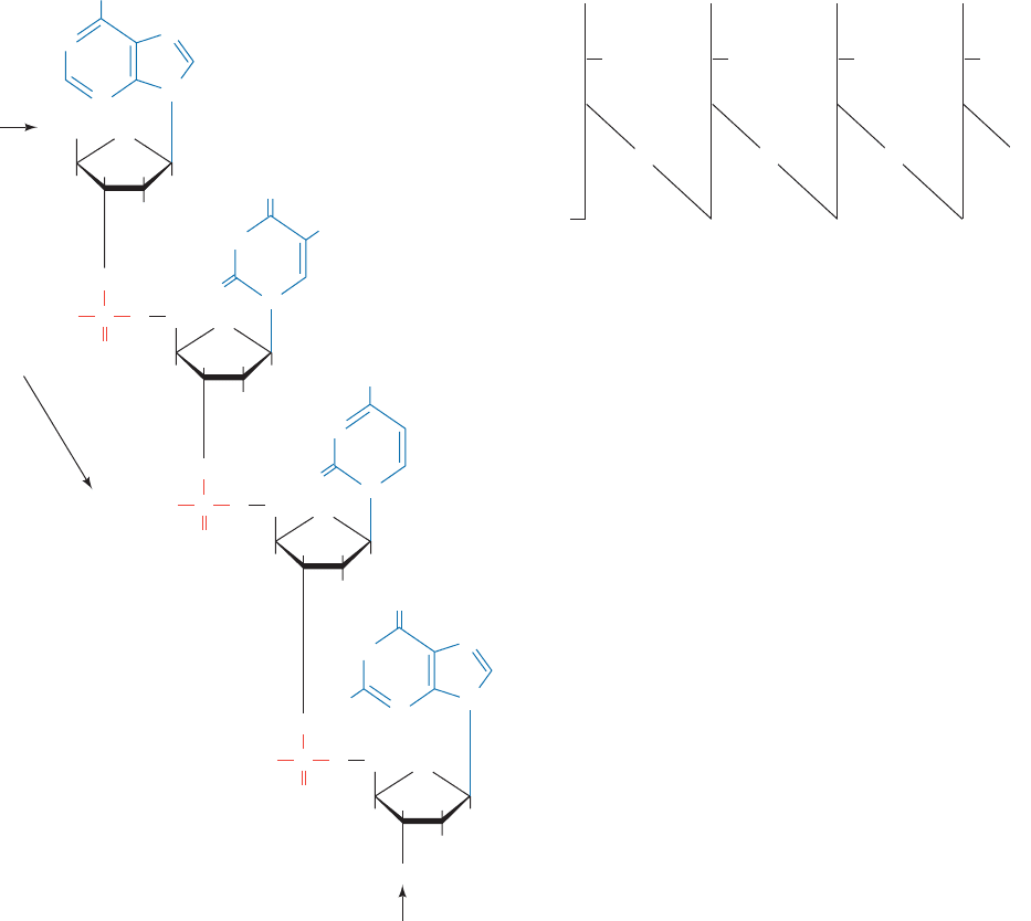
Figure 5-2 Chemical structure of a nucleic acid.
(a) The tetranucleotide adenyl-3¿,5¿-uridyl-3¿,5¿-cytidyl-
3¿,5¿-guanylyl-3¿-phosphate.The sugar atom numbers are
primed to distinguish them from the atomic positions of
the bases. By convention, a polynucleotide sequence is
written with its 5¿ end at the left and its 3¿ end to the
right.Thus, reading left to right, the phosphodiester
bond links neighboring ribose residues in the 5¿S3¿
direction.The above sequence may be abbreviated
ApUpCpGp or just AUCGp (where a “p” to the left
and/or right of a nucleoside symbol indicates a 5¿ and/or
a 3¿ phosphate group, respectively; see Table 5-1 for
other symbol definitions).The corresponding
deoxytetranucleotide, in which the 2¿-OH groups are
each replaced by H atoms and the base uracil (U) is
replaced by thymine (5-methyluracil; T), is abbreviated
d(ApTpCpGp) or d(ATCGp). (b) A schematic
representation of AUCGp. Here a vertical line denotes
a ribose residue, its attached base is indicated by the
corresponding one-letter abbreviation, and a diagonal
line flanking an optional “p” represents a phosphodiester
bond.The atom numbering of the ribose residues, which
is indicated here, is usually omitted.The equivalent
representation of deoxypolynucleotides differs only by
the absence of the 2¿-OH groups and the replacement
of U by T.
JWCL281_c05_082-128.qxd 5/31/10 2:01 PM Page 84
species of bacteria. It is, however, more or less constant
among related species; for example, in mammals G ⫹ C
ranges from 39% to 46%.
RNA, which usually occurs as single-stranded mole-
cules, has no apparent constraints on its base composition.
However, double-stranded RNA, which comprises the ge-
netic material of certain viruses, also obeys Chargaff’s rules
(here A base pairs with U in the same way it does with T in
DNA; Fig. 1-16). Conversely, single-stranded DNA, which
occurs in certain viruses, does not obey Chargaff’s rules. On
entering its host organism, however, such DNA is repli-
cated to form a double-stranded molecule, which then
obeys Chargaff’s rules.
b. Nucleic Acid Bases May Be Modified
Some DNAs contain bases that are chemical derivatives
of the standard set. For example, dA and dC in the DNAs
of many organisms are partially replaced by N
6
-methyl-dA
and 5-methyl-dC, respectively.
The altered bases are generated by the sequence-specific
enzymatic modification of normal DNA (Sections 5-5A and
30-7). The modified DNAs obey Chargaff’s rules if the de-
rivatized bases are taken as equivalent to their parent bases.
Likewise, many bases in RNAs and, in particular, those in
transfer RNAs (tRNAs; Section 32-2Aa) are derivatized.
c. RNA but Not DNA Is Susceptible to Base-
Catalyzed Hydrolysis
RNA is highly susceptible to base-catalyzed hydrolysis
by the reaction mechanism diagrammed in Fig. 5-3 so as to
yield a mixture of 2¿ and 3¿ nucleotides. In contrast, DNA,
which lacks 2¿-OH groups, is resistant to base-catalyzed hy-
drolysis and is therefore much more chemically stable than
RNA.This is probably why DNA rather than RNA evolved
to be the cellular genetic archive.
2 DNA IS THE CARRIER OF
GENETIC INFORMATION
Nucleic acids were first isolated in 1869 by Friedrich
Miescher and so named because he found them in the nuclei
of leukocytes (pus cells) from discarded surgical bandages.
The presence of nucleic acids in other cells was demon-
strated within a few years, but it was not until some 75 years
after their discovery that their biological function was elu-
cidated. Indeed, in the 1930s and 1940s it was widely held,
in what was termed the tetranucleotide hypothesis, that
nucleic acids have a monotonously repeating sequence of
all four bases, so that they were not suspected of having a
N
6
-Methyl-dA
N
N
N
N
N
dR
N
dR
CH
3
H
5-Methyl-dC
N
NH
2
N
CH
3
O
genetic function. Rather, it was generally assumed that
genes were proteins since proteins were the only biochem-
ical entities that, at that time, seemed capable of the re-
quired specificity. In this section, we outline the experi-
ments that established DNA’s genetic role.
A. Transforming Principle Is DNA
The virulent (capable of causing disease) form of pneumo-
coccus (Diplococcus pneumoniae), a bacterium that causes
pneumonia, is encapsulated by a gelatinous polysaccharide
coating that contains the binding sites (known as O-antigens;
Section 11-3Bc) through which it recognizes the cells it
infects. Mutant pneumococci that lack this coating, because
of a defect in an enzyme involved in its formation, are not
pathogenic (capable of causing disease). The virulent and
Section 5-2. DNA is the Carrier of Genetic Information 85
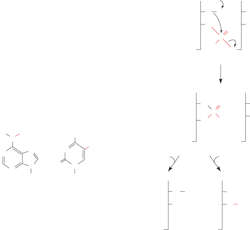
Figure 5-3 Mechanism of base-catalyzed RNA hydrolysis. The
base-induced deprotonation of the 2¿-OH group facilitates its
nucleophilic attack on the adjacent phosphorus atom, thereby
cleaving the RNA backbone.The resultant 2¿,3¿-cyclic phosphate
group subsequently hydrolyzes to either the 2¿ or the 3¿ phosphate.
RNA
2⬘
3⬘
...5⬘
OH
O
P
–
O
O
O
OH
O
...
B
n
B
n + 1
OH
–
..
...
B
n
O
O
P
O
O
–
OH
O
B
n + 1
...
HO
2ⴕ,3ⴕ-Cyclic nucleotide
OH
n
B
n
...
B
O
PO
3
2–
PO
3
2–
OH
O
...
2ⴕ-Nucleotide 3ⴕ-Nucleotide
or
H
2
OH
2
O
+
JWCL281_c05_082-128.qxd 2/19/10 4:46 PM Page 85
nonpathogenic pneumococci are known as the S and R
forms, respectively, because of the smooth and rough ap-
pearances of their colonies in culture (Fig. 5-4).
In 1928, Frederick Griffith made a startling discovery.
He injected mice with a mixture of live R and heat-killed S
pneumococci. This experiment resulted in the death of
most of the mice. More surprising yet was that the blood of
the dead mice contained live S pneumococci. The dead S
pneumococci initially injected into the mice had somehow
transformed the otherwise innocuous R pneumococci to
the virulent S form. Furthermore, the progeny of the trans-
formed pneumococci were also S; the transformation was
permanent. Eventually, it was shown that the transforma-
tion could also be made in vitro (outside a living organism;
literally “in glass”) by mixing R cells with a cell-free extract
of S cells. The question remained:What is the nature of the
transforming principle?
In 1944, Oswald Avery, Colin MacLeod, and Maclyn
McCarty, after a 10-year investigation, reported that trans-
forming principle is DNA.The conclusion was based on the
observations that the laboriously purified (few modern
fractionation techniques were then available) transforming
principle had all the physical and chemical properties of
DNA, contained no detectable protein, was unaffected by
enzymes that catalyze the hydrolysis of proteins and RNA,
and was totally inactivated by treatment with an enzyme
that catalyzes the hydrolysis of DNA. DNA must therefore
be the carrier of genetic information.
Avery’s discovery was another idea whose time had not
yet come. This seminal advance was initially greeted with
skepticism and then largely ignored. Indeed, even Avery did
not directly state that DNA is the hereditary material but
merely that it has “biological specificity.” His work, how-
ever, influenced several biochemists, including Erwin Char-
gaff, whose subsequent accurate determination of DNA
base ratios refuted the tetranucleotide hypothesis and
thereby indicated that DNA could be a complex molecule.
It was eventually demonstrated that eukaryotes are also
subject to transformation by DNA.Thus DNA, which cyto-
logical studies had shown resides in the chromosomes,
must also be the hereditary material of eukaryotes. In a
spectacular demonstration of eukaryotic transformation,
Ralph Brinster, in 1982, microinjected DNA bearing the
gene for rat growth hormone (a polypeptide) into the nu-
clei of fertilized mouse eggs (a technique discussed in Sec-
tion 5-5H) and implanted these eggs into the uteri of foster
mothers. The resulting “supermice” (Fig. 5-5), which had
high levels of rat growth hormone in their serum, grew to
nearly twice the weight of their normal littermates. Such
genetically altered animals are said to be transgenic.
B. The Hereditary Molecule of Many
Bacteriophages Is DNA
Electron micrographs of bacteria infected with bacterio-
phages show empty-headed phage “ghosts” attached to the
bacterial surface (Fig. 5-6). This observation led Roger
Herriott to suggest that “the virus may act like a little hypo-
dermic needle full of transforming principle,” which it in-
86 Chapter 5. Nucleic Acids, Gene Expression, and Recombinant DNA Technology
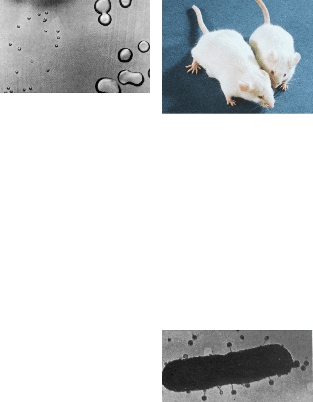
Figure 5-5 Transgenic mouse. The gigantic mouse (left) grew
from a fertilized ovum that had been microinjected with DNA
bearing the rat growth hormone gene. His normal-sized litter-
mate (right) is shown for comparison. [Courtesy of Ralph Brin-
ster, University of Pennsylvania.]
Figure 5-6 Bacteriophages attached to the surface of a
bacterium. This early electron micrograph shows an E. coli
cell to which bacteriophage T5 are adsorbed by their tails.
[Courtesy of Thomas F.Anderson, Fox Chase Cancer Center.]
Figure 5-4 Pneumococci. The large glistening colonies are
virulent S-type pneumococci that resulted from the transformation
of nonpathogenic R-type pneumococci (smaller colonies) by
DNA from heat-killed S pneumococci. [From Avery, O.T.,
MacLeod, C.M., and McCarty, M., J. Exp. Med. 79, 153 (1944).
Copyright © 1944 by Rockefeller University Press.]
JWCL281_c05_082-128.qxd 2/19/10 4:46 PM Page 86
jects into the bacterial host (Fig. 5-7). This proposal was
tested in 1952 by Alfred Hershey and Martha Chase as is
diagrammed in Fig. 5-8. Bacteriophage T2 was grown on E.
coli in a medium containing the radioactive isotopes
32
P
and
35
S. This labeled the phage capsid, which contains no P,
with
35
S, and its DNA, which contains no S, with
32
P. These
phages were added to an unlabeled culture of E. coli and,
after sufficient time was allowed for the phages to infect
the bacterial cells, the culture was agitated in a kitchen
blender so as to shear the phage ghosts from the bacterial
cells. This rough treatment neither injured the bacteria nor
altered the course of the phage infection. When the phage
ghosts were separated from the bacteria (by centrifugation;
Section 6-5), the ghosts were found to contain most of the
35
S, whereas the bacteria contained most of the
32
P. Further-
more, 30% of the
32
P appeared in the progeny phages but
only 1% of the
35
S did so. Hershey and Chase therefore
concluded that only the phage DNA was essential for the
production of progeny. DNA therefore must be the heredi-
tary material. In later years it was shown that, in a process
known as transfection, purified phage DNA can, by itself,
induce a normal phage infection of a properly treated bac-
terial host (transfection differs from transformation in that
the latter results from the recombination of the bacterial
chromosome with a fragment of homologous DNA).
In 1952, the state of knowledge of biochemistry was
such that Hershey’s discovery was much more readily ac-
cepted than Avery’s identification of the transforming prin-
ciple had been some 8 years earlier. Within a few months,
the first speculations arose as to the nature of the genetic
Section 5-2. DNA is the Carrier of Genetic Information 87
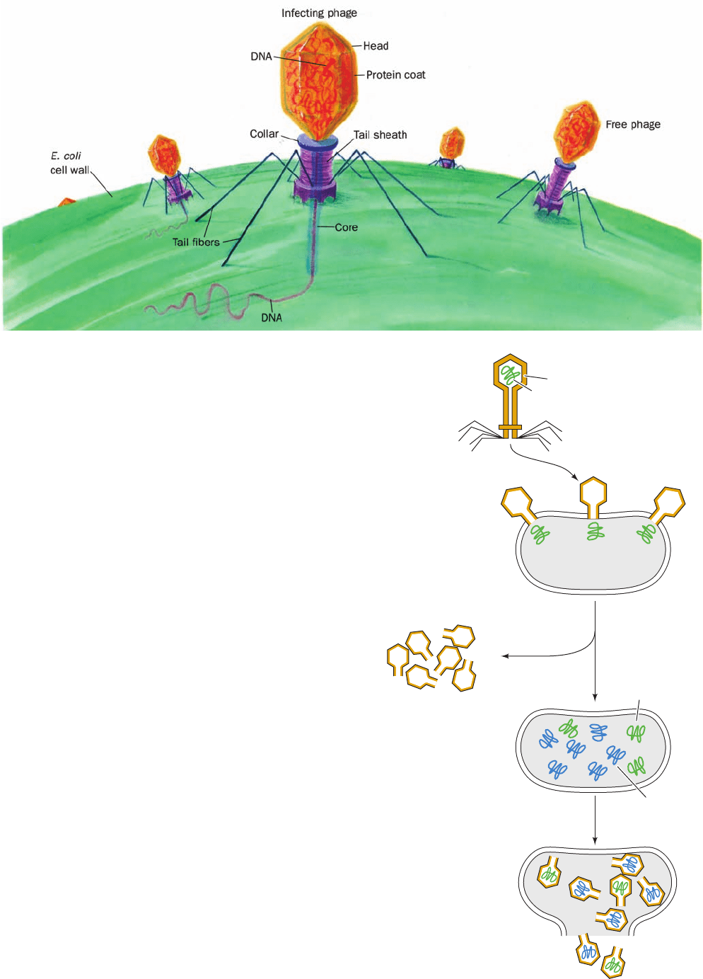
Figure 5-7 Diagram of T2 bacteriophage injecting its DNA
into an E. coli cell.
Figure 5-8 The Hershey–Chase experiment. This experiment
demonstrated that only the nucleic acid component of bacterio-
phages enters the bacterial host during phage infection.
Phage particle with
35
S-labeled shell
and
32
P-labeled
DNA
Phage infects E.coli;
only labeled DNA
enters cell
Parental
32
P-labeled
DNA replicates.
Replica DNA is unlabeled
Phages assemble:
only parental DNA
is
32
P-labeled.
Some progeny phages
are unlabeled.
No
35
S shell label remains
32
P labeled
DNA
Unlabeled
replica DNA
35
S
35
S phage shells
32
P
JWCL281_c05_082-128.qxd 2/19/10 4:46 PM Page 87
was prerequisite for the prediction of the correct hydrogen
bonding associations of the bases, was provided by Jerry
Donohue, an office mate of Watson and Crick and an expert
on the X-ray structures of small organic molecules.
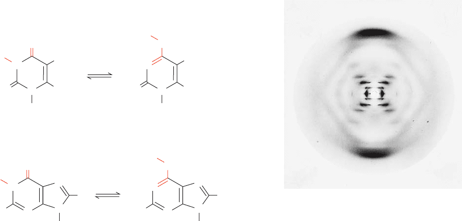
3. Information that DNA is a helical molecule. This was
provided by an X-ray diffraction photograph of a DNA fiber
taken by Rosalind Franklin (Fig. 5-10; DNA, being a thread-
like molecule, does not crystallize but, rather, can be drawn
out in fibers consisting of parallel bundles of molecules).
This photograph enabled Crick, an X-ray crystallographer
by training who had earlier derived the equations describing
diffraction by helical molecules, to deduce (a) that DNA is a
helical molecule and (b) that its planar aromatic bases form
a stack of parallel rings which is parallel to the fiber axis.
This information only provided a few crude landmarks
that guided the elucidation of the DNA structure. It mostly
sprang from Watson and Crick’s imaginations through model
building studies. Once the Watson–Crick model had been
published, however, its basic simplicity combined with its ob-
vious biological relevance led to its rapid acceptance. Later in-
vestigations have confirmed the essential correctness of the
Watson–Crick model,although its details have been modified.
A. The Watson–Crick Structure: B-DNA
Fibers of DNA assume the so-called B conformation, as in-
dicated by their X-ray diffraction patterns, when the coun-
terion is an alkali metal such as Na
⫹
and the relative humid-
ity is ⬎92%. B-DNA is regarded as the native (biologically
functional) form of DNA because, for example, its X-ray pat-
tern resembles that of the DNA in intact sperm heads.
88 Chapter 5. Nucleic Acids, Gene Expression, and Recombinant DNA Technology
code (the correspondence between the base sequence of
a gene and the amino acid sequence of a protein, Section
5-4Bb), and James Watson and Francis Crick were inspired
to investigate the structure of DNA. In 1955, it was shown
that the somatic cells of eukaryotes have twice the DNA of
the corresponding germ cells. When this observation was
proposed to be a further indicator of DNA’s genetic role,
there was little comment even though the same could be
said of any other chromosomal component.
3 DOUBLE HELICAL DNA
The determination of the structure of DNA by Watson and
Crick in 1953 is often said to mark the birth of modern mo-
lecular biology. The Watson–Crick structure of DNA is of
such importance because, in addition to providing the struc-
ture of what is arguably the central molecule of life, it sug-
gested the molecular mechanism of heredity. Watson and
Crick’s accomplishment, which is ranked as one of science’s
major intellectual achievements, tied together the less than
universally accepted results of several diverse studies:
1. Chargaff’s rules. At the time, the relationships A ⫽ T
and G ⫽ C were quite obscure because their significance was
not apparent. In fact, even Chargaff did not emphasize them.
2. Correct tautomeric forms of the bases. X-ray, nuclear
magnetic resonance (NMR), and spectroscopic investigations
have firmly established that the nucleic acid bases are over-
whelmingly in the keto tautomeric forms shown in Table 5-1.
In 1953, however, this was not generally appreciated. Indeed,
guanine and thymine were widely believed to be in their enol
forms (Fig. 5-9) because it was thought that the resonance sta-
bility of these aromatic molecules would thereby be maxi-
mized. Knowledge of the dominant tautomeric forms, which
Figure 5-9 Some possible tautomeric conversions for bases.
(a) Thymine and (b) guanine residues. Cytosine and adenine
residues can undergo similar proton shifts.
Figure 5-10 X-ray diffraction photograph of a vertically
oriented Na
ⴙ
DNA fiber in the B conformation taken by Rosalind
Franklin. This is the photograph that provided key information
for the elucidation of the Watson–Crick structure.The central
X-shaped pattern of spots is indicative of a helix, whereas the
heavy black arcs on the top and bottom of the diffraction pattern
correspond to a distance of 3.4 Å and indicate that the DNA
structure largely repeats every 3.4 Å along the fiber axis. [Courtesy
of Maurice Wilkins, King’s College, London.]
N
O
N
H
H
2
N
N
R
N
Guanine
(keto lactam form)or
H
N
O
N
H
H
2
N
N
R
N
H
Guanine
(enol lactim form)or
(b)
H
CH
3
CH
3
N
O
N
O
H
Thymine
(keto lactam form)
R
or
H
N
O
N
O
H
Thymine
(enol lactim form)
R
or
(a)
JWCL281_c05_082-128.qxd 2/19/10 4:46 PM Page 88
The Watson–Crick structure of B-DNA has the follow-
ing major features:
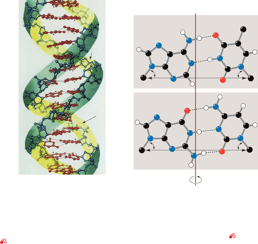
1. It consists of two polynucleotide strands that wind
about a common axis with a right-handed twist to form an
⬃20-Å-diameter double helix (Fig. 5-11). The two strands
are antiparallel (run in opposite directions) and wrap
around each other such that they cannot be separated with-
out unwinding the helix. The bases occupy the core of the
helix and the sugar–phosphate chains are coiled about its
periphery, thereby minimizing the repulsions between
charged phosphate groups.
2. The planes of the bases are nearly perpendicular to
the helix axis. Each base is hydrogen bonded to a base on
the opposite strand to form a planar base pair (Fig. 5-11). It
is these hydrogen bonding interactions, a phenomenon
known as complementary base pairing, that result in the
specific association of the two chains of the double helix.
3. The “ideal” B-DNA helix has 10 base pairs (bp) per
turn (a helical twist of 36° per bp) and, since the aromatic
bases have van der Waals thicknesses of 3.4 Å and are par-
tially stacked on each other (base stacking, Fig. 5-11), the
helix has a pitch (rise per turn) of 34 Å.
The most remarkable feature of the Watson–Crick
structure is that it can accommodate only two types of
base pairs: Each adenine residue must pair with a
thymine residue and vice versa, and each guanine residue
must pair with a cytosine residue and vice versa. The
geometries of these A ⴢ T and G ⴢ C base pairs, the so-called
Watson–Crick base pairs, are shown in Fig. 5-12. It can be
seen that both of these base pairs are interchangeable in
that they can replace each other in the double helix without
altering the positions of the sugar–phosphate backbone’s
Section 5-3. Double Helical DNA 89
Figure 5-11 Three-dimensional structure of B-DNA. The
repeating helix in this ball-and-stick drawing is based on
the X-ray structure of the self-complementary dodecamer
d(CGCGAATTCGCG) determined by Richard Dickerson and
Horace Drew. The view is perpendicular to the helix axis.The
sugar–phosphate backbones (blue with blue-green ribbon
outlines) wind about the periphery of the molecule in opposite
directions.The bases (red), which occupy its core, form hydrogen
bonded base pairs. H atoms have been omitted for clarity.
[Illustration, Irving Geis. Image from the Irving Geis Collection,
Howard Hughes Medical Institute. Reprinted with permission.]
See Interactive Exercise 1 and Kinemage 2-1
Minor
groove
Major
groove
B-DNA
51.5°
51.5°
AT
C
G
10.85 Å
10.85 Å
Major groove
Minor groove
6
6
5
4
3
2
1
7
8
9
4
4
5
6
3
2
1
2
1′
7
8
9
6
5
4
3
2
1
4
45
6
3
2
1
1′
Minor groove
Major groove
51.5°
51.5°
1′
1′
CH
3
2
6
2
Figure 5-12 Watson–Crick base pairs. The line joining the C1¿
atoms is the same length in both base pairs and makes equal an-
gles with the glycosidic bonds to the bases.This gives DNA a se-
ries of pseudo-twofold symmetry axes (often referred to as dyad
axes) that pass through the center of each base pair (red line)
and are perpendicular to the helix axis. Note that A ⴢ T base pairs
associate via two hydrogen bonds, whereas C ⴢ G base pairs are
joined by three hydrogen bonds. [After Arnott, S., Dover, S.D.,
and Wonacott, A.J., Acta Cryst. B25, 2192 (1969).]
See
Kinemages 2-2 and 17-2
JWCL281_c05_082-128.qxd 10/21/10 6:30 PM Page 89

C1¿ atoms. Likewise, the double helix is undisturbed by
exchanging the partners of a Watson–Crick base pair, that
is, by changing a G ⴢ C to a C ⴢ G or an A ⴢ T to a T ⴢ A. In
contrast, any other combination of bases (e.g., A ⴢ G or
A ⴢ C) would significantly distort the double helix since
the formation of a non-Watson–Crick base pair would re-
quire considerable reorientation of the sugar–phosphate
chain.
B-DNA has two deep exterior grooves that wind be-
tween its sugar–phosphate chains as a consequence of the
helix axis passing through the approximate center of each
base pair. However, the grooves are of unequal size (Fig.
5-11) because (1) the top edge of each base pair, as drawn
in Fig. 5-12, is structurally distinct from the bottom edge;
and (2) the deoxyribose residues are asymmetric.The minor
groove exposes that edge of a base pair from which its C1¿
atoms extend (opening toward the bottom in Fig. 5-12),
whereas the major groove exposes the opposite edge of
each base pair (the top of Fig. 5-12).
Although B-DNA is, by far, the most prevalent form of
DNA in the cell, double helical DNAs and RNAs can as-
sume several distinct structures. The structures of these
other double helical nucleic acids are discussed in Section
29-1B.
B. DNA Is Semiconservatively Replicated
The Watson–Crick structure can accommodate any se-
quence of bases on one polynucleotide strand if the oppo-
site strand has the complementary base sequence. This im-
mediately accounts for Chargaff’s rules. More importantly,
it suggests that hereditary information is encoded in the se-
quence of bases on either strand. Furthermore, each
polynucleotide strand can act as a template for the forma-
tion of its complementary strand through base pairing in-
teractions (Fig. 1-17). The two strands of the parent mole-
cule must therefore separate so that a complementary
daughter strand may be enzymatically synthesized on the
surface of each parent strand. This results in two molecules
of duplex (double-stranded) DNA, each consisting of one
polynucleotide strand from the parent molecule and a
newly synthesized complementary strand. Such a mode of
replication is termed semiconservative in contrast with
conservative replication, which, if it occurred, would result
in a newly synthesized duplex copy of the original DNA
molecule with the parent DNA molecule remaining intact.
The mechanism of DNA replication is the main subject of
Chapter 30.
The semiconservative nature of DNA replication was
elegantly demonstrated in 1958 by Matthew Meselson and
Franklin Stahl.The density of DNA was increased by label-
ing it with
15
N, a heavy isotope of nitrogen (
14
N is the natu-
rally abundant isotope).This was accomplished by growing
E. coli for 14 generations in a medium that contained
15
NH
4
Cl as the only nitrogen source. The labeled bacteria
were then abruptly transferred to an
14
N-containing
medium, and the density of their DNA was monitored as a
function of bacterial growth by equilibrium density gradi-
ent ultracentrifugation (a technique for separating macro-
molecules according to their densities, which Meselson,
Stahl, and Jerome Vinograd had developed for the purpose
of distinguishing
15
N-labeled DNA from unlabeled DNA;
Section 6-5Bb).
The results of the Meselson–Stahl experiment are dis-
played in Fig. 5-13. After one generation (doubling of the
cell population), all of the DNA had a density exactly
halfway between the densities of fully
15
N-labeled DNA
and unlabeled DNA. This DNA must therefore contain
equal amounts of
14
N and
15
N, as is expected after one gen-
eration of semiconservative replication. Conservative
DNA replication, in contrast, would result in the preserva-
tion of the parental DNA, so that it maintained its original
density, and the generation of an equal amount of unla-
beled DNA.After two generations, half of the DNA mole-
cules were unlabeled and the remainder were
14
N–
15
N hy-
brids. This is also in accord with the predictions of the
semiconservative replication model and in disagreement
with the conservative replication model. In succeeding
generations, the amount of unlabeled DNA increased rel-
ative to the amount of hybrid DNA, although the hybrid
never totally disappeared. This is again in harmony with
semiconservative replication but at odds with conservative
replication, which predicts that the fully labeled parental
DNA will always be present and that hybrid DNA never
forms.
C. Denaturation and Renaturation
When a solution of duplex DNA is heated above a charac-
teristic temperature, its native structure collapses and its
two complementary strands separate and assume a flexi-
ble and rapidly fluctuating conformational state known as
a random coil (Fig. 5-14). This denaturation process is
accompanied by a qualitative change in the DNA’s phys-
ical properties. For instance, the characteristic high vis-
cosity of native DNA solutions, which arises from the re-
sistance to deformation of its rigid and rodlike duplex
molecules, drastically decreases when the duplex DNA
decomposes (denatures) to two relatively freely jointed
single strands.
a. DNA Denaturation Is a Cooperative Process
The most convenient way of monitoring the amount of
nucleic acid present is by its ultraviolet (UV) absorbance
spectrum. A solution containing a solute that absorbs light
does so according to the Beer–Lambert law,
[5.1]
where A is the solute’s absorbance (alternatively, its optical
density), I
0
is the incident intensity of light at a given wave-
length l, I is its transmitted intensity at l, ε is the molar ex-
tinction coefficient of the solute at l, c is its molar concen-
tration, and l is the length of the light path in centimeters.
The value of ε varies with l; a plot of ε versus l for the
A ⫽⫺log
a
I
I
0
b⫽ εcl
90 Chapter 5. Nucleic Acids, Gene Expression, and Recombinant DNA Technology
JWCL281_c05_082-128.qxd 2/19/10 4:46 PM Page 90
