Voet D., Voet Ju.G. Biochemistry
Подождите немного. Документ загружается.

can be easily freed of ammonium sulfate by dissolving the
protein precipitate in a minimum volume of suitable buffer
and applying this solution to a column of gel with an exclu-
sion limit less than the molecular mass of the protein. On
elution of the column with buffer, the protein will precede
the ammonium sulfate through the column.
Dextran and agarose gels can be derivatized with
ionizable groups such as DEAE and CM to form ion
exchange gels (Section 6-3Ab). Substances that are chro-
matographed on these gels are therefore subject to separa-
tion according to their ionic charges as well as their sizes
and shapes.
c. Dialysis Is a Form of Molecular Filtration
Dialysis is a process that separates molecules according
to size through the use of semipermeable membranes con-
taining pores of less than macromolecular dimensions.
These pores allow small molecules, such as those of sol-
vents, salts, and small metabolites, to diffuse across the
membrane but block the passage of larger molecules. Cel-
lophane (cellulose acetate) is the most commonly used
dialysis material, although several other substances such as
cellulose and collodion are similarly employed. These are
available in a wide variety of molecular weight cutoff val-
ues (the size of the smallest particle that cannot penetrate
the membrane) that range from 0.1 to 500 kD.
Dialysis (which is not considered to be a form of chrom-
atography) is used to change the solvent in which macro-
molecules are dissolved. A macromolecular solution is
sealed inside a dialysis bag (usually made by knotting dialy-
sis membrane tubing at both ends), which is immersed in a
relatively large volume of the new solvent (Fig. 6-11a).After
several hours of stirring,the solutions will have equilibrated,
but with the macromolecules remaining inside the dialysis
bag (Fig. 6-11b). The process can be repeated several times
to replace one solvent system completely by another.
Dialysis has been largely supplanted by a related tech-
nique known as ultrafiltration in which a macromolecular
solution is forced, under pressure or by centrifugation,
through a semipermeable membranous disk, which can be
made from a variety of materials including cellulose ac-
etate, nylon, and polyvinylidene fluoride (PVDF). Solvent
and small solutes pass through the membrane, leaving
behind a more concentrated macromolecular solution. Ul-
trafiltration can thus be used to desalt a macromolecular
solution. Since ultrafiltration membranes with different
pore sizes are available, ultrafiltration can also be used to
separate different-sized macromolecules.
Solvent may also be removed from a sample solution
through lyophilization (freeze-drying), a process in which
the solution is frozen and the solvent sublimed away under
vacuum. Lyophilization is usually used to prepare biologi-
cal materials for long-term storage or transport.
C. Affinity Chromatography
A striking characteristic of many proteins is their ability to
bind specific molecules tightly but noncovalently. This
property can be used to purify such proteins by affinity
chromatography (Fig. 6-12). In this technique, a molecule,
Section 6-3. Chromatographic Separations 141
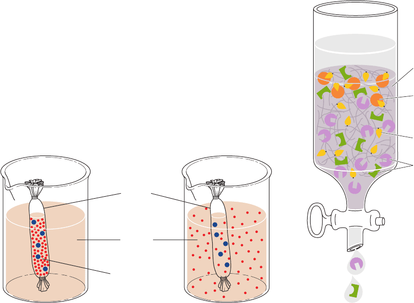
Figure 6-11 Use of dialysis to separate small and large
molecules. (a) Only small molecules can diffuse through the
pores in the bag, which is shown here as a tube knotted at both
ends. (b) At equilibrium, the concentrations of small molecules
are nearly the same inside and outside the bag, whereas the
macromolecules remain in the bag.
At equilibriumAt start of
dialysis
(a) (b)
Dialysis
membrane
Solvent
Concentrated
solution
Specific binding
of molecule to
matrix ligand
Macromolecules
with differing
ligand-binding
sites
Matrix-anchored
ligand
Solid resin
matrix
Figure 6-12 Affinity chromatography. A ligand (yellow) is
covalently anchored to a porous matrix.The sample mixture
(whose ligand-binding sites are represented by the cutout squares,
semicircles, and triangles) is passed through the column. Only
certain molecules (represented by orange circles) specifically bind
to the ligand; the others are washed through the column.
JWCL281_c06_129-162.qxd 2/22/10 2:25 PM Page 141
known as a ligand (in analogy with the ligands of coordina-
tion compounds), which specifically binds to the protein of
interest, is covalently attached to an inert and porous ma-
trix. When an impure protein solution is passed through this
chromatographic material, the desired protein binds to the
immobilized ligand, whereas other substances are washed
through the column with the buffer. The desired protein can
then be recovered in highly purified form by changing the
elution conditions such that the protein is released from the
chromatographic matrix. The great advantage of affinity
chromatography is its ability to exploit the desired pro-
tein’s unique biochemical properties rather than the small
differences in physicochemical properties between pro-
teins that other chromatographic methods must utilize.
The chromatographic matrix in affinity chromatography
must be chemically inert, have high porosity, and have large
numbers of functional groups capable of forming covalent
linkages to ligands. Of the few materials available that meet
these criteria, agarose, which has numerous free hydroxyl
groups, is by far the most widely used. If the ligand has a pri-
mary amino group that is not essential for its binding to the
protein of interest, the ligand can be covalently linked to
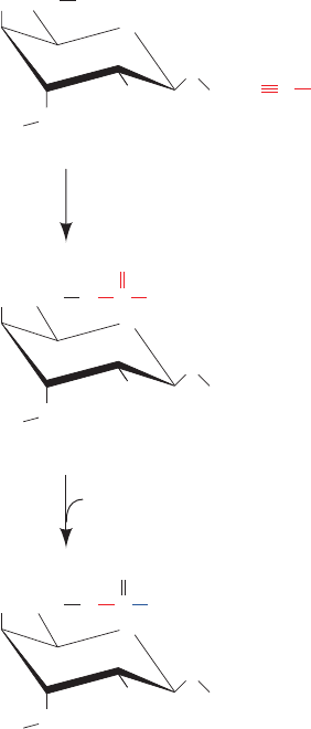
the agarose in a two-step process (Fig. 6-13):
1. Agarose is reacted with cyanogen bromide to form
an “activated” but stable intermediate (which is commer-
cially available).
2. Ligand reacts with the activated agarose to form co-
valently bound product.
Many proteins are unable to bind their cyanogen bro-
mide–coupled ligands due to steric interference with the
agarose matrix. This problem is alleviated by attaching the
ligand to the agarose by a flexible “spacer” group. This is
conveniently done through the use of commercially avail-
able activated resins. One such resin is “epoxy-activated”
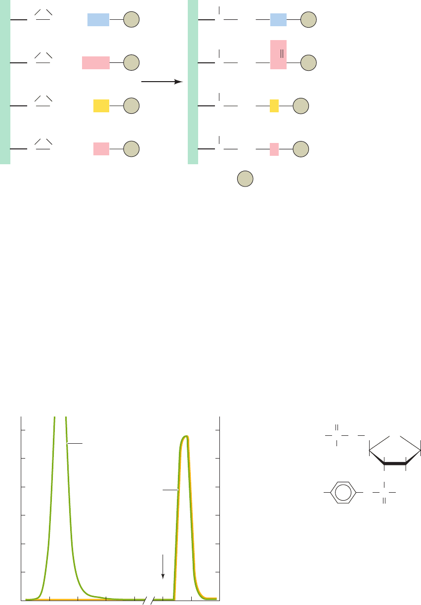
agarose, in which a spacer group (containing,e.g., a chain of
12 atoms) links the resin to a reactive epoxy group. The
epoxy group can react with many of the nucleophilic
groups on ligands, thereby permitting the ligand of choice
to be covalently linked to the agarose via a tether of de-
fined length (Fig. 6-14).
The ligand used in the affinity chromatography isola-
tion of a particular protein must have an affinity for the
protein high enough to immobilize it on the agarose gel
but not so high as to prevent its subsequent release. If the
ligand is a substrate for an enzyme being isolated, the
chromatography conditions must be such that the enzyme
does not function catalytically or the ligand will be de-
stroyed.
After a protein has been bound to an affinity chro-
matography column and washed free of impurities, it must
be released from the column. One method of doing so is to
elute the column with a solution of a compound that has
higher affinity for the protein-binding site than the bound
ligand.Another is to alter the solution conditions such that
the protein–ligand complex is no longer stable, for exam-
ple, by changes in pH, ionic strength, and/or temperature.
However, care must be taken that the solution conditions
are not so inhospitable to the protein being isolated that it
is irreversibly damaged. An example of protein purifica-
tion by affinity chromatography is shown in Fig. 6-15.
Affinity chromatography has been used to isolate such
substances as enzymes, antibodies, transport proteins, hor-
mone receptors, membranes, and even whole cells. For in-
stance, the protein hormone insulin (Section 7-1) has been
covalently attached to agarose and used to isolate insulin
receptor (Section 19-3Ac), a cell-surface protein whose
other properties were previously unknown and which is
present in tissues in only very small amounts. Genetic engin-
eering techniques (Section 5-5G) have permitted the
affinity purification of proteins for which there is no useful
ligand by forming a fusion protein with (linking them to) a
protein for which a useful ligand is available. For example,
fusion proteins whose N-terminal portions consist of the
enzyme glutathione-S-transferase (GST; Section 25-7Cb)
tightly bind the tripeptide glutathione (Section 21-2Ba)
and hence are readily purified by affinity chromatography
on glutathione–agarose.
The separation power of affinity chromatography for a
specific protein is often far greater than that of other chro-
matographic techniques. Indeed, the replacement of many
chromatographic steps in a tried-and-true protein isolation
142 Chapter 6. Techniques of Protein and Nucleic Acid Purification
Figure 6-13 Covalent linking of ligand to agarose. The
formation of cyanogen bromide–activated agarose (top) and its
reaction with a primary amine to form a covalently attached
ligand for affinity chromatography (bottom).
CH
2
HO
OH
OH
O
O
O
NC
Cyanogen
bromide
Agarose
1 OH
⫺
Br
⫹
NH
3
⫹
CH
2
HO
O C
NH
Br
OH
O
O
O
“Activated” agarose
2 RNH
2
H
2
O⫹
HBr⫹
CH
2
HO
O C
O
NHR
OH
O
O
O
JWCL281_c06_129-162.qxd 2/22/10 2:25 PM Page 142
protocol by a single affinity chromatographic step often re-
sults in purer protein in higher yield.
a. Immunoaffinity Chromatography Employs the
Binding Specificity of Monoclonal Antibodies
A melding of immunochemistry with affinity chro-
matography has generated a powerful method for purify-
ing biological molecules. Cross-linking monoclonal anti-
bodies (Section 6-1Da) to a suitable column material yields
a substance that will bind only the protein against which
the antibody has been raised. Such immunoaffinity chro-
matography can achieve a 10,000-fold purification in a
single step. Disadvantages of immunoaffinity chroma-
tography include the technical difficulty of producing mon-
oclonal antibodies and the harsh conditions that are often
required to elute the bound protein.
D. Other Chromatographic Techniques
A number of other chromatographic techniques are of bio-
chemical value. These are briefly discussed below.
a. Adsorption Chromatography Separates
Nonpolar Substances
In adsorption chromatography (the original chromato-
graphic method), molecules are physically adsorbed on the
Section 6-3. Chromatographic Separations 143
Figure 6-14 Derivatization of epoxy-
activated agarose. Various types of
nucleophilic groups can be covalently
attached to epoxy-activated agarose via
reaction with its epoxide groups.
CH
2
C
Spacer arm
Spacer arm
Spacer arm
Spacer arm
O
H
2
NR
CH
2
C
O
HOOC
Agarose
R
CH
2
C
O
HS R
CH
2
C
O
HO R
CH
2
C
OH
HN R
CH
2
C
OH
OC
Agarose
R
CH
2
C
OH
SR
CH
2
C
OH
OR
R = Ligand
O
0
Absorbance at 280 nm
3
2
1
0
10
Acetic acid
pH 3.1
Absorbance
20
Effluent (mL)
50 60
Nuclease activity
(a)
Nuclease
activity
HO
HH
CH
2
H
2
N
H
Thymine
H
PO
–
O
O
O
PO
–
O
O
–
(b)
O
Figure 6-15 (a) The purification of staphylococcal nuclease (a
DNA-hydrolyzing enzyme) by affinity chromatography. The
compound shown in Part b, whose bisphosphothymidine moiety
specifically binds to the enzyme, was covalently linked to
cyanogen bromide–activated agarose.The column was equilibrated
with 0.05M borate buffer, pH 8.0, containing 0.01M CaCl
2
, and
approximately 40 mg of partially purified material was applied to
the column.After 50 mL of buffer had been passed through the
column to wash away the unbound material, 0.1M acetic acid was
added to elute the enzyme. All of the original enzymatic activity,
comprising 8.2 mg of pure nuclease, was recovered. [After
Cuatrecasas, P., Wilchek, M., and Anfinsen, C.B., Proc. Natl. Acad.
Sci. 61, 636 (1968).]
JWCL281_c06_129-162.qxd 2/22/10 2:25 PM Page 143

surface of an insoluble substance such as alumina (Al
2
O
3
),
charcoal, diatomaceous earth (also called kieselguhr, the
siliceous fossils of unicellular organisms known as di-
atoms), finely powdered sucrose, or silica gel (silicic acid),
through van der Waals and hydrogen bonding associations.
The molecules are then eluted from the column by a pure
solvent such as chloroform, hexane, or ethyl ether or by a
mixture of such solvents. The separation process is based
on the partition of the various substances between the po-
lar column material and the nonpolar solvent. This proce-
dure is most often used to separate nonpolar molecules
rather than proteins.
b. Hydroxyapatite Chromatography
Separates Proteins
Proteins are adsorbed by gels of crystalline hydroxyap-
atite, an insoluble form of calcium phosphate with empiri-
cal formula Ca
5
(PO
4
)
3
OH. The separation of the proteins
occurs on gradient elution of the column with phosphate
buffer (the presence of other anions is unimportant). The
physicochemical basis of this fractionation procedure is not
fully understood but apparently involves the adsorption of
anions to the Ca
2⫹
sites and cations to the sites of the
hydroxyapatite crystalline lattice.
c. Paper Chromatography Separates Small
Polar Molecules
Paper chromatography, developed in 1941 by Archer
Martin and Richard Synge, played an indispensable role in
biochemical analysis due to its ability to efficiently sepa-
rate small molecules such as amino acids, oligopeptides, nu-
cleotides, and oligonucleotides and its requirement for
only the simplest of equipment. Although paper chro-
matography has been supplanted by the more modern
techniques discussed in this chapter, we briefly describe it
here because of its historical importance and because
many of its principles and ancillary techniques are directly
applicable to the more modern techniques.
In paper chromatography (Fig. 6-16), a few drops of so-
lution containing a mixture of the components to be sepa-
rated are applied (spotted) ⬃2 cm above one end of a strip
of filter paper.After drying, that end of the paper is dipped
into a solvent mixture consisting of aqueous and organic
components; for example, water/butanol/acetic acid in a
4:5:1 ratio, 77% aqueous ethanol, or 6:7:7 water/t-amyl al-
cohol/pyridine. The paper should also be in contact with
the equilibrium vapors of the solvent. The solvent soaks
into the paper by capillary action because of the fibrous na-
ture of the paper. The aqueous component of the solvent
binds to the cellulose of the paper and thereby forms a sta-
tionary gel-like phase with it. The organic component of
the solvent continues migrating, thus forming the mobile
phase.
The rates of migration of the various substances being
separated are governed by their relative solubilities in the
polar stationary phase and the nonpolar mobile phase. In a
single step of the separation process, a given solute is dis-
tributed between the mobile and stationary phases accord-
PO
3⫺
4
ing to its partition coefficient, an equilibrium constant de-
fined as
[6.4]
The molecules are therefore separated according to their po-
larities, with nonpolar molecules moving faster than polar
ones.
After the solvent front has migrated an appropriate dis-
tance, the chromatogram is removed from the solvent and
dried. The separated materials, if not colored, may be de-
tected by such means as their radioactivity, their fluores-
cence or ability to quench the normal fluorescence of pa-
per under UV light, or by spraying the chromatogram with
a solution of a substance that forms a colored product on
reaction with the substance(s) under investigation.
The migration rate of a substance may be expressed ac-
cording to the ratio
[6.5]
For a given solvent system and paper type, each substance
has a characteristic R
f
value.
A complex mixture that is incompletely separated in a
single paper chromatogram can often be fully resolved by
two-dimensional paper chromatography (Fig. 6-17). In this
technique, a chromatogram is made as previously de-
scribed except that the sample is spotted onto one corner
of a sheet of filter paper and the chromatogram is run par-
allel to an edge of the paper.After the chromatography has
been completed and the paper dried, the chromatogram is
R
f
⫽
distance traveled by substance
distance traveled by solvent front
K
p
⫽
concentration in stationary phase
concentration in mobile phase
144 Chapter 6. Techniques of Protein and Nucleic Acid Purification
Support
rod
Paper
Solvent
front
Solvent for
development
Sample
Clip
Glass
jar
Ascending paper
chromatography
Figure 6-16 Experimental arrangement for paper
chromatography.
JWCL281_c06_129-162.qxd 2/22/10 2:25 PM Page 144
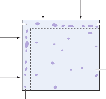
rotated 90° and is chromatographed parallel to the second
edge using another solvent system. Since each compound
migrates at a characteristic rate in a given solvent system,
the second chromatographic step should greatly enhance
the separation of the mixture into its components.
d. Thin Layer Chromatography Is Used to Separate
Organic Molecules
In thin layer chromatography (TLC), a thin (⬃0.25 mm)
coating of a solid material spread on a glass or plastic plate
is utilized in a manner similar to that of the paper in paper
chromatography. In the case of TLC, however, the chro-
matographic material can be a variety of substances such as
ion exchangers, gel filtration agents, and physical adsor-
bents. According to the choice of solvent for the mobile
phase, the separation may be based on adsorption, parti-
tion, gel filtration, ion exchange processes, or some combi-
nation of these. The advantages of thin layer chromatogra-
phy in convenience, rapidity, and high resolution have led
to its routine use in the analysis of organic molecules.
e. Reverse-Phase Chromatography Separates
Nonpolar Substances Including Denatured Proteins
Reverse-phase chromatography (RPC) is a form of liq-
uid–liquid partition chromatography in which the polar
character of the phases is reversed relative to that of paper
chromatography:The stationary phase typically consists of
a nonpolar liquid immobilized on silica substituted with n-
alkyl chains such as C
8
and C
18
, and the mobile phase is a
more polar liquid. Reverse-phase chromatography was
first developed to separate mixtures of nonpolar sub-
stances such as lipids but has also been found to be effec-
tive in separating polar substances such as oligonucleotides
and proteins, provided that they have exposed nonpolar
areas. Although nonpolar side chains tend to inhabit the
water-free interiors of native proteins (Section 8-3Bb),
denaturation results in the exposure of these side chains to
the solvent. Even when the protein is still in the native
state, a significant fraction of these hydrophobic groups
are at least partially exposed to the solvent at the protein
surface. Consequently, under suitable conditions, proteins
hydrophobically interact with the nonpolar groups on an
immobilized matrix.The hydrophobic interactions in RPC
are strong,so the eluting mobile phase must be highly non-
polar (containing high concentrations of organic solvents
such as acetonitrile) to dislodge adsorbed substances from
the stationary phase. RPC therefore usually denatures
proteins.
f. Hydrophobic Interaction Chromatography
Separates Native Proteins on the Basis
of Surface Hydrophobicity
Hydrophobic interactions form the basis not only of
RPC but of hydrophobic interaction chromatography
(HIC). However, whereas the stationary phase in RPC is
strongly hydrophobic in character, often resulting in pro-
tein denaturation, in HIC it is a hydrophilic substance, such
as an agarose gel, that is only lightly substituted with hy-
drophobic groups, usually octyl or phenyl residues. The re-
sulting hydrophobic interactions in HIC are therefore rela-
tively weak, so proteins maintain their native structures.
The eluants in HIC, whose gradients must progressively re-
duce these weak hydrophobic interactions, are aqueous
buffers with, for example, decreasing salt concentrations
(hydrophobic interactions are strengthened by increased
ionic strength; Section 6-2A), increasing concentrations of
detergents, or increasing pH. Thus, HIC separates native
proteins according to their degree of surface hydrophobic-
ity, a criterion that differs from those on which other types
of chromatography are based.
g. Metal Chelation Affinity Chromatography
Separates Proteins with Metal-Chelating Groups
In metal chelation affinity chromatography, a divalent
metal ion such as Mn
2⫹
,Zn
2⫹
, or Ni
2⫹
is attached to a chro-
matographic matrix such as agarose beads covalently
linked to metal-chelating groups under conditions that
proteins bearing metal-chelating groups (e.g., multiple His
or Cys side chains) are retained. Recombinant DNA tech-
niques (Section 5-5G) can be used to append a segment of
six consecutive His residues, known as a His Tag, to the N-
or C-terminus of the polypeptide to be isolated. This cre-
ates a metal ion-binding site that allows the recombinant
protein to be purified by metal chelation affinity chro-
matography. After the protein has been eluted, usually by
altering the pH, the His-Tag can be removed by the action
of a specific protease whose recognition sequence cleaves
the (His)
6
sequence from the rest of the protein.
h. HPLC Has Permitted Greatly
Improved Separations
In high-performance liquid chromatography (HPLC), a
separation may be based on adsorption, ion exchange, size
exclusion, HIC, or RPC as previously described. The sepa-
Section 6-3. Chromatographic Separations 145
Figure 6-17 Two-dimensional paper chromatography.
Origin
Direction
of flow
of second
solvent
Direction of flow
of first solvent
Separation
if only
first solvent
is used
Separation
if both solvents
are used
sequentially
Separation
if only second
solvent is used
JWCL281_c06_129-162.qxd 7/20/10 5:23 PM Page 145

rations are greatly improved, however, through the use of
high-resolution columns, and the column retention times
are much reduced.The narrow and relatively long columns
are packed with a noncompressible matrix of fine (1–10
m in diameter) silica beads, whose available hydroxyl
groups can be derivatized with many of the commonly used
functional groups of ion exchange chromatography, RPC,
HIC, or affinity chromatography. Alternatively, glass or
plastic beads may be coated with a thin layer of the station-
ary phase. The mobile phase is one of the solvent systems
previously discussed, including gradient elutions with bi-
nary or even ternary mixtures. In the case of HPLC, how-
ever, the mobile phase is forced through the tightly packed
column at pressures of up to 15,000 psi (pounds per square
inch), leading to greatly reduced analysis times. The elu-
tants are detected as they leave the column according to
their UV absorption, refractive index, or fluorescence.
The advantages of HPLC are
1. Its high resolution, which permits the routine purifi-
cation of mixtures that have defied separation by other
techniques.
2. Its speed, which permits separations to be accom-
plished in as little as a few minutes.
3. Its high sensitivity, which, in favorable cases, permits
the quantitative estimation of less than picomole quantities
of materials.
4. Its capacity for automation.
Thus, few biochemistry laboratories now function without
access to at least one HPLC system. HPLC is also often uti-
lized in the clinical analyses of body fluids because it can
rapidly, routinely, and automatically yield reliable quantita-
tive estimates of nanogram quantities of biological materi-
als such as vitamins, steroids, lipids, and drug metabolites.
4 ELECTROPHORESIS
Electrophoresis, the migration of ions in an electric field, is
widely used for the analytical separation of biological mol-
ecules. The laws of electrostatics state that the electrical
force, F
electric
, on an ion with charge q in an electric field of
strength E is expressed by
[6.6]
The resulting electrophoretic migration of the ion through
the solution is opposed by a frictional force
[6.7]
where v is the rate of migration (velocity) of the ion and f
is its frictional coefficient. The frictional coefficient is a
measure of the drag that the solution exerts on the moving
ion and is dependent on the size, shape, and state of solva-
tion of the ion as well as on the viscosity of the solution (Sec-
tion 6-5A). In a constant electric field, the forces on the ion
balance each other:
[6.8]qE ⫽ vf
F
friction
⫽ vf
F
electric
⫽ qE
so that each ion moves with a constant characteristic veloc-
ity. An ion’s electrophoretic mobility, , is defined
[6.9]
The electrophoretic (ionic) mobilities of several common
small ions in H
2
O at 25°C are listed in Table 2-2.
Equation [6.9] really applies only to ions at infinite dilu-
tion in a nonconducting solvent. In aqueous solutions, poly-
electrolytes such as proteins are surrounded by a cloud of
counterions, which impose an additional electric field of
such magnitude that Eq. [6.9] is, at best, a poor approxima-
tion of reality. Unfortunately, the complexities of ionic so-
lutions have, so far, precluded the development of a theory
that can accurately predict the mobilities of polyelec-
trolytes. Equation [6.9], however, correctly indicates that
molecules at their isoelectric points, pI, have zero elec-
trophoretic mobility. Furthermore, for proteins and other
polyelectrolytes that have acid–base properties, the ionic
charge, and hence the electrophoretic mobility, is a func-
tion of pH.
The use of electrophoresis to separate proteins was first
reported in 1937 by the Swedish biochemist Arne Tiselius.
The technique he introduced, moving boundary elec-
trophoresis, was one of the few powerful analytical tech-
niques available in the early years of protein chemistry.
However, since this method takes place entirely in solu-
tion, preventing the convective mixing of the migrating
proteins necessitates a cumbersome apparatus that re-
quires very large samples. Moving boundary electro-phore-
sis has therefore been supplanted by zone electrophoresis,
a technique in which the sample is constrained to move in
a solid support such as filter paper, cellulose acetate, or,
most commonly, a gel. This largely eliminates the convec-
tive mixing of the sample that limits the resolution achiev-
able by moving boundary electrophoresis. Moreover, in
zone electrophoresis, the various sample components mi-
grate as discrete bands (zones) and hence only small quan-
tities of materials are required.
A. Paper Electrophoresis
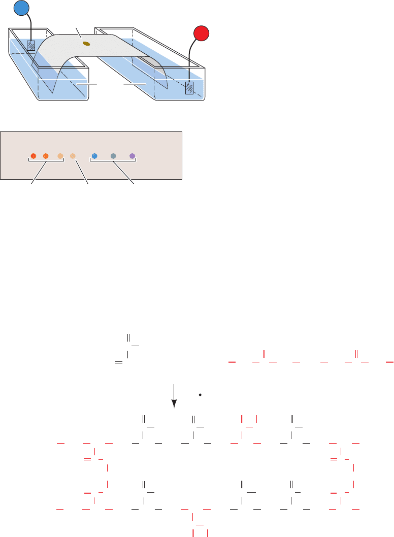
In paper electrophoresis, the sample is applied to a point
on a strip of filter paper or cellulose acetate moistened
with buffer solution. The ends of the strip are immersed in
separate reservoirs of buffer in which the electrodes are
placed (Fig. 6-18). On application of a direct current (often
of ⬃20 V ⴢ cm
–1
), the ions of the sample migrate toward the
electrodes of opposite polarity at characteristic rates to
eventually form discrete bands. An ion’s migration rate is
influenced, to some extent, by its interaction with the sup-
port matrix but is largely a function of its charge. On com-
pletion of the electrophoretogram (which usually takes
several hours), the strip is dried and the sample compo-
nents are located using the same detection methods em-
ployed in paper chromatography (Section 6-3D).
Paper electrophoresis and paper chromatography are su-
perficially similar. However, paper electrophoresis separates
⫽
v
E
⫽
q
f
146 Chapter 6. Techniques of Protein and Nucleic Acid Purification
JWCL281_c06_129-162.qxd 2/22/10 2:25 PM Page 146
ions largely on the basis of their ionic charges, whereas pa-
per chromatography separates molecules on the basis of
their polarities. The two methods can be combined in a two-
dimensional technique called fingerprinting in which a
sample is first treated as in two-dimensional paper chro-
matography (Section 6-3D) but is subjected to elec-
trophoresis in place of the second chromatographic step.
Molecules are thereby separated according to both their
charge and their polarity.
B. Gel Electrophoresis
Gel electrophoresis, which is among the most powerful and
yet conveniently used methods of macromolecular separa-
tion, has supplanted paper electrophoresis. The gels in
common use, polyacrylamide and agarose, have pores of
molecular dimensions whose sizes can be specified. The
molecular separations are therefore based on gel filtration
as well as the electrophoretic mobilities of the molecules be-
ing separated. The gels in gel electrophoresis, however, re-
tard large molecules relative to smaller ones, the reverse of
what occurs in gel filtration chromatography, because there
is no solvent space in gel electrophoresis analogous to that
between the gel beads in gel filtration chromatography
(electrophoretic gels are often directly cast in the elec-
trophoresis device, although precast gels are also widely
used). Since the molecules in a sample cannot leave the gel,
the electrophoretic movement of larger molecules is im-
peded relative to that of smaller molecules.
In polyacrylamide gel electrophoresis (PAGE), gels are
made by the free radical–induced polymerization of acryl-
amide and N,Nⴕ-methylenebisacrylamide in the buffer of
choice (Fig. 6-19).The gel is usually cast as a thin rectangu-
lar slab in which several samples can be simultaneously
Section 6-4. Electrophoresis 147
Figure 6-18 Paper electrophoresis. (a) A diagram of the apparatus
used. The sample is applied to a point on the buffer-moistened
paper. The ends of the paper are dipped into reservoirs of buffer
in which the electrodes are immersed, and an electric field is
applied. (b) The completed paper electrophoretogram. Note that
positive ions (cations) have migrated toward the cathode and
negative ions (anions) have migrated toward the anode. Uncharged
molecules remain at the point of sample application.
+
–
Buffer
Paper
(a)
+–
Negative
ions
Positive
ions
Point of
sample
application
Paper strip
(b)
CH
2
NH
2
C
CH
⫹
Acrylamide N,Nⴕ-Methylenebisacrylamide
O
O
CH
2
CH
2
NH
2
C
CH
O
CH
CH
2
CH
2
CHCH
CH
2
CH
CH
CH
2
CH
CH
2
NH
C
CH
2
CH
2
CH CH
2
CH
2
CHCH CH
2
CH CH
O
NH
2
C
O
NH
2
C
O
NH
C
O
CH
2
CH
2
CH
2
CH C
O
NH C CH
NH
O
O
C
CH
2
NH
NH
CH
2
O
C NH
O
C
NH
2
C
O
NH
2
C
O
NH
2
C NHC
O
SO
4
⫺
Figure 6-19 Polymerization of acrylamide and
N,Nⴕ-methylenebisacrylamide to form a cross-linked
polyacrylamide gel. The polymerization is induced by free radicals
resulting from the chemical decomposition of ammonium
persulfate or the photodecomposition of
riboflavin in the presence of traces of O
2
. In either case,
-tetramethylethylenediamine (TEMED), a free N,N,Nⴕ,Nⴕ
(S
2
O
2⫺
8
S
2SO
⫺
4
ⴢ)
radical stabilizer, is usually added to the gel mixture. The physical
properties of the gel and its pore size are controlled by the
proportion of polyacrylamide in the gel and its degree of
cross-linking.The most commonly used polyacrylamide
concentrations are in the range 3–15%, with the amount of
N, N¿-methylenebisacrylamide usually fixed at 5% of the
total acrylamide present.
JWCL281_c06_129-162.qxd 6/3/10 10:47 AM Page 147
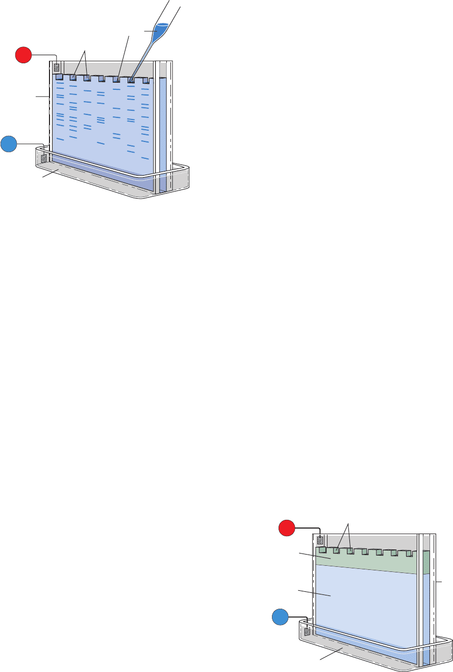
analyzed in parallel lanes (Fig. 6-20), a good way of com-
paring similar samples. The buffer, which is the same in
both reservoirs and the gel, has a pH (usually ⬃9 for pro-
teins) such that the macromolecules have net negative
charges and hence migrate to the anode in the lower reser-
voir. Each sample, which can contain as little as 10 g of
macromolecular material, is dissolved in a minimal
amount of a relatively dense glycerol or sucrose solution
to prevent it from mixing with the buffer in the upper
reservoir and is applied in preformed slots at the top of the
gel (Fig. 6-20). Alternatively, the sample may be contained
in a short length of “sample gel,” whose pores are too large
to impede macromolecular migration. A direct current of
⬃300 V is passed through the gel for a time sufficient to
separate the macromolecular components into a series of
discrete bands (30–90 min), the gel is removed from its
holder, and the bands are visualized by an appropriate
method (see below). Using this technique, a protein mix-
ture of 0.1 to 0.2 mg can be resolved into as many as 20 dis-
crete bands.
a. Disc Electrophoresis Has Improved Resolution
The narrowness of the bands in the foregoing method,
and therefore the resolution of the separations, is limited
by the length of the sample column as it enters the gel. The
bands are greatly sharpened by an ingenious technique
known as discontinuous pH or disc electrophoresis, which
requires a two-gel system and several different buffers
(Fig. 6-21).The “running gel,” in which the separation takes
place, is prepared as described previously and then over-
layered by a short (1 cm), large-pored “stacking” or “spacer
gel.” The buffer in the lower reservoir and in the running
gel is as described before, while that in the sample solution
(or gel) and in the stacking gel has a pH about two units
less than that of the lower reservoir.The pH of the buffer in
the upper reservoir, which must contain a weak acid (usu-
ally glycine, pK
2
⫽ 9.78), is adjusted to a pH near that of the
lower reservoir.
When the current is switched on, the buffer ions from
the upper reservoir migrate into the stacking gel as the
stacking gel buffer ions migrate ahead of them. As this oc-
curs, the upper reservoir buffer ions encounter a pH that is
much lower than their pK.They therefore assume their un-
charged (or, in the case of glycine, zwitterionic) form and
become electrophoretically immobile. This causes a defi-
ciency of charge carriers, that is, a high electrical resistance
R, in this region which, because of the requirement of a
constant current, I, throughout the electrical circuit, results,
according to Ohm’s law (E ⫽ IR), in a highly localized in-
crease in the electric field, E. In response to this increased
field, the macromolecular anions migrate rapidly until they
reach the region containing the stacking gel buffer ions,
where they slow down because at that point there is no ion
deficiency.This effect causes the macromolecular ions to ap-
proach the running gel as stacks of very narrow (⬃0.01 mm
thick) bands or disks that are ordered according to their mo-
bilities and lie between the migrating ions of the upper
reservoir and those of the stacking gel. As the macromo-
lecular ions enter the running gel, they slow down as a re-
sult of gel filtration effects.This permits the upper reservoir
buffer ions to overtake the macromolecular bands and, be-
cause of the running gel’s higher pH, assume their fully
charged form as they too enter the gel. The charge carrier
deficiency therefore disappears and from this point on the
electrophoretic separation proceeds normally. However,
the compactness of the macromolecular bands entering the
running gel greatly increases the resolution of the macro-
molecular separations (e.g., Fig. 6-22).
b. Agarose Gels Are Used to Separate Large
Molecules Electrophoretically
The very large pores needed for the PAGE of large mo-
lecular mass compounds (⬎200 kD) requires gels with such
low polyacrylamide concentrations (⬍2.5%) that they are
too soft to be usable. This difficulty is circumvented by us-
ing agarose (Fig. 6-13). For example, a 0.8% agarose gel is
148 Chapter 6. Techniques of Protein and Nucleic Acid Purification
Figure 6-20 Apparatus for slab gel electrophoresis. Samples,
applied in slots that have been cast in the top of the gel, are
electrophoresed in parallel lanes.
Figure 6-21 Diagram of a disc electrophoresis apparatus.
+
–
Cathode
Sample
Buffer
Buffer
Gel
Sample
wells
Anode
Plastic
frame
+
–
Buffer
Buffer
Sample
wells
Running
gel
Stacking
gel
Plastic
frame
Cathode
Anode
JWCL281_c06_129-162.qxd 2/22/10 2:25 PM Page 148
used for the electrophoretic separation of nucleic acids
with molecular masses of up to 50,000 kD.
c. Gel Bands May Be Detected by Staining,
Radioactive Counting, or Immunoblotting
Bands resulting from a gel electrophoretic separation
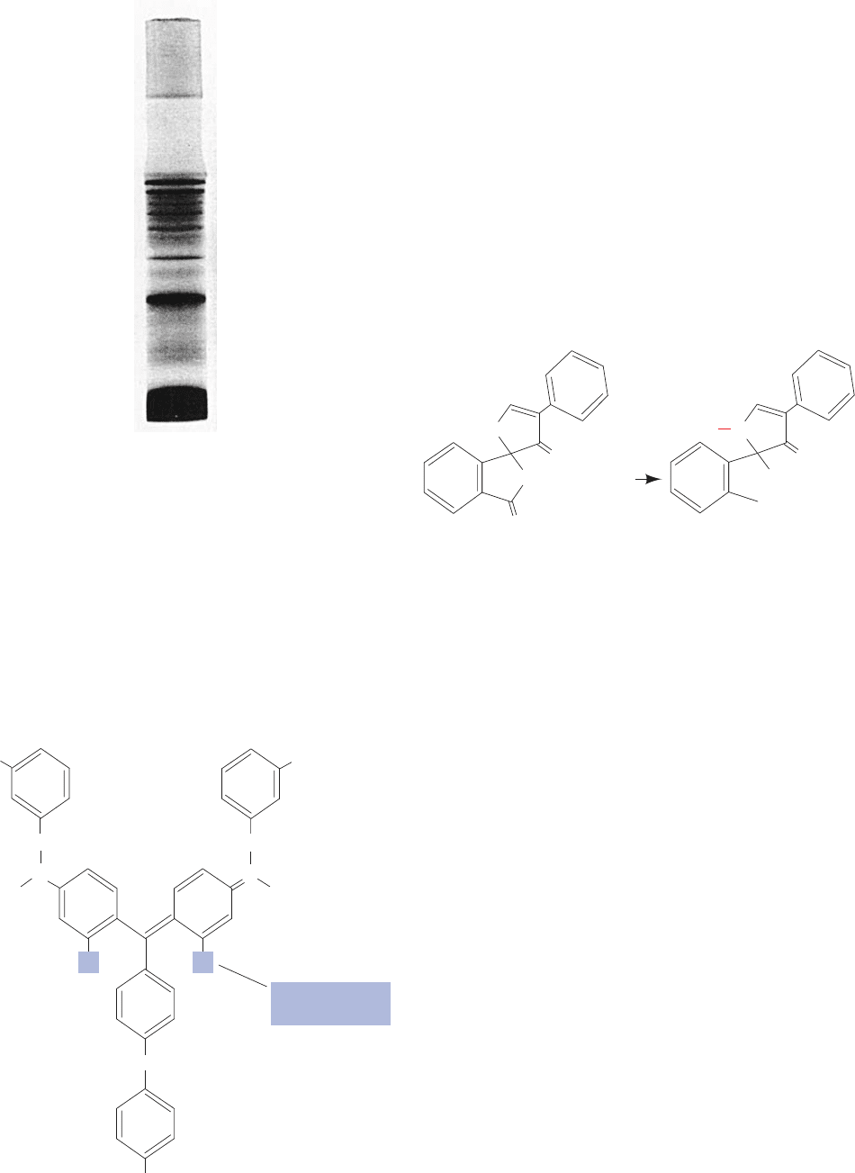
can be located by a variety of techniques. Proteins are often
visualized by staining. Coomassie brilliant blue,
N
CH
2
H
5
C
2
C
2
H
5
OC
2
H
5
⫺
O
3
S
SO
3
⫺
Coomassie brilliant blue
N
⫹
CH
2
RR
NH
R250: R ⫽ H
G250: R ⫽ CH
3
which is the most widely used dye for this purpose, is ap-
plied by soaking the gel in an acidic, alcoholic solution con-
taining the dye.This fixes the protein in the gel by denatur-
ing it and complexes the dye to the protein. Excess dye is
removed by extensively washing the gel with an acidic so-
lution or by electrophoretic destaining. Protein bands con-
taining as little as 0.1 g can thereby be detected. Gel
bands containing less than this amount of protein may be
visualized with silver stain, which is ⬃50 times more sensi-
tive but more difficult to apply. The recently developed
SYPRO dyes, which strongly fluoresce under UV light
when bound to protein, are equally sensitive as silver stain
but easier to apply. Fluorescamine, a widely used protein
stain, is a nonfluorescent molecule that reacts with primary
amines, such as lysine residues, to yield an addition product
that is highly UV-fluorescent.
Proteins, as well as other substances, can be detected
through the UV absorption of a gel along its length. If the
sample is radioactive, the gel may be dried under vacuum
to form a cellophane-like material or, alternatively, covered
with plastic wrap, and then clamped over a sheet of X-ray
film. After a time (from a few minutes to many weeks
depending on the radiation intensity) the film is developed,
and the resulting autoradiograph shows the positions of
the radioactive components by a blackening of the film [al-
ternatively, a phosphorimager (Section 5-5D) can be used
to reveal the locations of the radioactive components
within even a few seconds]. A gel may also be sectioned
widthwise into many slices and the level of radioactivity in
each slice determined using a scintillation counter. The lat-
ter method yields quantitatively more accurate results than
autoradiography. Sample materials can also be eluted from
gel slices for identification and/or further treatment.
If an antibody to the protein of interest is available, it is
possible to specifically detect this protein on a gel in the
presence of many other proteins by an immunoblot (also
known as a Western blot). This procedure is a variation of
Southern blotting (Section 5-5D) that uses a technique
similar to ELISA (Section 6-1Da) to detect the protein(s)
of interest (Fig. 6-23):
1. A completed gel electrophoretogram is blotted onto
a sheet of nitrocellulose (much like in Fig. 5-48), which
strongly and nonspecifically binds proteins [nylon or
polyvinylidene fluoride (PVDF) membranes may also be
used].
O
⫹
RNH
2
O
O
O
Fluorescamine adduct
(highly fluorescent)
Fluorescamine
(nonfluorescent)
NR
OH
COOH
O
Section 6-4. Electrophoresis 149
Figure 6-22 Disc electrophoresis of human serum in a 0.5 ⫻
4.0–cm polyacrylamide gel column. The proteins were visualized
by staining them with amido black. [From B. J. Davis,Annals of
the New York Academy of Science 121, 404 (1964), Fig. 8.]
JWCL281_c06_129-162.qxd 2/22/10 2:25 PM Page 149

2. The excess adsorption sites on the nitrocellulose are
blocked with a nonspecific protein such as casein (milk
protein; nonfat milk itself is often used) to prevent the non-
specific adsorption of the antibodies (which are also pro-
teins) used in Steps 3 and 4.
3. The blot is treated with antibody to the protein of in-
terest (the primary antibody). This is usually a rabbit anti-
body.
4. After washing away the unbound primary antibody,
the blot is incubated with a goat antibody, directed against
all rabbit antibodies, to which an easily assayed enzyme has
been covalently linked (the secondary antibody).
5. After washing away the unbound secondary anti-
body, the enzyme in the bound secondary antibody is as-
sayed with a color-producing reaction, causing colored
bands to appear on the nitrocellulose where the protein of
interest is bound.
Alternatively, the primary antibody used in Step 3 may be
labeled with the radioactive isotope
125
I, the unbound anti-
body washed away, and the position of the bound protein
on the blot revealed by autoradiography.
C. SDS–PAGE
Soaps and detergents are amphipathic molecules (Section
2-1Ba) that are strong protein denaturing agents for rea-
sons explained in Section 8-4E. Sodium dodecyl sulfate
(SDS),
Sodium dodecyl sulfate (SDS)
a detergent that is often used in biochemical preparations,
binds quite tenaciously to proteins, causing them to assume
a rodlike shape. Most proteins bind SDS in the same ratio,
1.4 g of SDS per gram of protein (about one SDS molecule
for every two amino acid residues). The large negative
[CH
3
⫺ (CH
2
)
10
⫺ CH
2
⫺ O ⫺ SO
3
⫺
]Na
⫹
charge that the SDS imparts masks the protein’s intrinsic
charge so that SDS-treated proteins tend to have identical
charge-to-mass ratios and similar shapes. Consequently, the
electrophoresis of proteins in an SDS-containing polyacryl-
amide gel separates them in order of their molecular masses
because of gel filtration effects. Figure 6-24 provides an ex-
ample of the resolving power and the reproducibility of
SDS–PAGE.
The molecular masses of “normal” proteins are rou-
tinely determined to an accuracy of 5 to 10% through
SDS–PAGE. The relative mobilities of proteins on such
gels vary linearly with the logarithm of their molecular
masses (Fig. 6-25). In practice, a protein’s molecular mass is
determined by electrophoresing it together with several
“marker” proteins of known molecular masses which
bracket that of the protein of interest.
Many proteins consist of more than one polypeptide
chain (Section 8-5A). SDS treatment disrupts the noncova-
lent interactions between these subunits. Therefore,
SDS–PAGE yields the molecular masses of the protein’s
subunits rather than that of the intact protein unless the
subunits are disulfide linked. However, mercaptoethanol is
usually added to SDS–PAGE gels so as to reductively
cleave these disulfide bonds (Section 7-1B).
D. Isoelectric Focusing
A protein has charged groups of both polarities and there-
fore has an isoelectric point, pI, the pH at which it is immo-
bile in an electric field (Section 4-1D). If a mixture of pro-
teins is electrophoresed through a solution having a stable
pH gradient in which the pH smoothly increases from anode
to cathode, each protein will migrate to the position in the
pH gradient corresponding to its isoelectric point. If a pro-
tein molecule diffuses away from this position, its net
charge will change as it moves into a region of different
pH, and the resulting electrophoretic forces will move it
back to its isoelectric position. Each species of protein is
150 Chapter 6. Techniques of Protein and Nucleic Acid Purification
Figure 6-23 Detection of proteins by immunoblotting.
3. 4. 5.Perform gel electrophoresis
on a sample containing the
protein of interest
1.
Blot the proteins from the gel
onto nitrocellulose
Incubate with rabbit
antibody to the protein
of interest
2. Block the unoccupied
binding sites on the
nitrocellulose with
casein
Weight
Nitrocellulose
replica of gel
electrophoretogram
Binding of
primary
antibody
Paper towels
Nitrocellulose
sheet
Wick
Buffer
solution
Gel electrophoretogram
containing the protein
of interest
Wash and incubate
with an enzyme-linked
goat anti-rabbit
antibody
Assay the
linked enzyme
with a colori-
metric reaction
Binding of enzyme-
linked secondary
antibody
Immunoblot
Voet/Voet
Biochemistry
JWCL281_c06_129-162.qxd 2/22/10 2:25 PM Page 150
