Shani G. Radiation Dosimetry: Instrumentation and Methods
Подождите немного. Документ загружается.

50
Radiation Dosimetry: Instrumentation and Methods
therapeutic uses, especially in connection with so-called
endovascular (or intravascular) brachytherapy. Since accu-
rate dose estimation is necessary for the success of such
applications, some problems in beta-ray dosimetry need
further study. Among these problems is the effect of elec-
tron backscattering on dose, which has significance not
only for accurate dose estimation but also for new source
design. In that study, an empirical measure of electron
backscattering, known as the dose backscatter factor, was
calculated using EGS4 Monte Carlo calculations for
monoenergetic electrons and various scattering materials.
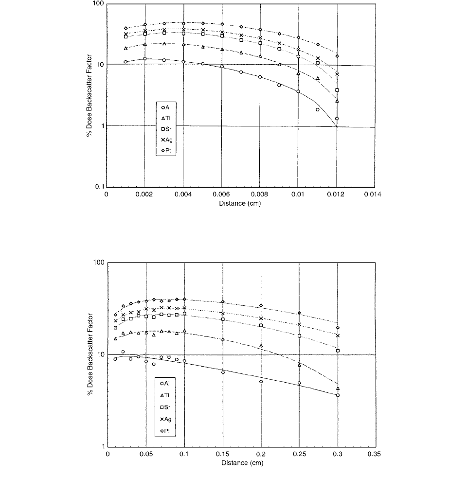
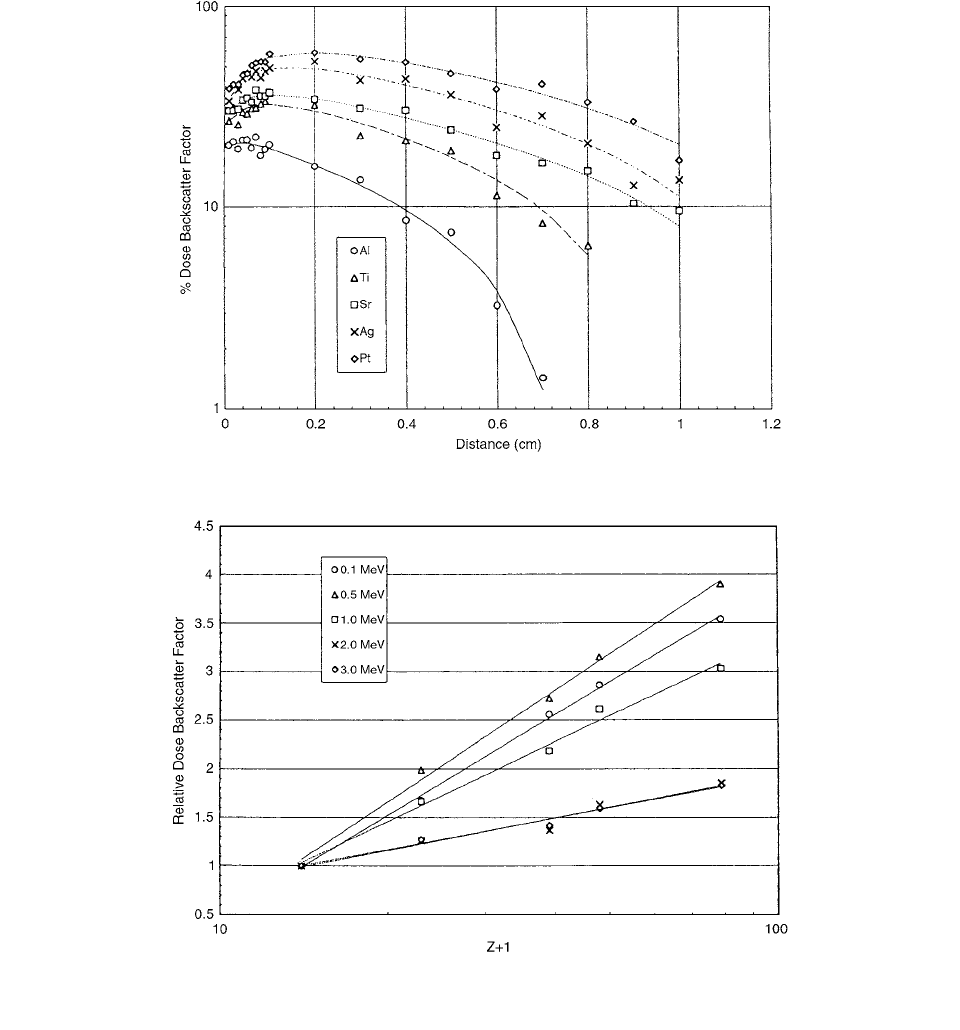
Electron energies were 0.1, 0.5, 1.0, 2.0, and 3.0 MeV in
combination with Al (
Z
13), Ti (
Z
22), Sr (
Z
38),
Ag (
Z
47), and Pt (
Z
78) scatterers. The dose back-
scatter factor ranged from 10% to 60%, depending on
electron energy and material, and was found to increase
with the atomic number
by a log (
1) relationship.
Results are shown in Figures 2.29 to 2.31.
In Figure 2.32, dose backscatter factors at 0.001-cm
depth for 0.1 MeV and 0.01-cm depth for other energies
are plotted against log (
Z
1) of the scatterers used in
this study. Data were fitted well by straight lines in a semi-
log scale, showing that the dose backscatter factor is pro-
portional to log (
Z
1).
FIGURE 2.29
Dose backscatter factor depth profile for
E
0
0.1 MeV. Note, in Figures 2.29 to 2.31, that the statistical errors in
the data are comparable with the size of the symbols. (From Reference [33]. With permission.)
FIGURE 2.30
Dose backscatter factor depth profile for . (From Reference [33]. With permission.)
E
0
1.0 MeV
Ch-02.fm Page 50 Friday, November 10, 2000 10:52 AM
Theoretical Aspects of Radiation Dosimetry
51
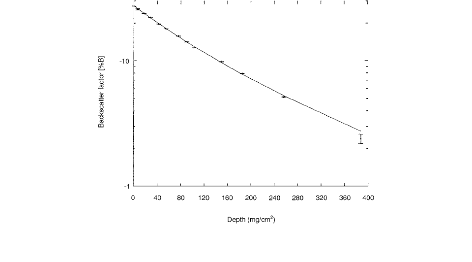
Beta-ray dose backscatter factors with respect to soft
tissue were measured by Nunes et al. [34] using an
extrapolation chamber. The beta-ray dose backscatter
factor is a measure of the change effected in absorbed
dose to a soft-tissue medium when part of the medium
is replaced by a material other than soft tissue (i.e., a
scatterer); the source is located at the boundary between
the two media.
The variation of backscatter factor with distance from
the boundary is well represented analytically by sums of
exponentials. Therefore, the rate of decrease of backscatter
factor with distance can be specified by a relaxation
length, defined as the depth through which the backscatter
factor is reduced by 1/
e
, where
e
is the base of the natural
logarithm. With a
32
P planar source, relaxation lengths in
Mylar are 588 mg/cm
2
and 238 mg/cm
2
for bismuth and
aluminum scatterers, respectively.
The backscatter factor is given by ,
where is the dose measured when both slabs are soft
tissue; this is the “homogeneous dose,” and is the dose
measured when the replaceable slab is substituted with a
scatterer—a material that is not soft tissue; this is the “inho-
mogeneous dose.”
B
is usually multiplied by 100 and
referred to as a percentage. is called the dose ratio;
FIGURE 2.31
Dose backscatter factor depth profile for . (From reference [33]. With permission.)
FIGURE 2.32
Log (
Z
l) dependence of the dose backscatter factor data for each electron energy. (From Reference [33]. With
permission.)
E
0
3.0 Me
V
BD
i
D
h
1
D
h
D
i
D
i
D
h
Ch-02.fm Page 51 Friday, November 10, 2000 10:52 AM
52
Radiation Dosimetry: Instrumentation and Methods
if it is larger than unity, it is a dose enhancement factor, and
if it is smaller than unity, it is a dose reduction factor.
The extrapolation chamber, model EIC-I, is 35 cm
long, including the connectors. The stem is 29.2 cm long
and the head, about 2.3 cm long, with a diameter of
3.81 cm. It weighs 150 g. The air gap spacing is contin-
uously variable, from 0.3 mm to 4.5 mm; the space is
between the entrance window, which is one electrode, and
the collecting electrode. The entrance window is made of
graphite-coated polypropylene and is 0.2 mg/cm
2
thick.
The collecting electrode and guard ring are made of
A-150 (Shonka) plastic, which is a soft-tissue equiv-
alent material with an effective atomic number of 5.49.
The sensitive region of the chamber is a right circular
cylinder with a diameter of 1.05 cm and height equivalent
to the interelectrode spacing. Phosphorus-32 point source,
with a diameter about 2 mm, was prepared by depositing
5
l of stock solution onto a Mylar substrate and allowing
it to evaporate in air. The activity of the source at the time
of preparation was about 92.5 MBq. Both point and dis-
tributed or planar sources were supported by, and covered
with, Mylar, 0.35 mg/cm
2
thick.
The variation of backscatter factor with distance from
an interface where a
32
P source is located is found to be
well represented by sums of exponentials. Specifically,
The air interface depth profile, Figure 2.33, demonstrates
that the dose reduction, which is largest at the interface, falls
off in roughly two stages. Close to the boundary, the dose
reduction decreases sharply; at larger distances, the falloff
is less rapid.
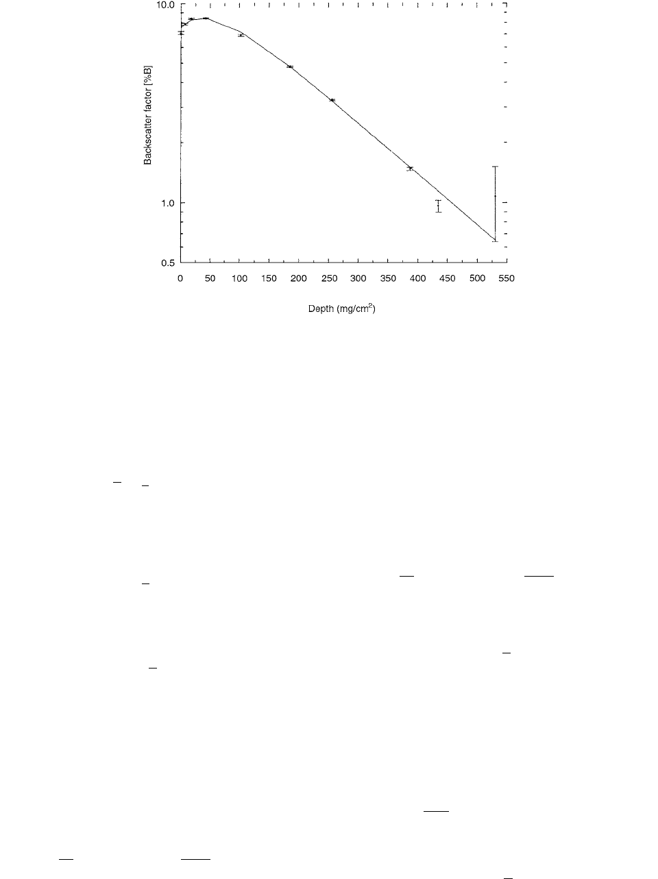
The aluminum and bismuth interface depth profiles,
Figure 2.34 for aluminum, exhibit gentle maxima that are
located away from the interface.
VII. DOSIMETER PERTURBATION
Perturbation effects are defined as departures from ideal
large-detector or Bragg-Gray cavity behavior. Perturba-
tions are involved in the determination of the absorbed
dose to a medium irradiated by the photon and electron
beams used in external-beam radiotherapy. Kilovoltage x-
ray beams and electron beams were discussed by Nahum.
[35,36] Many correction factors are involved in radiation
dosimetry, including, for an ionization chamber, correc-
tions for differences between the temperature, pressure,
and humidity of the air in an ion chamber at measurement
and at calibration, the recombination of ions before they
can contribute to the measured charge, and possible polar-
ity effects.
Assuming the absorbed dose to the sensitive material
of the detector, , is known, one can generally write
(2.141)
where is the desired quantity, the absorbed dose in
the undisturbed medium, it is the task of cavity theory to
evaluate the factor
f
. In general, there will be a perturbation
FIGURE 2.33
Backscatter factor depth profile for a
32
P distributed source located at an air/Mylar interface. Symbols denote
experimental data and the solid curve is a fit to these data. (From Reference [34]. With permission.)
%Bx() A
i
e
xv
i
for solid material interfaces and
i
%Bx() A
i
e
xv
i
i
for air interfaces.
D
de
t
D
med
fD
det
D
med
Ch-02.fm Page 52 Friday, November 10, 2000 10:52 AM
Theoretical Aspects of Radiation Dosimetry
53
due to differences in atomic composition, density, or both
between the detector material and the undisturbed medium,
and to correct for this, a perturbation correction factor,
p
,
is introduced.
In the case of a photon detector,
p
can be defined by
(2.142)
where it is the photon (energy) fluence that is perturbed.
In the case of a Bragg-Gray cavity or electron detector,
p
is defined by
(2.143)
where it is the electron fluence that is perturbed.
A simple rearrangement yields
(2.144)
The quantities and
s
med
,
det
can be expres-
sed as a cavity integral using the Spencer-Attix formula-
tion. The ratio can be written as
(2.145)
where is the standard nomenclature for the electron
fluence differential in energy
E
, with the subscripts
med
and
det
referring to its value at a reference point
z
in the
undisturbed medium and its value averaged over the sen-
sitive detectors volume, respectively. The other quantities
have their usual meaning. The term outside the integral is
the dose due to track ends below energy
. The expression
for
p
is
(2.146)
This is equivalent to writing
(2.147)
where is the detector dose that would result from
ideal Bragg-Gray behavior an is the actual detec-
tor dose.
p
is unity if in the medium at
z
is identical to that
avenged over the detector. In the case of the difference
being only one of magnitude and not of spectral shape,
i.e., if
(2.148)
where
k
is a constant for all
E
, then
(2.149)
FIGURE 2.34
Backscatter factor depth profile for a
32
P point source located at an aluminum/Mylar interface. Symbols denote
experimental data, and the solid curve is a fit to these data. (From Reference [34]. With permission.)
D
med
D
det
en
()
med,det
p
Dz()
med
D
def
s
med,det
p
pDz()
med
D
det
s
med,det
p
D
med
z()D
det
,
D
med
D
det
D
med
z()
D
det
-------------------
E
()
med
z
L
()
med
E ()
med
z
S ()
()
med
[]d
E
()
det
L
()
det
E ()
det
S ()
()
det
[]d
--------------------------------------------------------------------------------------------------------------------------
E
p
E
()
med
z
L
()
det
E ()
med
z
S ()
()
det
[]d
E
()
det
L
()
det
E ()
det
S ()
()
det
[]d
-----------------------------------------------------------------------------------------------------------------------
pD
det
()
BG
D
det
()
meas
D
det
()
BG
D
det
()
meas
E
E
()
det
k
E
()
med
z
p
E
med
z
det
Ch-02.fm Page 53 Friday, November 10, 2000 10:52 AM
54 Radiation Dosimetry: Instrumentation and Methods
The perturbation (displacement) correction factor for
chamber radius r in beam energy E was defined as
(2.150)
where the
K(E, 0) were obtained by extrapolation of the
K(E, r) to zero radius. This was done by a weighted least-
squares fit of the straight line to the 1/K(E, r) values as a
function of r. The kerma K(E, r) was computed by inte-
grating the photon fluence in the cavity.
Figure 2.35, taken from Seuntjens et al. [37], shows
that is appreciably below unity at 20 and
30 keV, which can be explained by the virtually scatter-
free attenuation of the beam at these energies. At higher
energies the deviations from unity are very small. These
were then integrated over the primary photon
fluence spectrum to yield for different x-ray spec-
tra. Their results are shown in Figure 2.36 as a function
of HVL in both mm of copper and mm of aluminium. For
the range 2 mm Al HVL 5 mm Cu and for the Farmer
chamber (r 3.5 mm), the factor lay between 0.99
and 1.01. Thus, the authors concluded that the original
assumption made by ICRU 23 [77] that the perturbation
due to displacement could be neglected was justified. They
were unable to find any support in their calculations for
the values given by IAEA [64], which exceed unity by
several percent.
The overall perturbation factor is denoted by , it is
now given by
(2.151)
where fs indicates free space.
For a cylinder of height
t and radius R irradiated
parallel to the axis, i.e., a plane-parallel chamber, the
Harder expression can be written in terms of a perturbation
factor p as: [38]
(2.152)
where is the linear electron scattering power, ,
for the medium surrounding the cavity. Harder [71] also
gave an expression for the more practically useful geom-
etry of a long cylinder irradiated perpendicular to its axis,
i.e., a cylindrical or thimble chamber. This can be written
as
(2.153)
where r has been used for the chamber radius to distin-
guish it from R for the case of a coin-shaped cavity.
The dependence of the perturbation correction factor
on the radius of the chamber is
(2.154)
where
r is the cavity radius and k is energy dependent.
Harders theoretical treatment [Equation (2.153)] had pre-
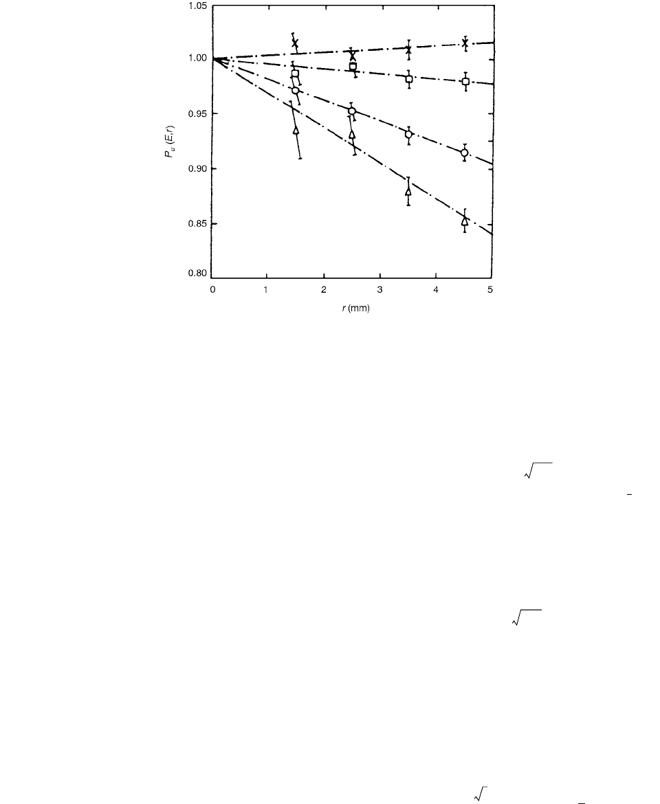
dicted a dependence on . Figure 2.37 shows their exper-
imental results, at mean energy at depth
2.5 MeV,
as a function of cavity radius.
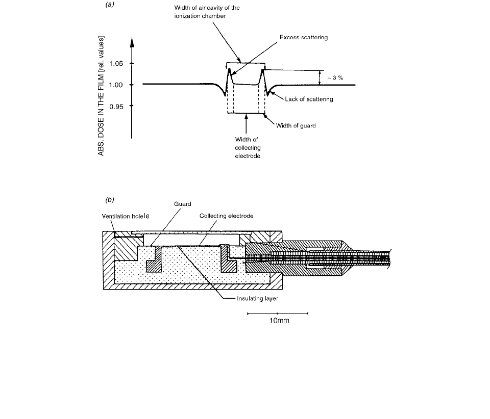
Figure 2.38a shows how the perturbation is confined
to the edges of a coin-shaped cavity. The guard ring in the
NACP chamber was designed to prevent the signal due to
this edge perturbation being collected.
FIGURE 2.35 The dependence of the mono-energetic perturbation correction factor on the chamber radius r for monoenergetic
photon beams with energies
E 20 (), 30 (), 75 (), and 200 keV (). (From Reference [35]. With permission.)
p
dis
Er,() KE0,()KEr,()
p
dis
Er,()
p
dis
Er,()
p
dis
r()
p
dis
p
u
p
u
p
u
K
air
med
K
air
fs
()M
fs
M
med
()
p 1 0.13 tR()tT
m
T
m
d
2
ds
p 1 0.040 rT
m
p
wg,
1 kr
r
E
Ch-02.fm Page 54 Friday, November 10, 2000 10:52 AM
Theoretical Aspects of Radiation Dosimetry 55
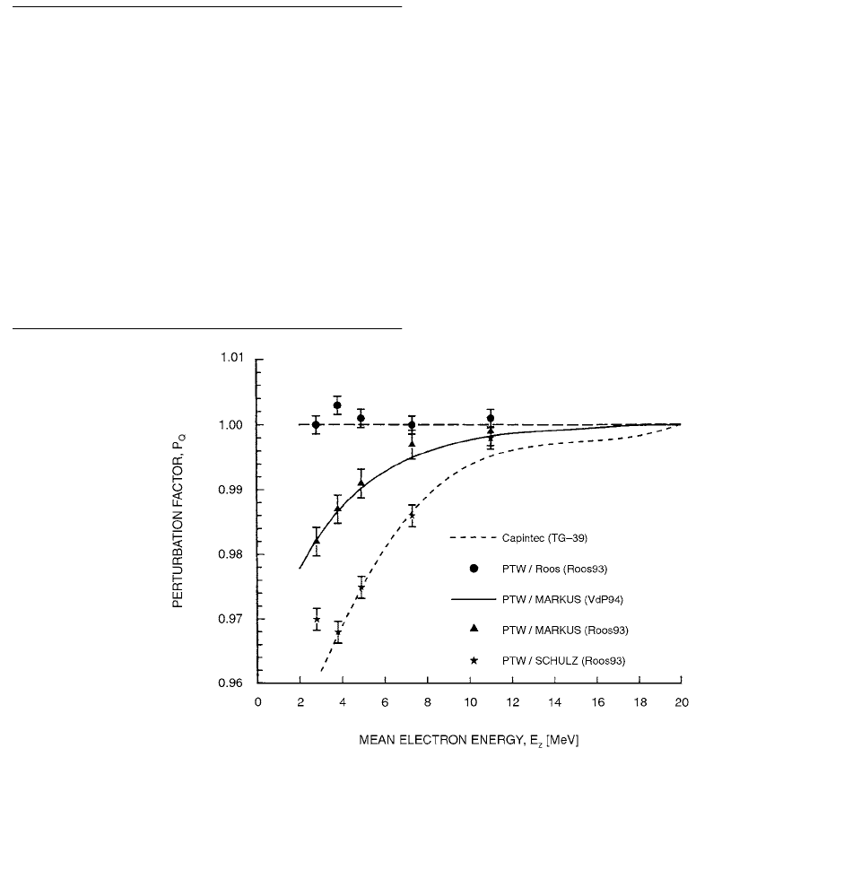
In a series of measurements, Roos [39] compared the
responses of the NACP, PTW/Markus, and PTW/Schul
(M23346) designs with the Roos chamber (PTW 34001)
which has a 4-mm guard. The latter chamber was spe-
cifically designed to exhibit negligible perturbation in
low-energy electron beams. It was found that there was
negligible difference between the responses of the Roos
and NACP chambers. However, for the other two chambers
investigated, the PTW/Marklis and PTW/Schulz models,
the perturbation factor began to decrease below unity for
energies of MeV and below (Figure 2.39). The
ratio of the response of the PTW/Markus and NACP
chambers that Roos obtained was in excellent agreement
with the measurements of Van der Piaetsen et al. [40];
the latter’s fit to the ratio is shown as the
full line in Figure 2.39.
FIGURE 2.36 Variation of the perturbation correction factor with HVL (mm Al) for radiation qualities with tube potential 100
(
) and 120 () and with HVL (mm Cu) for 120 (), 150 (O), 200 () and 250 kV (). The factor is independent of the wall
material and central electrode. (From Reference [35]. With permission.)
FIGURE 2.37 Perturbation correction factors for cylindrical chambers for electron radiation (
z
2.5 MeV) as a function of
the chamber radius. Experimental values were obtained from measurements using plane-parallel and cylindrical chambers. Theoretical
values calculated according to Harder. (From Reference [35]. With permission.)
p
u
p
wg,
E
E
z
10
P
MARKUS
P
NACP
Ch-02.fm Page 55 Friday, November 10, 2000 10:52 AM
56 Radiation Dosimetry: Instrumentation and Methods
If backscatter plays a role in the response of certain
plane-parallel chambers, then this should be taken into
account in the perturbation factor. It is suggested, there-
fore, that a total or overall perturbation factor for
plane-parallel chambers should be written as the product
of the cavity factor and two separate wall factors:
(2.155)
where the component of the wall correction due to back
scatter effects has been written as . Naturally, this
expression can also be applied to cylindrical chambers.
VIII. DOSIMETRY FOR BRACHYTHERAPY
In brachytherapy treatment planning, dose distributions are
most commonly calculated from point-source approxima-
tions where correction factors are applied to account for the
geometrical shape of a non-pointlike source, the radial
dependence and angular anisotropy of the photon absorption
and scatter in the source, encapsulation, and the surrounding
medium. The functions describing the radial dependence of
the total dose distribution, both primary and scatter, for a
pointlike source are usually polynomial functions of the
radial distance from the center of the source and along the
perpendicular bisector to the long axis of the source.
A model based on the EGS4 Monte Carlo system was
developed by Luxton [41] for calculating dose rate to water
from an embedded low-energy brachytherapy source,
given measurement data of dose rate to water within a
water-substitute solid phantom for a source of given
strength. The EGS4-based model was used to calculate
point source dose rate distributions per unit source
strength for water and for several species of solid phantoms.
The Monte Carlo system was used to calculate dose rate
to a thin spherical shell of water contained within the solid
phantom at various distances centered on the source. Cor-
rection factors were calculated for polymethylmethacrylate
FIGURE 2.38 (a) Film measurements across the front surface of an air cavity in a PMMA phantom at the depth of maximum dose
in an electron beam: the effect of the guard ring in reducing the perturbation is clearly demonstrated. (b) A design
for a plane-parallel chamber (the “NACP” chamber) which follows the specification set out in the NACP [72] protocol. The very
narrow (2-mm) air gap and the presence of the guard ring minimize perturbation effects. The collecting electrode is very thin
(
0.1 mm) and is mounted on a thin insulating layer (0.2 mm) in order to give a negligible polarity effect. The front wall (0.5 mm
thick to enable measurements to be made at small depths) and back wall are made of one single material (in this case, graphite).
(From Reference [35]. With permission.)
E
0
6 MeV
p
Q
p
Q
p
cav
p
wall
p
wall
sc
p
wall
sc
Ch-02.fm Page 56 Friday, November 10, 2000 10:52 AM
Theoretical Aspects of Radiation Dosimetry 57
(PMMA or acrylic), solid water (WTI), and RW-I, a mate-
rial optimized for low-energy dosimetry, with photon
spectra from Pd-103 and from two commercial models of
1-125 seed used as input. For model 6711 1-125 seeds
at 1 cm in PMMA and WTI, the calculated ratios of dose
rate to water to dose rate to water in the solid phantom
are 0.893 and 1.038, respectively. Atomic composition of
phantom materials is given in Table 2.5.
The dose rate in an arbitrary medium is written in the
point source model as
(2.156)
where is the dose rate to the medium in cGy
h
1
at the radial distance r in cm from the center of a point
source; is the reference distance; is the air-
kerma strength of the source in units of cGy cm
2
h
1
,
numerically equal to the air-kerma strength in the recom-
mended unit U 1
Gy m
2
h
1
; and is the dose rate
constant for the source in that medium, defined here as
the dose rate to the medium at 1 cm from the center of the
point source per U of air-kerma strength. The units of
are cGy h
1
U
1
. will be understood to refer
to a 1 cm distance along the transverse axis when reference
is made to any physical nonpoint source that possesses
cylindrical symmetry. g(r) is defined by this equation as the
dimensionless radial dose function for that source/medium
combination, normalized to g(r) 1 at r r
0
1 cm.
The dose rate to infinitesimal water substance in the
medium may be written as
(2.157)
where is the dimensionless ratio of dose rate at
distance r from the point source to water as compared to
the substance of the medium. The Monte Carlo method
was used to calculate dose rate distribution for as
well as for because it is conventional and
clinically more pertinent to express dose rate and dose
rate distributions in terms of water substance, whether in
water or in a water substitute phantom. distributions
were calculated per unit primary photon emission per unit
TABLE 2.5
Percent Atomic Composition (by weight) and
Densities of Phantoms
Atomic Species Water PMMA WT1 RW-1
H 11.2 8.0 8.1 13.2
C … 60.0 67.2 79.4
N … … 2.4 ...
O 88.8 32.0 19.9 3.8
Ca … … 2.3 2.7
Mg … … … 0.9
Cl … … 0.1 ...
Density (g cm
3
) 1.00 1.17 1.015 0.97
Source: From Reference [41]. With permission.
D
med,point
r()
med
S
K
gr()
rr
0
()
2
----------------
D
med,point
r()
r
0
1cm
S
K
med
med
med
D
med
w
r() f
med
w
r()D
med point,
r()
f
med
w
r()
D
med
w
r()
D
med point,
r()
D
med
w
r()
FIGURE 2.39 The variation of the overall perturbation factor for several different plane-parallel chambers in common use,
relative to the NACP chamber, indicated by the dashed line drawn at
p
Q
1.000. All the measurements were made at the depth of
dose maximum and normalized to the quotient test chamber/NACP in a high-energy electron beam. The full line is a fit to three
separate measurement series on different accelerators using the PTW/Markus chamber. [40] The individual data points are measure-
ments on three different PTW designs taken from Roos [39] and re-normalized so that
p
Q
1.000 for the NACP chamber; the dashed
curve is for the Capintee-PS-003 chamber as given by AAPM. (From Reference [35]. With permission.)
p
Q
Ch-02.fm Page 57 Friday, November 10, 2000 10:52 AM
58
Radiation Dosimetry: Instrumentation and Methods
time. The magnitude of primary photon emission per unit
time is proportional to for each brachytherapy source.
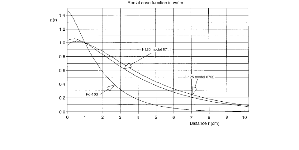
The radial dose distribution in water for I
125
and Pd
103
is shown in Figure 2.40. The dose rate distributions per
unit activity to water in the solid phantoms are obtained
from the
g
(
r
) for the homogeneous media by multiplica-
tion by the product of corresponding ratios. The results
are given in Figure 2.41.
Computer algorithms for electron-binding correction to
Compton scattering and for detailed simulation of
K
-edge
characteristic x-ray production were incorporated into EGS4
unix version 2.0 by Wang and Sloboda. [42] Based on
detailed modelling of the internal structures of sources, the
modified version was used to calculate dose rate constants,
radial dose functions, and anisotropy functions on the long
axis for an
125
I model 6711 source,
169
Yb Type 5 and Type
8 sources, and a stainless steel clad (SS)
192
Ir source. The
geometry of these sources is cylindrically symmetric.
Following the dose calculation formalism proposed
originally by the Interstitial Collaborative Working Group
(ICWG) for interstitial brachytherapy sources, and
expanded to all brachytherapy sources by TG-43, dose
rate in medium at distance
r
(cm) from the source center
and angle
relative to its long axis is expressed as
(2.158)
where is the air-kerma strength of the source,
is the
dose rate constant,
G
(
r
,
) is the geometry factor,
F
(
r
,
)
is the anisotropy function, and
g
(
r
) is the radial dose
function. With and calculated,
,
g
(
r
), and
F
(
r
,
) can be formulated as follows:
(2.159)
where is the dose rate at 1 cm from the source
center on the transverse axis, and
(2.160)
where is the dose rate at distance
r
on the trans-
verse axis and and are geometry
factors for the source at 1 cm and at a distance
r
on the
transverse axis. Finally
(2.161)
The electron binding correction added to EGSU was
carried out for both total and differential Compton scat-
tering cross sections. Data for the total cross sections and
for the incoherent scattering function were taken from
photon cross-section library DLC-99.
To explore the influence of the binding effect of
Compton scattering, the modified and original EGS4
codes were used to calculate dose distributions for a real-
istic
125
I 6711 source in unbounded water phantoms. For
simplicity, call the doses evaluated in the two cases bound
(Compton) dose and free (Compton) dose. Figure 2.42
shows ratios of bound-to-free doses on the transverse axis,
delivered by the source in water. At distances less than or
equal to 7 cm, the influence of the binding correction is
small, less than or equal to 1%. However, the bound dose
increases by a factor of 1.02 to 1.04 at distances from 8 to
12 cm and by a factor of up to 1.14 at further distances
from 13 to 20 cm.
Figure 2.43 compares the radial dose function
g
(
r
)
calculated for an
125
I Model 6711 source in unbounded
water with corresponding Monte Carlo results obtained
by Williamson (using his photon transport code’3”4),
FIGURE 2.40
Calculated radial dose functions in water for point sources of
103
Pd and two models of
125
I seed. (From Reference [41].
With permission.)
S
K
D
˙
r
,() S
k
Gr
,()
G 1
2,()
------------------------
Fr
,()gr()
S
k
S
k
D
˙
r
,()
D
˙
1
2,()
S
k
------------------------
D
˙
1
2,()
gr()
D
˙
r
2,()G 1
2,()
D
˙
1
2,() Gr
2,()
----------------------------------------------------------
D
˙
r
2,(
)
G 1
2,()
Gr
2,()
Fr
,()
D
˙
r
,()Gr
2,()
D
˙
r
2,()Gr
,()
------------------------------------------
Ch-02.fm Page 58 Friday, November 10, 2000 10:53 AM
Theoretical Aspects of Radiation Dosimetry
59
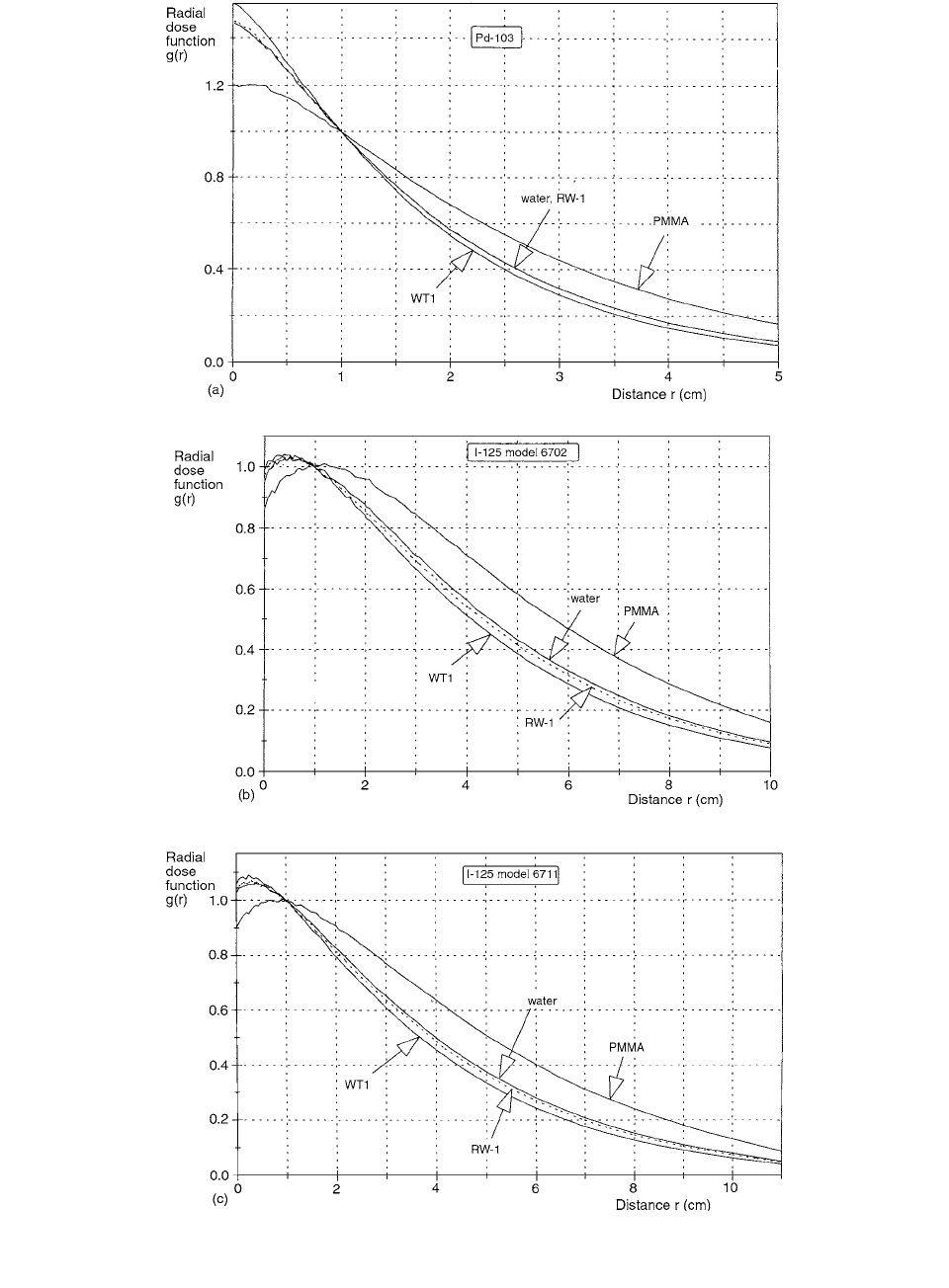
FIGURE 2.41
Calculated radial dose functions for PMMA, WTI, RW-1, and water, respectively, from point sources of (a)
103
Pd, (b)
model
125
I, and (c) model 6702
125
I. (From Reference [41]. With permission.)
Ch-02.fm Page 59 Friday, November 10, 2000 10:53 AM