Reed S.J.B. Electron microprobe analysis and scanning electron microscopy in geology
Подождите немного. Документ загружается.

Jacobson, C. E. (1989) Estimation of Fe
3+
from electron microprobe analyses:
observations on calcic amphibole and chlorite. J. Metamorphic Geol. 7 507–13.
Jarosewich, E. and Boat ner, L. A. (1991) Rare-earth element reference samples for
electron microprobe analysis. Geostand. Newslett. 15 397–9.
Jarosewich, E., Nelen, J. A. and Norberg, J. A. (1979) Electron microprobe reference
samples for mineral analyses. Smith son. Contrib. Earth Sci. 22 68–72.
(1980) Reference samples for electron microprobe analysis. Geostand. Newslett.
4 43–8.
Joy, D. C. (1995) Monte Carlo Modeling for Electron Microscopy and Microanalysis
(New York: Oxford University Press).
Joy, D. C., Romig, A. D. and Goldstein, J. I., eds. (1986) Principles of Analytical
Electron Microscopy (New York: Plenum Press).
Jurek, K., Renner, O. and Krousky, E. (1994) The role of coating densities in X-ray
microanalysis. Mikrochim. Acta 114/115 323–6.
Kanaya, K. and Okayama, S. (1972) Penetration and energy-loss theory of electrons
in solid targets. J. Phys. D 5 43–58.
Kerrick, D. M., Eminhizer, L. B. and Villaume, J. F. (1973) The role of carbon film
thickness in electron microprobe analysis. Amer. Mineral. 58 920–5.
Kloprogge, J. T., Bostro
¨
m, T. E. and Weier, M. L. (2004) In situ observation of the
thermal decomposition of weddelite by heating stage environmental scanning
electron microscopy. Amer. Mineral. 89 245–8.
Knowles, C. R. (1987) A BASIC program to recast garnet end members. Computers
Geosci. 13 655–8.
Krinsley, D. H., Pye, K., Boggs, S. and Tovey, N. K. (1998) Backscattered Scanning
Electron Microscopy and Image Analysis of Sediments and Sedimentary Rocks
(Cambridge: Cambridge University Press).
Labar, J. L. (1995) Standardless electron probe X-ray analysis of non-biological
samples. Microbeam Anal. 4 65–83.
Laflamme, J. H. G. (1990) The preparation of materials for microsocopic study.
In Advanced Microscopic Studies of Ore Minerals, ed. J. L. Jambor and
D. J. Vaughan (Ottawa: Mineralogical Association of Canada) pp. 37–68.
Lane, S. J. and Dalton, J. A. (1994) Electron microprobe analysis of geological
carbonates. Amer. Mineral. 79 745–9.
Lastra, R., Petruk, W. and Wilson, J. (1998). Image-analysis techniques and
applications to mineral processing. In Modern Approaches to Ore and
Environmental Mineralogy, ed. L. J. Cabri and D. J. Vaughan, Short Course
Series, vol. 27 (Ottawa: Mineralogical Association of Canada) pp. 327–66.
Laubach, S. E., Reed, R. M., Olson, J. E., Lander, R. H. and Bonnell, L. M. (2004)
Coevolution of crack–seal texture and fracture porosity in sedimentary rocks:
cathodoluminescence observations of regional fractures. J. Struct. Geol. 26 967–82.
Llovet, X. and Galan, G. (2003) Correction of secondary X-ray fluorescence
near grain boundaries in electron microprobe analysis: application to
thermobarometry of spinel lherzolites. Amer. Mineral. 88 121–30.
Lloyd, G. E. (1987) Atomic number and crystallographic contrast images in the SEM:
a review of backscattered electron techniques. Miner al. Mag. 51 3–19.
Lloyd, G. E., Hall, M. G., Cockayne, B. and Jones, D. W. (1981) Selected-area
channelling patterns from geological materials: specimen preparation, indexing
and representation of patterns, and applications. Canad. Mineral. 19 505–18.
Lohnes, R. A. and Demirel, T. (1978) SEM applications in soil mechanics. Scanning
Electron Microsc. 1978/I 643–54.
References 185
Maaskant, P. and Kaper, H. (1991) Fluorescence effects at phase boundaries:
petrological implications for Fe–Ti oxides. Mineral. Mag. 55 277–9.
Marinenko, R. B. (1991) Standards for electron probe microanalysis. In Electron
Probe Quantitation, ed. K. F. J. Heinrich and D. E. Newbury (New York: Plenum
Press) pp. 251–60.
Markowitz, A. and Milliken, K. L. (2003) Quantification of brittle deformation in
burial compaction, Frio and Mount Simon Formation sandstones. J. Sed. Res.
73 1007–21.
Marshall, D. L. (1988) Cathodoluminescence of Geological Materials (Boston: Unwin
Hyman).
Matthews, S. J., Moncrieff, D. H. S. and Carroll, M. R. (1999) Empirical calibration
of the sulphur valence oxygen barometer from natural and experimental glasses:
method an d applications. Mineral. Mag. 63 421–31.
McGee, J. J. and Anovitz, L. M. (1996) Electron microprobe analysis of geologic
materials for boron. In Boron: Mineralogy, Petrology and Geochemistry, ed.
E. S. Grew and L. M. Anovitz, Reviews of Minera logy, vol. 33 (Washington:
Mineralogical Society of America) pp. 771–88.
McGuire, A. V., Francis, C. A. and Dyar, M. D. (1992) Mineral standards for electron
microprobe analysis of oxygen. Amer. Mineral. 77 1087–91.
McHardy, W. J., Wilson, M. J. and Tait, J. M. (1982) Electron microscope and X-ray
diffraction studies of filamentous illitic clay from sandstones of the Magnus
Field. Clay Mineral. 17 23–39.
McMahon, G. and Cabri, L. J. (1998) The SIMS technique in ore mineralogy. In
Modern Approaches to Ore and Environmental Mineralogy, ed. L. J. Cabri and
D. J. Vaughan, Short Course Series, vol. 27 (Ottawa: Mineralogical Association
of Canada) pp. 153–80.
Metzger, F. W., Kelly, W. C., Nesbitt, B. E. and Essene, E. J. (1977) Scanning electron
microscopy of daughter minerals in fluid inclusions. Econ. Geol . 72 141–52.
Miller, J. (1988) Microscopical techniques: 1. Slices, slides, stains and peels. In
Techniques in Sedimentology, ed. M. Tucker (Oxford: Blackwells) pp. 86–107.
Mills, A. A. (1988) Silver as a removable conductive coating for scanning electron
microscopy. Scanning Microsc. 2 1265–71.
Mohr, D. W., Fritz, S. J. and Eckert, J. O. (1990) Estimation of elemental
microvariation within minerals analyzed by the microprobe: use of model
population estimates. Amer. Mineral. 75 1406–14.
Morgan, G. B. and London, D. (1996) Optimizing the electron microprobe analysis of
hydrous alkali aluminosilicate glass. Amer. Mineral. 81 1176–85.
Moskowitz, B. M., Halgedahl, S. L. and Lawson, C. A. (1988) Magnetic domains on
unpolished and polished surfaces of titanium-rich tit anomagnetites. J. Geophys.
Res. 93 3372–86.
Nash, W. P. (1992) Analysis of oxygen with the electron microprobe: applications to
hydrated glasses and minerals. Amer. Mineral. 77 453–7.
Newbury, D. E. (2002) X-ray microanalysis in the variable pressure (environmental)
scanning electron microscope. J. Res . Nat. Inst. Stand. Technol. 107 567–603.
Newbury, D. E., Joy, D. C., Echlin, P., Fiori, C. E. and Goldstein, J. I. (1986)
Advanced Scanning Electron Microscopy and X-Ray Microanalysis (New York:
Plenum Press).
Nicholls, J. and Stout, M. Z. (1986) Electron beam analytical instruments and the
determination of modes, spatial variations in minerals and textural features of
rocks in polished section. Contrib. Mineral. Petrol. 94 395–404.
186 References
Nielsen, C. H. and Sigurdsson, H. (1981) Quantitative methods for electron microprobe
analysis of sodium in natural and synthetic glasses. Amer. Mineral. 66 547–52.
Oliveira, D. P. S. de, Reed, R. M., Milliken, K. L. et al. (2003) (Meta)cherts,
(meta)lydites, (meta)phthanites and quartzites of the se
´
rie negra (Crato-S.
Martinho), E. Portugal: towards a correct nomenclature based on mineralogy
and cathodoluminescence studies. Cie
ˆ
ncias da Terra, special issue no. V, 29.
Pagel, M., Barbin, V., Blanc, P. and Ohnenstetter, D. (2000) Cathodoluminescence in
Geosciences (Berlin: Springer ).
Patsoules, M. G. and Cripps, J. C. (1983) A quantitative analysis of chalk pore
geochemistry using resin casts. Energy Sources 7 15–31.
Perkins, W. T. and Pearce, N. J. G. (1995) Mineral microanalysis by laserprobe
inductively coupled plasma mass spectrometry. In Microprobe Techniques in the
Earth Sciences, ed. P. J. Potts, J. F. W. Bowles, S. J. B. Reed and M. R. Cave
(London: Chapman and Hall) pp. 291–325.
Pingitore, N. E., Meitzner, G. and Love, K. M. (1997) Discrimination of sulfate from
sulfide in carbonates by electron probe microanalysis. Carbonates Evaporites 12
130–3.
Potts, P. J. and Tindle, A. G. (1989) Analytical characteristics of a multilayer
dispersion element (2d =60A
˚
) in the determination of fluorine in minerals by
electron microprobe. Mineral. Mag. 53 357–62.
Potts, P. J., Tindle, A. G. and Isaacs, M. C. (1983) On the precision of electron
microprobe data: a new test for the homogeneity of mineral standards. Amer.
Mineral. 68 1237–42.
Prior, D. J., Boyle, A. P., Brenker, F. et al. (1999) The application of electron
backscatter diffraction and orientation contrast imaging in the scanning electron
microscope to textural problems. Amer. Mineral. 84 1741–59.
Prior, D. J., Trimby, P. W., Weber U. D. and Dingley, D. J. (1996) Orientation
contrast imaging of microstructures in rocks using forescatter de tectors in the
scanning electron microscope. Mineral. Mag. 60 859–69.
Purvis, K. (1991) Fibrous clay mineral collapse produced by beam damage during
scanning electron microscopy. Clay Mineral. 26 141–5.
Pyman, M. A. F., Hillyer, J. W. and Posner, A. M. (1978) The conversion of X-ray
intensity ratios to compositional ratios in the electron probe analysis of small
peaks using mineral standards. Clays Clay Mineral. 26 296–8.
Reay, A., Johnstone, R. A. and Kawachi, Y. (1989) Kaersutite, a possible
international microprobe standard. Geostand. Newslett. 13 187–90.
Reed, R. M. and Milliken, K. L. (2003) How to overcome imaging problems
associated with carbonate minerals on SEM-based cathodoluminescence
systems. J. Sed. Res. 73 328–32.
Reed, S. J. B. (2000) Quantitative trace analysis by wavelength-dispersive EPMA.
Mikrochim. Acta 132 145–51.
Reed, S. J. B. and Buckley, A. (1996) Virtual WDS. Mikrochim. Acta Suppl. 13,
479–83.
(1998) Rare-earth element determination in minerals by electron-probe
microanalysis: application of spectrum synthes is. Mineral. Mag. 62 1–8.
Rehbach, W. P. and Karduck, P. (1992) Mikrochim. Acta Suppl. 121 153–60.
Reid, A. F., Gottlieb, P., MacDonald, K. J. and Miller P. R. (1985) QEM*SEM image
analysis of ore minerals: volume fraction, liberation, and observational
variances. In Applied Mineralogy, ed. W. C. Park, W. M. Hausen and R. D. Hagni
(New York: Metallurgical Society AIME) pp. 191–204.
References 187
Richard, L. R. and Clarke, D. B. (1990) AMPHIBOL: a program for calculating
structural formulae and for classifying and plotting chemical analyses of
amphiboles. Amer. Mineral. 75 421–3.
Robinson, B. W. (1998) The ‘‘Geosem’’ (low-vacuum SEM): an under-utilized tool for
mineralogy. In Modern Approaches to Ore and Environmental Mineralogy, ed.
L. J. Cabri and D. J. Vaughan, Short Course Series, vol. 27 (Ottawa:
Mineralogical Association of Canada) pp. 139–51.
Robinson, B. W., Ware, N. G. and Smith, D. G. W. (1998) Modern electron
microprobe trace element analysis in mineralogy. In Modern Approaches to Ore
and Environmental Mineralogy, ed. L. J. Cabri and D. J. Vaughan, Short Course
Series, vol. 27 (Ottawa: Mineralogical Association of Canada) pp. 153–80.
Roeder, P. L (1985) Electron-microprobe analysis of minerals for rare-earth elem ents:
use of calculated peak-overlap corrections. Canad. Mineral. 23 263–71.
Schumacher, J. C. (1991) Empirical ferric iron corrections: necessity, assumptions,
and effects on selected geothermobarometers. Mineral. Mag . 55 3–18.
Schwartz, A. J., Kumar, M. and Adams, B. L. (2000) Electron Backscatter Diffraction
in Materials Science (New York: Kluwer).
Sela, J. and Boyde, A. (1977) Cyanide removal of gold from SEM specimens.
J. Microsc. 111 229–31.
Small, J. A., Newbury, D. E. and Myklebust, R. L. (1979) Analysis of particles and
rough samples by FRAME P, a ZAF method incorporating peak-to-background
measurements. In Microbeam Analysis – 1979, ed. D. E Newbury (San Francisco,
CA: San Francisco Press) pp. 243–6.
Smart, P. and Tovey, N. K. (1982) Electron Micro scopy of Soils and Sediments:
Techniques (Oxford: Oxford University Press).
Smith, D. G. W. and Leibowitz, D. (1986) MinIdent: a database for minerals and a
computer program for their identification. Canad. Mineral. 24 695–708.
Smith, J. V. and Rivers, M. L. (1995) Synchrotron X-ray microanalysis. In Microprobe
Techniques in the Earth Sciences, ed. P. J. Potts, J. F. W. Bowles, S. J. B. Reed and
M. R. Cave (London: Chapman and Hall) pp. 163–233.
Smith, M. P. (1986) Silver coating inhibits electron microprobe beam damage of
carbonates. J. Sed. Petrol . 56 560–1.
Spear, F. S. and Daniel, C. G. (1998). 3-Dimensional imaging of garnet porphyroblast
sizes and chemical zoning: nucleation and growth history in the garnet zone. Geol.
Mater Res. 1 1–44.
Statham, P. J. and Pawley, J. (1977) A new method for particle X-ray microanalysis based
on peak-to-backround measurement. Scanning Electron Microsc. 1978/I 445–54.
Stormer, J. C., Pierson, M. L. and Tacker, R. C. (1993) Variation of F and Cl X-ray
intensity due to anisotropic diffusion in apatite during electron microprobe
analysis. Amer. Mineral. 78 641–8.
Tindle, A. G. and Webb, P. C. (1990) Estimation of lithium content s in trioctahedral
micas using microprobe data: application to micas from granitic rocks. Eur.
J. Mineral. 2 595–610.
(1994) PROBE-AMPH – a spreadsheet program to classify microprobe-derived
amphibole analyses. Computers Geosci. 20 1201–28.
Uwins, P. J. R., Baker, J. C. and Mackinnon, I. D. R. (1993) Imaging fluid/solid
interactions in hydrocarbon reservoir rocks. Microsc. Res. Tech. 25 465–73.
Waldron, K., Lee, M. R. and Parsons, I. (1994) The microstructures of perthitic alkali
feldspars revealed by hydrofluoric acid etching. Cont rib. Mineral. Petrol.
116 360–4.
188 References
Walker, B. M. (1978) Chalk pore geometry using resin pore casts. In Scanning Electron
Microscopy in the Study of Sediments, ed. W. B. Whalley (Norwich: Geo
Abstracts).
Wallace, P. J. and Carmichael, I. S. E. (1994) S speciation in submarine basaltic glasses
as determined by measurements of S Ka X-ray wavelength shifts. Amer. Mineral.
79 161–7.
Ware, N. G. (1991) Combined energy-dispersive–wavelength-dispersive quantitative
electron microprobe analysis. X-Ray Spectrom. 20 73–9.
Watt, G. R., Griffin, B. J. and Kinny, P. D. (2000) Charge contrast imaging of
geological materials in the environmental scanning electron microscope. Amer.
Mineral. 85 1784–94.
Watt, G. R., Oliver, N. H. S. and Griffin, B. J. (2000) Evidence for reaction-induced
microfracturing in granulite facies migmatites. Geology 28 327–30.
Watt, G. R., Wright, P., Galloway, S. and McLean, C. (1997) Cathodoluminescence
and trace element zoning in quartz phenocrysts and xenocrysts. Geochim.
Cosmochim. Acta 61 4337–48.
Wiens, R. C., Burnett, D. S., Armstrong, J. T. and Johnson, M. L. (1994) A simple
method to recognize and correct for surface roughness in scanning electron
microscope energy-dispersive spectroscopy. Microbeam Anal. 3 117–24.
Willich, P. and Obertop, D. (1990) Quantitative EPMA of ultra-light elements in
non-conducting materials. In Proc. 12th ICXOM, Cracow, ed. S. Jaslen
´
ska and
J. Maksymowicz (Krako
´
w: Academy of Mining Metallurgy) pp. 100–3.
References 189
Index
aberrations, of electron lenses, 25
absorption coefficient, 16, 121, 123, 137
absorption edge, 16, 116, 123
absorption, of X-rays, 16
alignment, of column, 28
alpha coefficients, 124
analysis, accuracy of, 125
Auger, 37
broad-beam, 146
energy-dispersive, 117
light-element, 135
low-voltage, 138
modal, 104
particle, 147
qualitative, 109
quantitative, 109, 117
spatial resolution of, 138, 145
standardless, 134
trace-element, 141
wavelength-dispersive, 114
apertures, 26, 28
applications of EMPA, 2
applications of SEM, 2
artefacts, in ED spectra, 85
astigmatism, 25, 62
atomic number, mean, 53, 56, 121
Auger effect, 16, 37, 78
background, 115, 139
backscattering, of electrons, 10, 50, 56
beam current, 27, 28, 115
drift, 28, 115
regulation of, 29
beam damage, 138, 142
beam diameter, 27, 41, 50
Bence–Albee, 124
Bragg reflection, 87, 89
bremsstrahlung,seecontinuum
Castaing, 117
casts, 44
cathodoluminescence, 31, 37, 70
characteristic X-rays, 12, 109
charging, of specimen, 34, 53, 61, 115, 123
chemical bonding effects, 115, 128, 136
coating,
removal of, 161
artefacts, 62
conductive, 141, 158
metal, 159, 160
sputter, 160
coating carbon, 61, 159
collimator, for ED detector, 86
contamination, carbon, 27, 33, 135
continuous X-ray spectrum, see
continuum
continuum, 11, 101, 115, 119
fluorescence, 123
contrast, image, 56, 63
correction,
absorption, 121, 137
atomic number, 121
background, 101, 115, 119
backscattering, 121
fluorescence, 123
matrix, 121, 125
overlap, 102, 116
phi–rho–z, 123, 137
programs, 126
ZAF, 121, 122, 137
counter,
flow, 94
proportional, 135
sealed, 94
counting precision, 139
crystals, for WD spectrometers, 88
dead-time,
of ED spectrometer, 82
of WD spectrometer, 96
defocussing, in WD spectrometers, 90
depth of focus, 51
detection efficiency, of ED detector, 80
detection limits, 120, 138, 141
detector,
backscattered-electron, 35, 53
190
cathodoluminescence, 37
Everhart–Thornley, 34, 42
germanium, 78
lithium-drifted silicon, 78, 85
Robinson, 35
scintillation, 34
secondary-electron, 34
silicon drift, 78
Duane–Hunt limit, 115, 125
electron-backscatter diffraction, 10,
39, 68
electron gun, 21
high-brightness, 23, 32
electron-optical column, 21, 28
electron–specimen interactions, 7, 20
energy, of X-rays, 13
energy levels, 13
environmental SEM, 34, 35, 52, 73, 150
escape peak, 85, 95
etching, 59, 157
excitation energy, critical, 13
expert system, 139
Faraday cup, 28
field emission, 23, 50
filament, tungsten, 21
fluid inclusions, 149
fluorescence,
at boundary, 123
yield, 17
focussing, of SEM image, 41
gamma function, 63
grain boundaries, 145
grain mount 104, 155
grid,seewehnelt
heating, of specimen, 20, 142
high order 87, 113
homogeneity, 140
identification,
of elements, 112
of minerals, 113
image,
absorbed current, 68
Auger, 74
backscattered electron, 44, 53, 56, 106
cathodoluminescence, 70
charge contrast, 73
colour, 66
compositional, 44, 53
digital, 51, 60, 63
enhancement, 63
magnetic, 68
magnification of, 40
processing of, 63
resolution of, 41, 56
scanning, 29
secondary-electron, 42
stereoscopic, 51
topographic, 42, 44
intensity, of X-rays 16
ionisation cross-section, 13
Johann geometry, 90
Johansson geometry, 89
Kramers’ law, 11, 119
lanthanum hexaboride, 23, 50
lead stearate, 88, 135
lens,
condenser, 24, 28
electron, 23
objective, 24
line scans, 108
maps,
age, 103
colour, 103
digital, 98
EDS, 99
element, 98, 107
of specimen, 162
orientation, 70
quantitative, 101
three-dimensional, 108
WDS, 101
microscope, optical, 31
migration, of alkalies, 143
minerals,
ED spectra of, 164
formulae of, 129
Monte Carlo method, 9
multi-channel analyser, 83
multilayers, 88, 135, 137
noise,
in ED systems, 80
in element maps, 103
in scanning images, 60
oxide ratios, 127
peak-to-background ratio, 90, 97,
119, 147
peak-seek procedure, 114
polishing, 157
porosity, effect of, 148
pulse counting, 95
pulse-height analysis, 94, 95
range, of electrons, 8
ratemeter, 95
replicas, 152
resolution,
energy, 79, 83
of SEM, 48
wavelength, 88
results, treatment of, 126
Index 191
roughness, effect of, 148
Rowland circle, 89, 91
sample preparation, 151, 163
scanning Auger microscope, 37, 74
scanning electron microscopy, 40, 76
scattering,
elastic, 8
inelastic, 7
secondary electrons, 10, 34, 42
specimen,
cleaning, 151
drying, 151
embedding, 154
holder, 30
impregnation, 152
marking, 162
mounting, 154
spectra, simulation of, 139
spectrometer,
energy-dispersive, 77
parallel beam, 93
semi-focussing, 92
wavelength-dispersive, 87
X-ray, 77
stage, specimen, 30
standard deviation, 139
standards, 131
preparation of, 156
stigmator, 25
stopping power, electron, 7, 121
stray field, 61
sum peaks, 85
thin sections, preparation of, 155
thin specimens, 148
tilted specimens, 146
top-hat filter, 119
vacuum system, 32
valency, 127
determination of, 129
of Fe, 128
vibration, 61
wavelengths, X-ray, 13
wehnelt, 21
window,
beryllium, 78, 81
thin, 78
working distance, 41
X-ray spectra, 13
X-ray take-off angle, 92
192 Index

(a) (b)
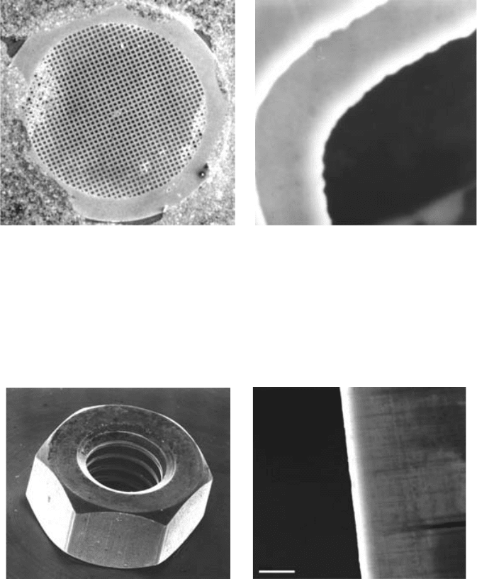
Plate 4.2. Secondary electron images showing: (a) three-dimensional effect
resulting from dependence of SE emission on angle of surface to beam;
(b) edge effect (razor blade); scale bar – 5 mm.
(a) (b)
Plate 4.1. Electron microscope grid (3mm diameter); magnification (a) 10,
(b) 10000.

(a)
(b)
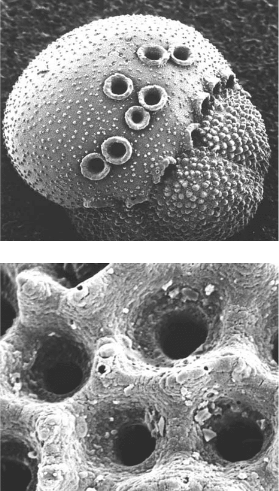
Plate 4.3. Secondary electron images of Globigerina: (a) 200; (b) 2000.
(By courtesy of P. Pearson.)
