Ortiz de Montellano Paul R.(Ed.) Cytochrome P450. Structure, Mechanism, and Biochemistry
Подождите немного. Документ загружается.

104
Thomas
L.
Poulos and Eric F. Johnson
F/G
helix
B' heli
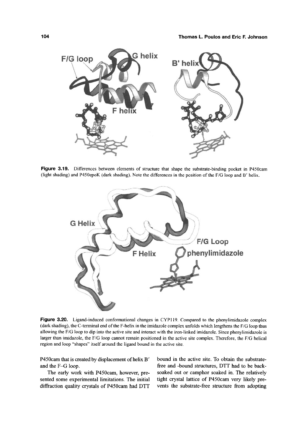
Figure
3.19.
Differences between elements
of
structure that shape
the
substrate-binding pocket
in
P450cam
(light shading) and P450epoK (dark shading). Note the differences
in
the position
of
the F/G loop
and B'
helix.
G Helix
F/G Loop
phenylimidazole
Figure
3.20.
Ligand-induced conformational changes
in
CYP119. Compared
to the
phenylimidazole complex
(dark shading), the C-terminal end of the F-helix in the imidazole complex unfolds which lengthens the F/G loop thus
allowing the F/G loop to dip into the active site and interact with the iron-linked imidazole. Since phenylimidazole
is
larger than imidazole,
the F/G
loop cannot remain positioned
in the
active site complex. Therefore,
the
F/G helical
region and loop "shapes" itself around the ligand bound
in
the active site.
P450cam that is created by displacement of helix B'
and the F-G loop.
The early work with P450cam, however, pre-
sented some experimental limitations. The initial
diffraction quality crystals of P450cam had DTT
bound in the active site. To obtain the substrate-
free and -bound structures, DTT had to be back-
soaked out or camphor soaked in. The relatively
tight crystal lattice of P450cam very likely pre-
vents the substrate-free structure from adopting
structures of Cytochrome P450 Enzymes
105
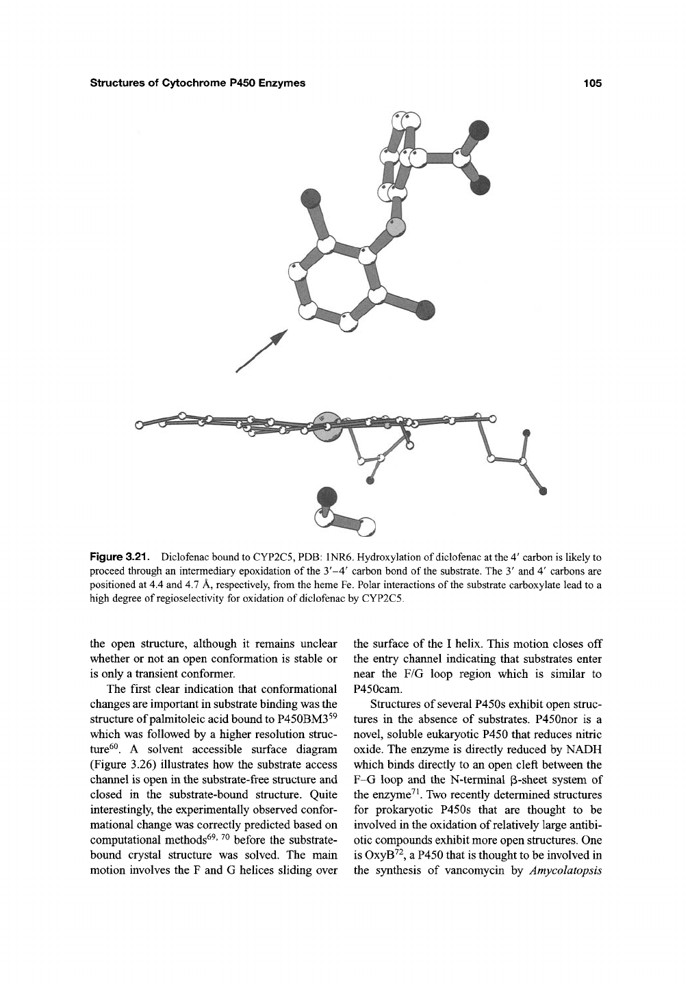
Figure 3.21. Diclofenac bound to CYP2C5, PDB: 1NR6. Hydroxylation of diclofenac at the 4' carbon is likely to
proceed through an intermediary epoxidation of the 3'-4' carbon bond of the substrate. The 3' and 4' carbons are
positioned at 4.4 and 4.7 A, respectively, from the heme Fe. Polar interactions of the substrate carboxylate lead to a
high degree of regioselectivity for oxidation of diclofenac by CYP2C5.
the open structure, although it remains unclear
whether or not an open conformation is stable or
is only a transient conformer.
The first clear indication that conformational
changes are important in substrate binding was the
structure of palmitoleic acid bound to P450BM3^^
which was followed by a higher resolution struc-
ture^^.
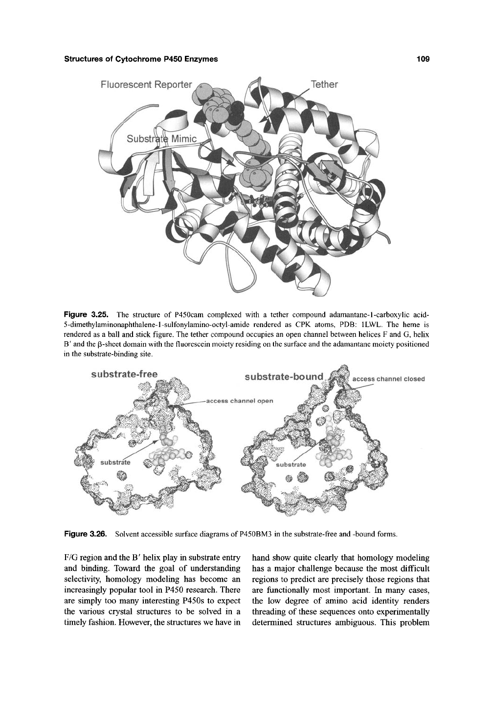
A solvent accessible surface diagram
(Figure 3.26) illustrates how the substrate access
channel is open in the substrate-free structure and
closed in the substrate-bound structure. Quite
interestingly, the experimentally observed confor-
mational change was correctly predicted based on
computational methods^^' ^^ before the substrate-
bound crystal structure was solved. The main
motion involves the F and G helices sliding over
the surface of the I helix. This motion closes off
the entry channel indicating that substrates enter
near the F/G loop region which is similar to
P450cam.
Structures of several P450s exhibit open struc-
tures in the absence of substrates. P450nor is a
novel, soluble eukaryotic P450 that reduces nitric
oxide. The enzyme is directly reduced by NADH
which binds directly to an open cleft between the
F-G loop and the N-terminal (B-sheet system of
the enzyme^ ^ Two recently determined structures
for prokaryotic P450s that are thought to be
involved in the oxidation of relatively large antibi-
otic compounds exhibit more open structures. One
is OxyB''^, a P450 that is thought to be involved in
the synthesis of vancomycin by Amycolatopsis
106 Thomas L. Poulos and Eric F. Johnson
<1°/c
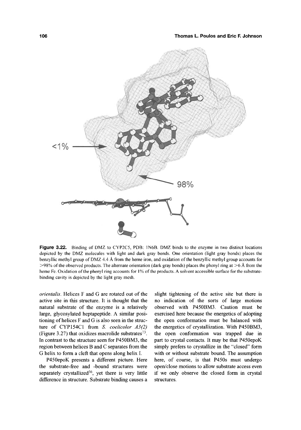
Figure 3.22. Binding of DMZ to CYP2C5, PDB: 1N6B. DMZ binds to the enzyme in two distinct locations
depicted by the DMZ molecules with light and dark gray bonds. One orientation (light gray bonds) places the
benzyllic methyl group of
DMZ
4.4
A
from the heme iron, and oxidation of the benzyllic methyl group accounts for
>98%
of the observed
products.
The alternate orientation (dark gray bonds) places the phenyl ring at >6
A
from the
heme
Fe.
Oxidation of the phenyl ring accounts for
1%
of the
products.
A
solvent accessible surface for
the
substrate-
binding cavity is depicted by the light gray mesh.
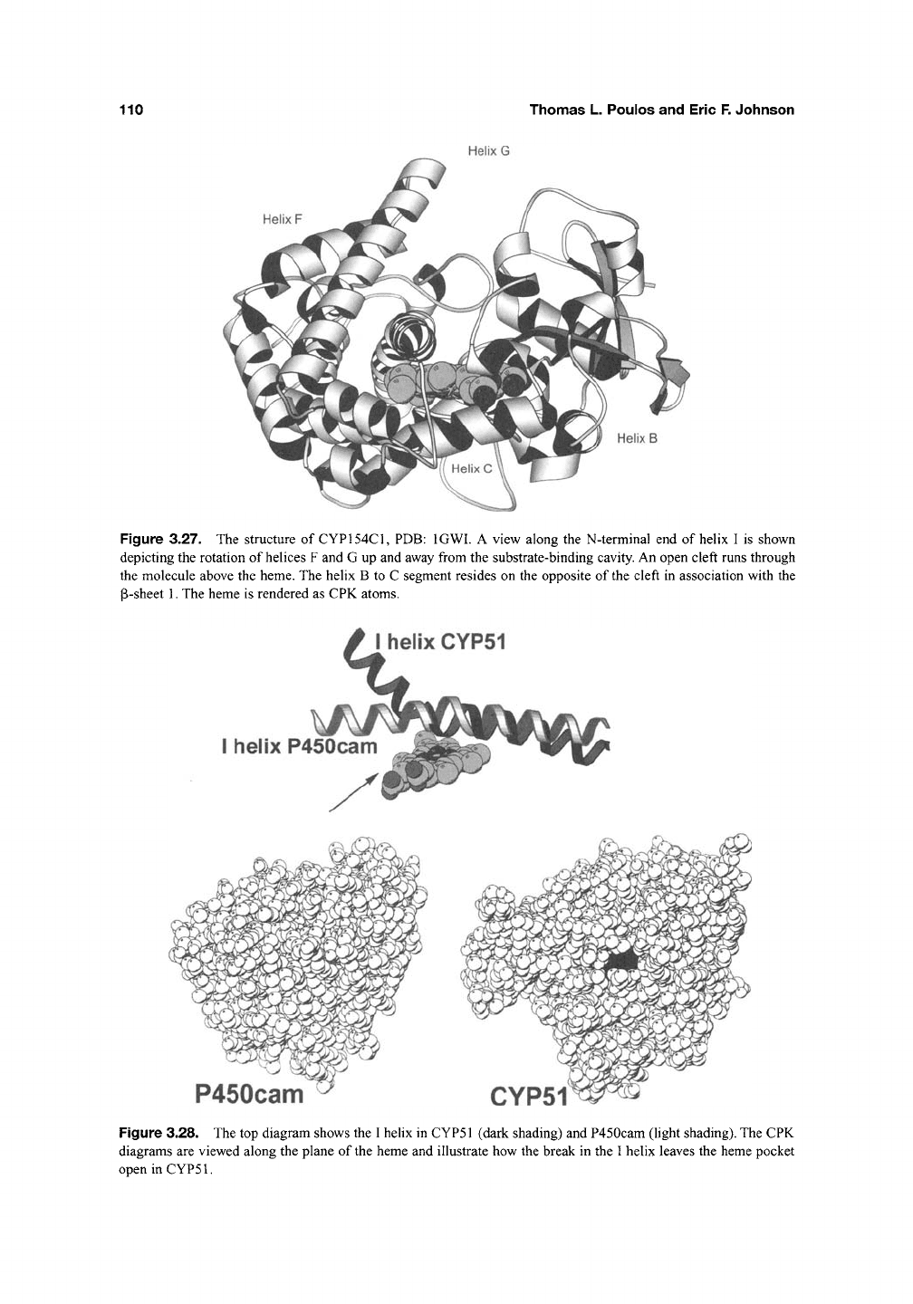
orientalis. Helices F and G are rotated out of the
active site in this structure. It is thought that the
natural substrate of the enzyme is a relatively
large, glycosylated heptapeptide. A similar posi-
tioning of helices F and G is also seen in the struc-
ture of CYP154C1 from S. coelicolor A3(2)
(Figure 3.27) that oxidizes macrolide substrates^^.
In contrast to the structure seen for P450BM3, the
region between helices B and C separates from the
G helix to form a cleft that opens along helix I.
P450epoK presents a different picture. Here
the substrate-free and -bound structures were
separately crystallized^^, yet there is very little
difference in structure. Substrate binding causes a
slight tightening of the active site but there is
no indication of the sorts of large motions
observed with P450BM3. Caution must be
exercised here because the energetics of adopting
the open conformation must be balanced with
the energetics of crystallization. With P450BM3,
the open conformation was trapped due in
part to crystal contacts. It may be that P450epoK
simply prefers to crystallize in the "closed" form
with or without substrate bound. The assumption
here,
of course, is that P450s must undergo
open/close motions to allow substrate access even
if we only observe the closed form in crystal
structures.
structures of Cytochrome P450 Enzymes
107
Helix B'
Val 106
Helix I
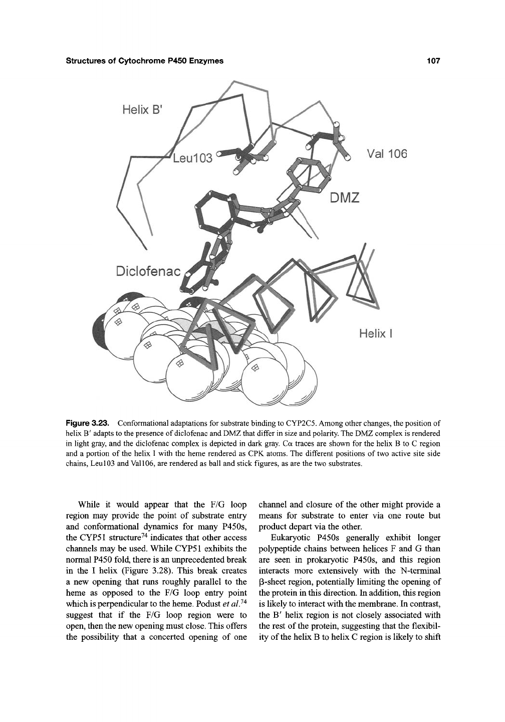
Figure 3.23. Conformational adaptations for substrate binding to CYP2C5. Among other changes, the position of
hehx B' adapts to the presence of diclofenac and DMZ that differ in size and polarity. The DMZ complex is rendered
in light gray, and the diclofenac complex is depicted in dark gray. Ca traces are shown for the helix B to C region
and a portion of the helix I with the heme rendered as CPK atoms. The different positions of two active site side
chains. Leu 103 and Val
106,
are rendered as ball and stick figures, as are the two substrates.
While it would appear that the F/G loop
region may provide the point of substrate entry
and conformational dynamics for many P450s,
the CYP51 structure^"^ indicates that other access
channels may be used. While CYP51 exhibits the
normal P450 fold, there is an unprecedented break
in the I helix (Figure 3.28). This break creates
a new opening that runs roughly parallel to the
heme as opposed to the F/G loop entry point
which is perpendicular to the heme. Podust et al?^
suggest that if the F/G loop region were to
open, then the new opening must
close.
This offers
the possibility that a concerted opening of one
channel and closure of the other might provide a
means for substrate to enter via one route but
product depart via the other.
Eukaryotic P450s generally exhibit longer
polypeptide chains between helices F and G than
are seen in prokaryotic P450s, and this region
interacts more extensively with the N-terminal
P-sheet region, potentially limiting the opening of
the protein in this direction. In addition, this region
is likely to interact with the membrane. In contrast,
the B' helix region is not closely associated with
the rest of
the
protein, suggesting that the flexibil-
ity of the helix B to helix C region is likely to shift
108
Thomas L. Poulos and Eric F. Johnson
B' helix unfolded
B' helix
camphor
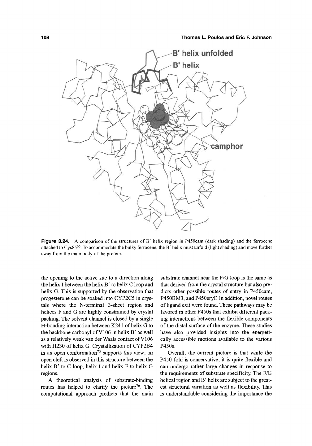
Figure 3.24. A comparison of the structures of B' helix region in P450cam (dark shading) and the ferrocene
attached
to
Cys85^^.
To
accommodate the bulky ferrocene, the B' helix must unfold (light shading) and move fiirther
away from the main body of
the
protein.
the opening to the active site to a direction along
the helix I between the helix B' to helix C loop and
helix G. This is supported by the observation that
progesterone can be soaked into CYP2C5 in crys-
tals where the N-terminal p-sheet region and
helices F and G are highly constrained by crystal
packing. The solvent channel is closed by a single
H-bonding interaction between K241 of helix G to
the backbone carbonyl of VI06 in helix B' as well
as a relatively weak van der Waals contact of VI06
with H230 of helix G. Crystallization of CYP2B4
in an open conformation^^ supports this view; an
open cleft is observed in this structure between the
helix B' to C loop, helix I and helix F to helix G
regions.
A theoretical analysis of substrate-binding
routes has helped to clarify the picture^^. The
computational approach predicts that the main
substrate channel near the F/G loop is the same as
that derived from the crystal structure but also pre-
dicts other possible routes of entry in P450cam,
P450BM3,
and P450eryF In addition, novel routes
of ligand exit were found. These pathways may be
favored in other P450s that exhibit different pack-
ing interactions between the flexible components
of the distal surface of the enzyme. These studies
have also provided insights into the energeti-
cally accessible motions available to the various
P450s.
Overall, the current picture is that while the
P450 fold is conservative, it is quite flexible and
can undergo rather large changes in response to
the requirements of substrate specificity. The F/G
helical region and B' helix are subject to the great-
est structural variation as well as flexibility. This
is understandable considering the importance the
structures of Cytochrome P450 Enzymes
109
Fluorescent Reporter
Tether
Figure 3.25. The structure of P450cam complexed with a tether compound adamantane-1-carboxyhc acid-
5-dimethylaminonaphthalene-l-sulfonylamino-octyl-amide rendered as CPK atoms, PDB: ILWL. The heme is
rendered as a ball and stick figure. The tether compound occupies an open channel between helices F and G, helix
B'
and the p-sheet domain with the fluorescein moiety residing on the surface and the adamantane moiety positioned
in the substrate-binding site.
substrate-free
substrate-bound,
access channel open
access channel closed
Figure 3.26. Solvent accessible surface diagrams of P450BM3 in the substrate-free and -bound forms.
F/G region and the B' helix play in substrate entry
and binding. Toward the goal of understanding
selectivity, homology modeling has become an
increasingly popular tool in P450 research. There
are simply too many interesting P450s to expect
the various crystal structures to be solved in a
timely fashion. However, the structures we have in
hand show quite clearly that homology modeling
has a major challenge because the most difficult
regions to predict are precisely those regions that
are functionally most important. In many cases,
the low degree of amino acid identity renders
threading of these sequences onto experimentally
determined structures ambiguous. This problem
110
Thomas L. Poulos and Eric F. Johnson
Helix G
Helix F
Helix B
Figure 3.27. The structure of CYP154C1, PDB: IGWl. A view along the N-terminal end of helix I is shown
depicting the rotation of helices F and G up and away from the substrate-binding cavity. An open cleft runs through
the molecule above the heme. The helix B to C segment resides on the opposite of the cleft in association with the
P-sheet 1. The heme is rendered as CPK atoms.
I helix CYP51
I helix P450cam
P450cam
CYP51
Figure 3.28. The top diagram shows the I helix in CYP51 (dark shading) and P450cam (light shading). The CPK
diagrams are viewed along the plane of the heme and illustrate how the break in the I helix leaves the heme pocket
openinCYPSl.
structures
of
Cytochrome P450 Enzymes 111
has been largely overcome for the relatively large
number of drug metabolizing enzymes in family 2
because alignments with known structures such
as those of CYP2C and CYP2B families are
relatively straightforward. However, the experi-
mentally determined structures of the family 2
enzymes exhibit significant differences in the con-
formations of the flexible portions of the catalytic
sites that underlie their functional diversity. Thus, a
critical need to determine structures of specific
drug metabolizing enzymes remains in order to
accurately model substrate enzyme interactions.
Acknowledgments
TLP would like to thank members of the UCI
P450 group, Huiying Li, Shingo Nagano, and
Irina Sevrioukova, and NIH grant GM33688.
EFJ would like to thank his colleagues at TSRI,
Guillaume Schoch, Mike Wester, Jason
Yano,
and
C. David Stout, as well as NIH Grant GM31001.
References
1.
Poulos,
T.L., B.C.
Finzel,
and
A.J. Howard (1987).
High-resolution crystal structure
of
cytochrome
P450cam. J.
Mol
Biol. 195, 687-700.
2.
Ravichandran,
K.G., S.S.
Boddupalli,
C.A. Hasermann,
J.A.
Peterson,
and J.
Deisenhofer
(1993).
Crystal structure
of
hemoprotein domain
of
P450BM-3,
a
prototype
for
microsomal P450's.
5c/e«ce 261, 731-736.
3.
Sundaramoorthy,
M., J.
Temer,
and T.L.
Poulos
(1995).
The
crystal structure
of
chloroperoxidase:
A
heme peroxidase-cytochrome P450 functional
hybrid. Structure 3, 1367-1377.
4.
Crane,
B.R., A.S.
Arvai,
D.K.
Ghosh,
C. Wu,
E.D.
Getzoff,
D.J.
Stuehr
et al
(1998). Structure
of
nitric oxide synthase oxygenase dimer with pterin
and substrate. Science 119, 2121-2126.
5.
Fischmann, T.O.,
A.
Hruza, X.D.
Niu,
J.D. Fossetta,
C.A. Lunn,
E.
Dolphin
et al
(1999). Structural char-
acterization of nitric oxide synthase isoforms reveals
striking active-site conservation. Nat. Struct. Biol.
6,
233-242.
6. Raman,
C.S., H. Li, R
Martasek,
V
Krai,
B.S.
Masters, and T.L. Poulos (1998). Crystal struc-
ture
of
constitutive endothelial nitric oxide synthase:
A paradigm
for
pterin function involving
a
novel
metal center. Cell 95, 939-950.
7.
Adman,
E., K.D.
Watenpaugh,
and L.H.
Jensen
(1975).
NH-S hydrogen bonds in Peptococcus aero-
genes ferredoxin, Clostridium pasteurianum rubre-
doxin,
and
Chromatium high potential iron protein.
Proc. Natl.
Acad.
Sci. USA 12, 4854^858.
8. Ueyama,
N., T.
Terakawa,
M.
Nakata,
and
A. Nakamura (1983). Positive shift
of
redox
potential
of
[Fe2S4(Z-cys-Gly-Ala-OMe)4]2-
in dichloromethane.
J. Am.
Chem.
Soc. 105,
7098-7102.
9. Ueyama, N., N. Nishikawa,
Y.
Yamada, T. Okamura,
and
A.
Nakamura (1996). Cytochrome P-450 model
(porphinato)(thiolatio)iron(III) complexes with
and
double NH-S hydrogen bonds.
J. Am.
Chem.
Soc.
118,
1286-1287.
10.
Poulos, T.L.
and
B.C. Finzel (1984). Heme enzyme
structure and function.
In
M.T. Meam (ed.). Peptide
and Protein Reviews, Vol.
4.
Marcel Dekker,
New
York, pp.
115-171.
11.
Chang, C.K. andT.G. Traylor (1973). Proximal base
influence
on the
binding
of
oxygen
and
carbon monoxide
to
heme.
J.
Am. Chem.
Soc. 95,
8477-8479.
12.
Doef,
M.M., D.A. Sweigart, and
P
O'Brien (1983).
Hydrogen bonding from coordinated imidazole
in ferric porphyrin complexes. Effect
on the
Fe(III)/Fe(n) reduction potential. Inorg. Chem.
22,
851-852.
13.
Nappa,
M.,
J.S. Valentine,
and
PA. Snyder (1977).
Imidazolate complexes
of
ferric porphyrins. J.
Am.
Chem.
Soc. 99, 5799-5800.
14.
Valentine,
J.S., R.P
Sheridan,
L.C.
Allen,
and
PC.
Kahn (1979). Coupling between oxidation state
and hydrogen bond conformation
in
heme proteins.
Proc. Nat. Acad Sci. USA 16, 1009-1013.
15.
Banci,
L., LI.
Bertin,
E.A.
Pease,
M.
Tien,
and
P.
Turano (1992).
^H NMR
investigation
of
man-
ganese peroxidase from Phanerochaete chrysospo-
rium.
A
comparison with other peroxidases.
Biochemistry 31, 10009-10017.
16.
Vidakovic,
M., S.G
SHgar,
H. Li, and
T.L. Poulos
(1998).
Understanding
the
role
of the
essential
Asp251
in
cytochrome p450cam using site-directed
mutagenesis, crystallography,
and
kinetic solvent
isotope effect. Biochemistry 37, 9211-9219.
17.
Benson,
D.E., K.S.
Suslick,
and S.G.
Sligar
(1997).
Reduced
oxy
intermediate observed
in
D251N cytochrome P450cam. Biochemistry
36,
5104-5107.
18.
Schlichting,
L, J.
Berendzen,
K.
Chu, A.M. Stock,
S.A. Maves, D.E. Benson etal. (2000). The catalytic
pathway
of
cytochrome p450cam
at
atomic resolu-
tion. Science 287, 1615-1622.
19.
Cupp-Vickery,
J.R., O. Han, C.R.
Hutchinson,
and
T.L.
Poulos (1996). Substrate-assisted cataly-sis
in cytochrome P450eryF.
Nat.
Struc. Biol.
3,
632-637.
112
Thomas L. Poulos and Eric F. Johnson
20.
Wright, R.L., K. Harris, B. Solow, R.H. White, and
P.J. Kennelly (1996). Cloning of a potential
cytochrome P450 from the archaeon Sulfolobus
solfataricus. FEES Lett. 384, 235-239.
21.
McLean, M.A., S.A. Maves, K.E. Weiss, S. Krepich,
and S.G. Sligar (1998). Characterization of a
cytochrome P450 from the acidothermophilic
archaea Sulfolobus solfataricus. Biochem. Biophys.
Res.
Comm. 252, 166-172.
22.
Yano, J.K., L.S. Koo, D.J. Schuller, H. Li, PR. Ortiz
de Montellano, and T.L. Poulos (2000). Crystal
structure of a thermophilic cytochrome P450 from
the archaeon Sulfolobus solfataricus. J. Biol. Chem.
275,31086-31092.
23.
Park, S.Y., K. Yamane, S. Adachi, Y. Shiro,
S.A. Maves, K.E. Weiss et al. (2002). Thermophilic
Cytochrome P450 (CYP119) from Sulfolobus
solfataricus: High resolution structural origin of its
thermostability and functional properties. J. Inorg.
Biochem. 91,491-501.
24.
Puchkaev, A.V., L.S. Koo, and PR. Ortiz de
Montellano (2003). Aromatic stacking as a determi-
nant of the thermal stability of CYP119 from
Sulfolobus solfataricus. Arch. Biochem. Biophys.
409,
52-58.
25.
Yano, J.K.,
F.
Blasco, H. Li, R.D. Schmid, A. Henne,
and T.L. Poulos (2003). Preliminary characteriza-
tion and crystal structure of a thermostable
cytochrome P450 from Thermus thermophilus.
J. Biol. Chem. 278, 608-616.
26.
Omura,
T.
and
A.
Ito (1991). Biosynthesis and intra-
cellular sorting of mitochondrial forms of
cytochrome P450. Meth. Enzymol. 206,
75-81.
27.
Sakaguchi, M. and T. Omura (1993). Topology and
biogenesis of microsomal cytochrome P-450s. In K.
Ruckpaul and H. Rein (eds.). Medicinal
Implications in Cytochrome P-450 Catalyzed
Biotransformations. Akademie Verlag, Berlin.
28.
Von Wachenfeldt, C, T.H. Richardson, J. Cosme,
and E.F. Johnson (1997). Microsomal P450 2C3 is
expressed as a soluble dimer in Escherichia coli fol-
lowing modifications of its N-terminus. Arch.
Biochem. Biophys. 339, 107-114.
29.
Cosme, J. and E.E Johnson (2000). Engineering
microsomal cytochrome P450 2C5 to be a soluble,
monomeric enzyme. Mutations that alter aggrega-
tion, phospholipid dependence of catalysis, and
membrane binding. J. Biol. Chem. 28, 2545-2553.
30.
Li,
YC. and J.Y.L. Chiang (1991). The expression of
a catalytically active cholesterol 7alpha-hydroxylase
cytochrome P450 in Escherichia coli. J. Biol. Chem.
266,
19186-19191.
31.
Sagara, Y, H.J. Barnes, and M.R. Waterman (1993).
Expression in Escherichia coli of functional
cytochrome P450cl7 lacking its hydrophobic
amino-terminal signal anchor. Arch. Biochem.
Biophys. 304, 272-278.
32.
Pernecky, S.J., J.R. Larson, R.M. Philpot, and
M.J. Coon (1993). Expression of truncated forms of
liver microsomal P450 cytochromes 2B4 and
2E1
in
Escherichia coli: Influence of NH2-terminal region
on localization in cytosol and membranes. Proc.
Natl. Acad Sci. USA 90, 2651-2655.
33.
Williams, PA., J. Cosme, V Sridhar, E.R Johnson,
and D.E. McRee (2000). The crystallographic struc-
ture of a mammalian microsomal cytochrome P450
monooxygenase: Structural adaptations for mem-
brane binding and functional diversity. Mol. Cell 5,
121-132.
34.
De Lemos-Chiarandini, C,
A.B.
Frey, D.D. Sabatini,
and G. Kreibich (1987). Determination of the
membrane topology of the phenobarbital-inducible
rat liver cytochrome P-450 isoenzyme PB-4
using site-specific antibodies. J. Cell Biol. 104,
209-219.
35.
Von Wachenfeldt, C. and E.R Johnson (1995).
Structures of eukaryotic cytochrome P450 enzymes.
In PR. Ortiz de Montellano (ed.), Cytochrome
P450: Structure, Mechanism, and Biochemistry.
Plenum Press, New York.
36.
Ohta, Y, S. Kawato, H. Tagashira, S. Takemori, and
S. Kominami (1992). Dynamic structures of adreno-
cortical cytochrome P-450 in proteoliposomes and
microsomes: Protein rotation study. Biochemistry
31,
12680-12687.
37.
Bayburt, TH. and S.G. Sligar (2002). Single-
molecule height measurements on microsomal
cytochrome P450 in nanometer-scale phospholipid
bilayer disks. Proc. Natl.
Acad.
Sci. USA 99,
6725-6730.
38.
Shank-Retzlaff,
M.L., G.M. Raner, M.J. Coon, and
S.G. Sligar (1998). Membrane topology of
cytochrome P450 2B4 in langmuir-blodgett mono-
layers. Arch. Biochem. Biophys. 359, 82-88.
39.
White, S.H., A.S. Ladokhin, S. Jayasinghe, and
K. Hristova (2001). How membranes shape protein
structure. J. Biol. Chem. 276, 32395-32398.
40.
Bridges, A., L. Gruenke, YT Chang, l.A. Vakser,
G. Loew, and L. Waskell (1998). Identification of
the binding site on cytochrome P450 2B4 for
cytochrome b5 and cytochrome P450 reductase.
J. Biol. Chem. 273, 17036-17049.
41.
Wang, M., D.L. Roberts, R. Paschke, TM. Shea,
B.S.
Masters, and J.J. Kim (1997). Three-
dimensional structure of NADPH-cytochrome P450
reductase: Prototype for FMN- and FAD-
containing enzymes. Proc. Natl.
Acad.
Sci. USA 94,
8411-8416.
42.
Sevrioukova, I.E., H. Li, H. Zhang, J.A. Peterson,
and T.L. Poulos (1999). Structure of a cytochrome
P450-redox partner electron-transfer complex.
Proc. Natl. Acad Sci. USA 96, 1863-1868.
43.
Gruez, A., D. Pignol, M.
Zeghouf,
J. Coves,
M. Fontecave, J.-L. Ferrer et al. (2000). Four crystal
structures of Cytochrome P450 Enzymes
113
structures of the 60kDa falvoprotein monomer of
the sulfite reductase indicate a disordered flavo-
doxin-Hke module. J. Mol. Biol. 299, 199-212.
44.
Sevrioukova, I.F., J.T. Hazzard, G. Tollin, and
T.L. Poulos (1999). The FMN to heme electron
transfer in cytochrome P450BM-3. Effect of
chemical modification of cysteines engineered at
the FMN-heme domain interaction site. J. Biol.
Chem.
274, 36097-36106.
45.
Sevrioukova, I.E., C.E. Immoos, T.L. Poulos, and
P.
Farmer (2000). Electron transfer in the ruthenated
heme domain of cytochrome P450BM-3. Isr. J.
Chem.
40,
47-53.
46.
Sevrioukova, I.E., C. Gracia, H. Li, B. Bhaskar,
T.L. Poulos (2003). Crystal structure of putidare-
doxin, the [2Fe-2S] component of the P450cam
monooxygenase system from Pseudomonas putida.
J. Molec. Biol 333, 377-392.
47.
Sevrioukova, LE, H. Li, T.L. Poulos (2004). Crystal
structure of putidaredoxin reductase from
pseudomonas putida, the final structural component
of the P450cam monooxygenase system. J. Mol.
Biol. 236, 889-902.
48.
Muller, J.J., A. Lapko, G. Bourenkov, K. Ruckpaul,
and U. Heinemann (2000). Adrenodoxin reductase-
adrenodoxin complex structure suggests electron
transfer path in steroid biosynthesis. J. Biol. Chem.
116, 2786-2789.
49.
Mlier, A., J.J. MUer, Y.A. Muller, H. Uhlmann,
R. Bernhardt, and U. Heinemann (1998). New
aspects of electron transfer revealed by the crystal
structure of a truncated bovine adrenodoxin, Adx(4-
108).
Structure 6, 269-280.
50.
Ziegler, G.A. and G.E. Schulz (2000). Crystal struc-
tures of adrenodoxin reductase in complex with
NADP(+) and NADPH suggesting a mechanism
for the electron transfer of an enzyme family.
Biochemistry'^9, 10986-10995.
51.
Mittl, RR.E. and G.E. Schulz (1994). The structure
of glutathione reductase from Escherichia coli at
1.86A resolution: Comparison with the enzyme
from human erythrocytes. Protein Sci. 3, 799-809.
52.
Sevrioukova, LE and T.L. Poulos (2002).
Putidaredoxin reductase, a new function for an old
protein. J. Biol. Chem. 277, 25831-25839.
53.
Aoki, M., K. Ishimori, and L Morishima (1998).
Roles of negatively charged surface residues of
puti-
daredoxin in interactions with redox partners in
P450cam monooxygenase system. Biochim.
Biophys.Acta 1386, 157-167.
54.
Sevrioukova, LE, XT. Hazzard, G. Tollin, and
T.L. Poulos (2001). Laser flash induced electron
transfer in P450cam monooxygenase: Putidaredoxin
reductase-putidaredoxin interaction. Biochemistry
40,
10592-10600.
55.
Pochapsky, T.C., X.M. Ye, G. Ratnaswamy, and
T.A. Lyons (1994). An NMR-derived model for the
solution structure of oxidized putidaredoxin, a 2-Fe,
2-S ferredoxin from Pseudomonas. Biochemistry
33,
6424-6432.
56.
Nagano, S., H. Li, H. Shimizu, C. Nishida,
H. Ogura, PR. Ortiz de Montellano et al. (2003).
Crystal structures of epothilone-D bound,
epothilone-B bound, and substrate-free forms of
cytochrome P450epoK. J. Biol. Chem. 278,
44886-^4893.
57.
Tang, L., S. Shah, L. Chung, J. Carney, L. Kaz,
C. Khosla et al. (2000). Cloning and heterologous
expression of the epothilone gene cluster. Science
287,
640-642.
58.
Lee, D.-S., A. Yamada, H. Sugimoto, L Matsunaga,
H. Ogura, K. Ichihara et al. (2003). Substrate recog-
nition and molecular mechanism of fatty acid
hydroxylation by cytochrome P450 from Bacillus
subtilis. J. Biol. Chem. 278, 9761-9767.
59.
Li, H. and T.L. Poulos (1997). The structure of the
cytochrome p450BM-3 haem domain complexed
with the fatty acid substrate, palmitoleic acid. Nat.
Struct. Biol. 4, 140-146.
60.
Haines, D.C., D.R. Tomchick, M. Machius, and
J.A. Peterson (2001). Pivotal role of water in the
mechanism of P450BM-3. Biochemistry 40,
13456-13465.
61.
Modi, S., M.J. Sutchffe, W.U. Primrose, L.Y. Lian,
and G.C. Roberts (1996). The catalytic mechanism
of cytochrome P450 BM3 involves a 6 A movement
of the bound substrate on reduction. Nat. Struc.
Biol. 3, 414-417.
62.
Wester, M.R., E.E Johnson, C. Marques-Soares,
S. Dijols, PM. Dansette, D. Mansuy (2003). The
structure of mammalian cytochrome P450 2C5
complexed with diclofenac at 2.1 A
resolution: Evidence for an induced fit model of
substrate binding. Biochemistry 42, 9335-9345.
63.
Wester, M.R., E.E Johnson, C. Marques-Soares,
PM. Dansette, D. Mansuy, and CD. Stout
(2003,
submitted). The structure of a substrate complex of
mammalian cytochrome P450 2C5 at 2.3 A resolu-
tion: Evidence for multiple substrate binding
modes. Biochemistry 42, 6370-6379.
64.
Marques-Soares, C, S. Dijols, A. Macherey,
M.R. Wester, E.E Johnson, P.M. Dansette et al.
(2003,
submitted). Sulfaphenazole derivatives as
tools for comparing cytochrome P450 2C5 and
human cytochrome P450 2Cs: Identification of a
new high affinity substrate common to those
enzymes. Biochemistry 42, 6363-6369.
65.
Poulos, T.L., B.C. Finzel, and A.J. Howard
(1986).
Crystal structure of substrate-free Pseudo-
monas putida cytochrome P450. Biochemistry 25,
5314-5322.
66.
DiGleria, K., D.R Nickerson, H.A.O. Hill,
L.L. Wong, and V Fulop (1998). Covalent attach-
ment of an electroactive sulfhydryl reagent in the
