N?lting B. Methods in Modern Biophysics
Подождите немного. Документ загружается.

7.1 Atomic force microscope (AFM) 123
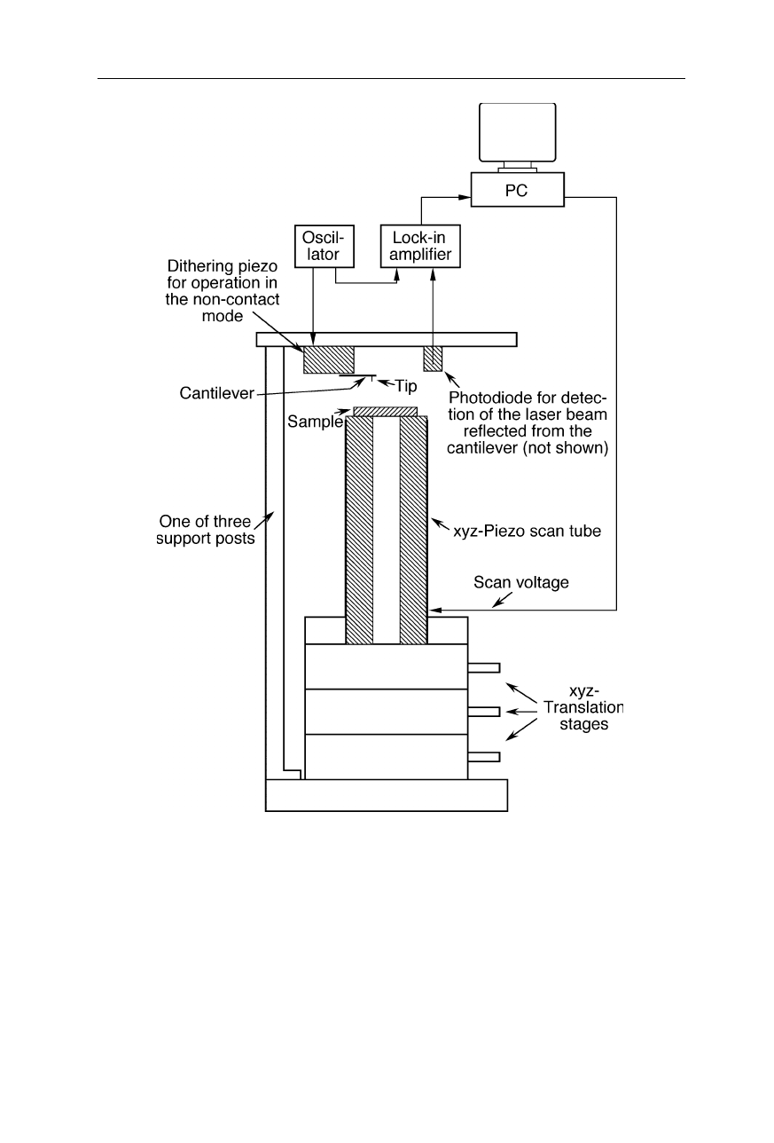
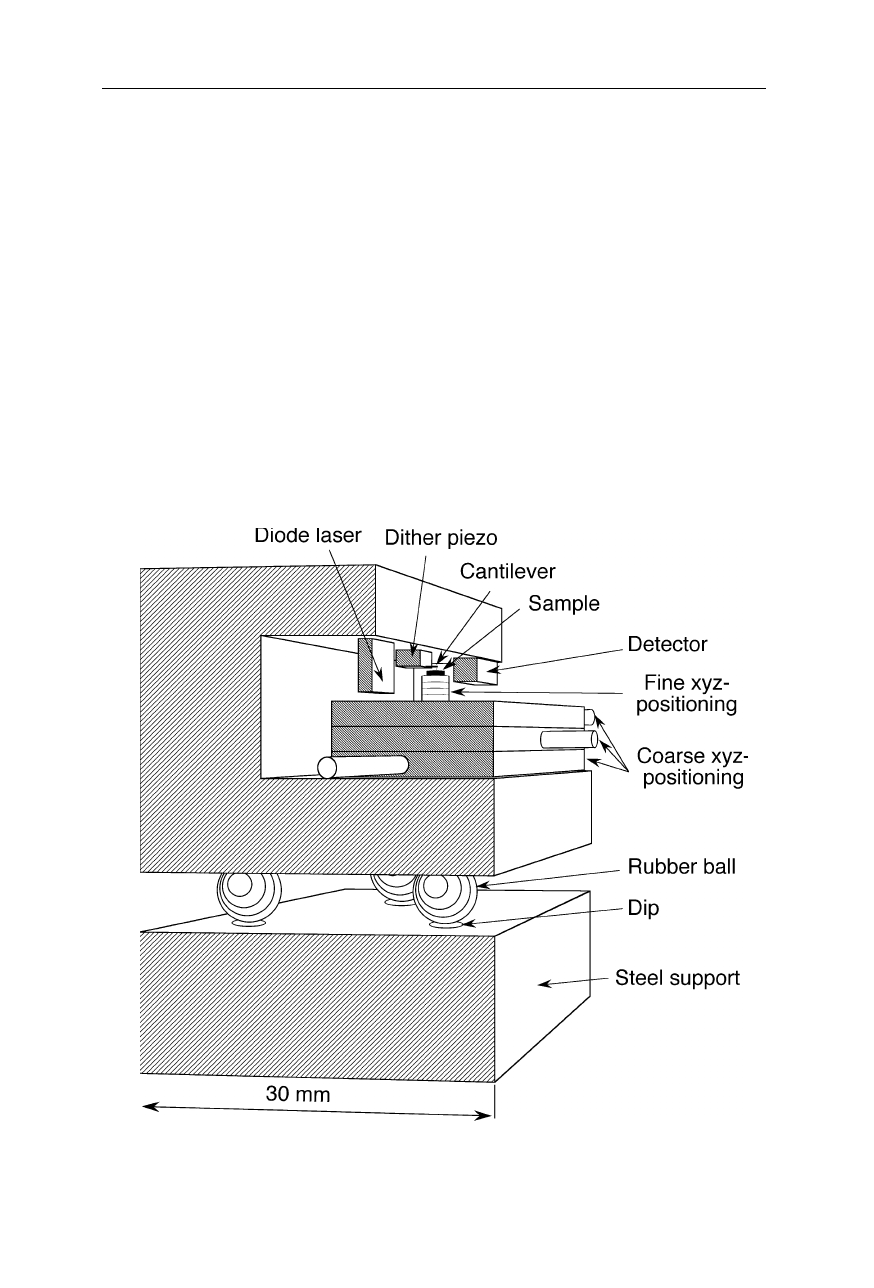
weight-bearing parts of the AFM (Figs. 7.4 and 7.5) have to be rigid and equipped
with a good vibrational damping. Three support posts in the design of Fig. 7.4
reduce wobbling. The xyz-translation stages for coarse adjustment of the piezo-
electric scanner with the sample on top are engineered for little wobbling as well.
The whole AFM is placed on a rubber support preventing transmission of high
frequency vibrations from the laboratory (not shown). A low force of interaction
is crucial for high resolution force microscopy on soft biological specimens. Low
spring constants of the cantilever may facilitate this purpose at the expense of
resolution, but the most common way of gentle measurement is to reduce the
intensity and duration of contact by oscillating the cantilever, as will be explained
later (Fig. 7.13).
Fig. 7.3
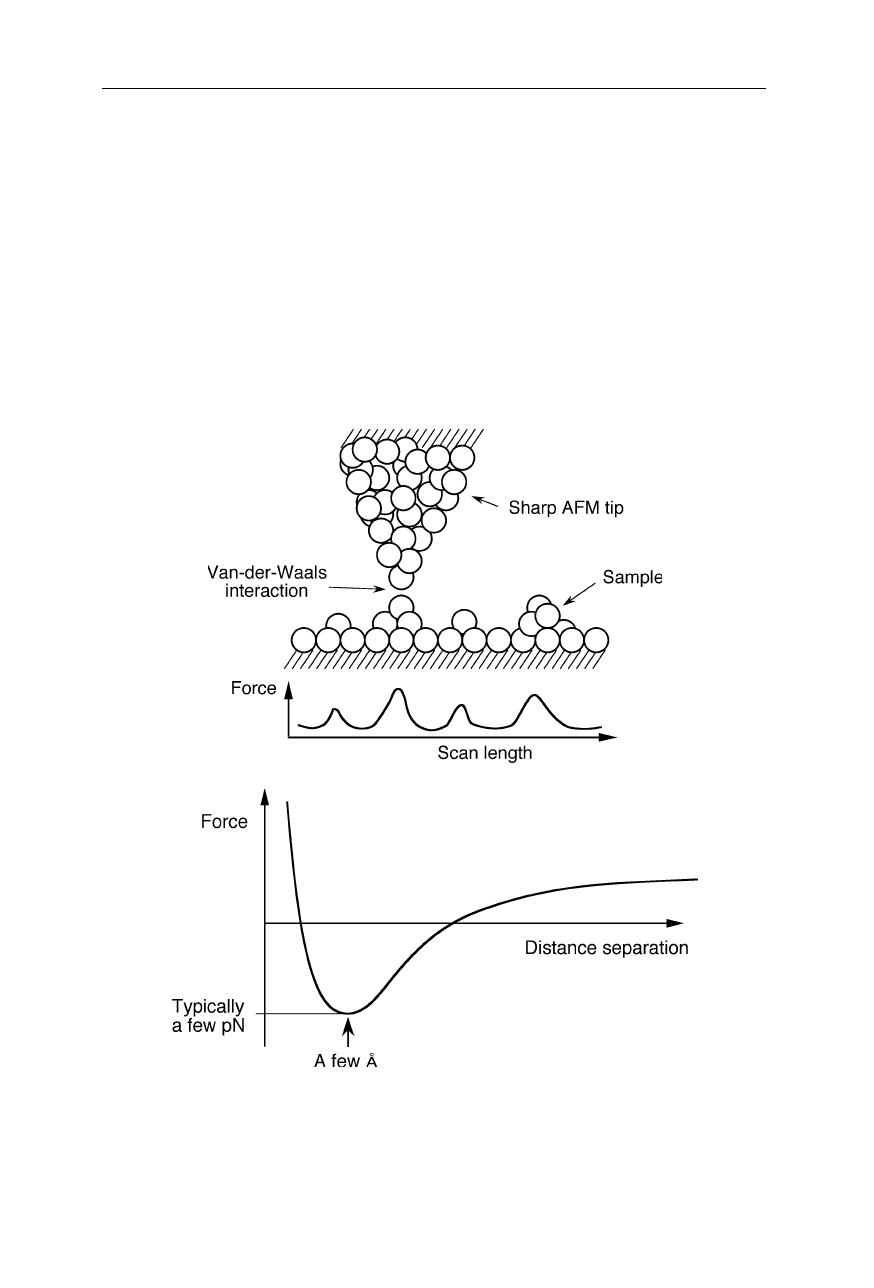
When a small tip approaches a surface, it experiences the van-der-Waals force
which is attractive at a distance of a few Å, but repulsive at very short distances (see, e.g.,
Chap. 3 in Nölting, 2005). Additional Coulomb forces may play a role when the AFM tip
was charged
124 7 Scanning probe microscopy
Fig. 7.4
Design of an AFM. The sample spot of interest is positioned near the tip by
coarse xyz-translation stages. The piezoelectric scanner (see also Fig. 7.5) then heightens
the sample position further till the tip starts to interact with the sample. It allows motion
control of the tip with subnanometer precision. A photodiode detects the reflection
changes of a laser beam from the cantilever upon approach of tip to sample. In this
example, the cantilever is mounted to a dithering piezo element which excites oscillations
of the cantilever. The lock-in amplifier detects changes of these oscillations due to tip-
sample interactions. The sample surface is scanned by sample movement in horizontal
direction by the piezoelectric scanner. The scanner also adjusts the relative height of the
cantilever during scanning to avoid crashes of the tip with the sample surface. Such
crashes can damage the tip and then cause artifacts (see Fig. 7.6)
7.1 Atomic force microscope (AFM) 125
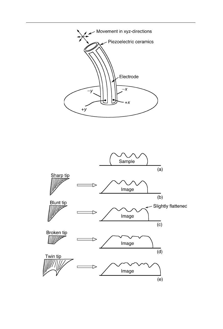
Fig. 7.5
Principle of operation of the xyz-piezoelectric scanner, a ceramic positioning
device which changes its size in response to a change in applied voltage. A voltage change
at the x- or y- electrodes causes bending in the horizontal plane; contraction and expansion
are generated by simultaneous application of x- and y-voltage
Fig. 7.6
Artifacts caused by different shapes of the AFM tip. Blunt tips and broken tips
give rise to an seemingly flattened sample relief which may be difficult to recognize as an
artifact
126 7 Scanning probe microscopy
Fig. 7.7
Tungsten tip, made by sharpening a tungsten wire by dragging it over a plate
coated with alumina. This self-made tip may also be used for STMs (see Sect. 7.2)
Obviously a robust and sharp single tip is essential for this method. Typical
apex radii of commercial tips are
≈
10–20 nm. Fig. 7.6 depicts common types of
artifacts observed when using worn out tips, broken tips, or probes with more than
one tip.
One can make tips themselve by grinding a tungsten wire on a sheet covered
with alumina (Fig. 7.7). These tips are also suitable for STM (Sect. 7.2), but the
tip shape is not very reproducible and tungsten is not very hard.
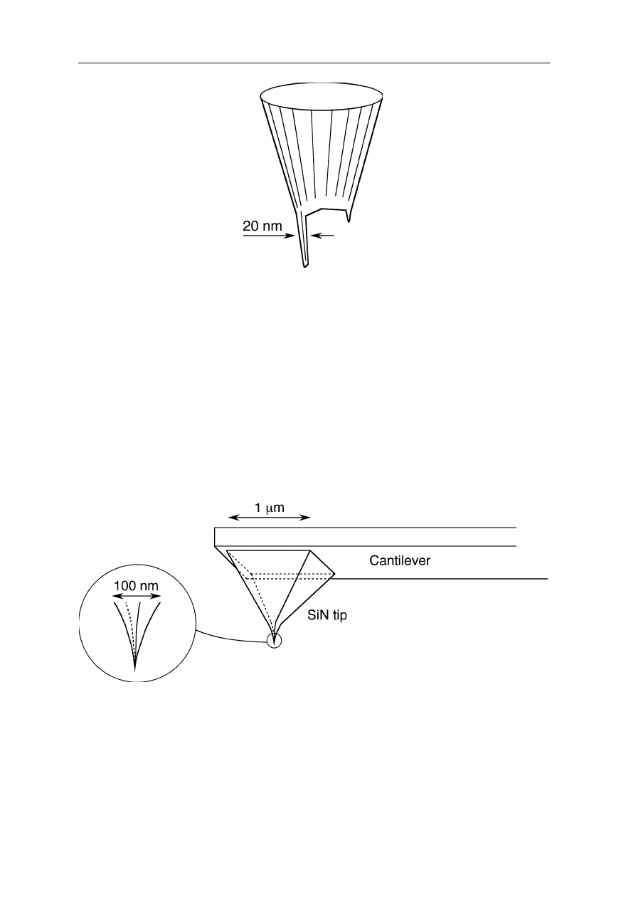
Fig. 7.8
Sharpened pyramidal silicon nitride tip. SiN is extremely hard, and tips can be
engineered with radii of only a few nm
Silicon nitride tips are the currently available tips with highest robustness
(Fig. 7.8). Sharper tips with quite reproducible shape are made from silicon which
is relatively fragile, however (Fig. 7.9a). Even sharper tips for application on
samples with particularly deep structures are manufactured by attaching a high
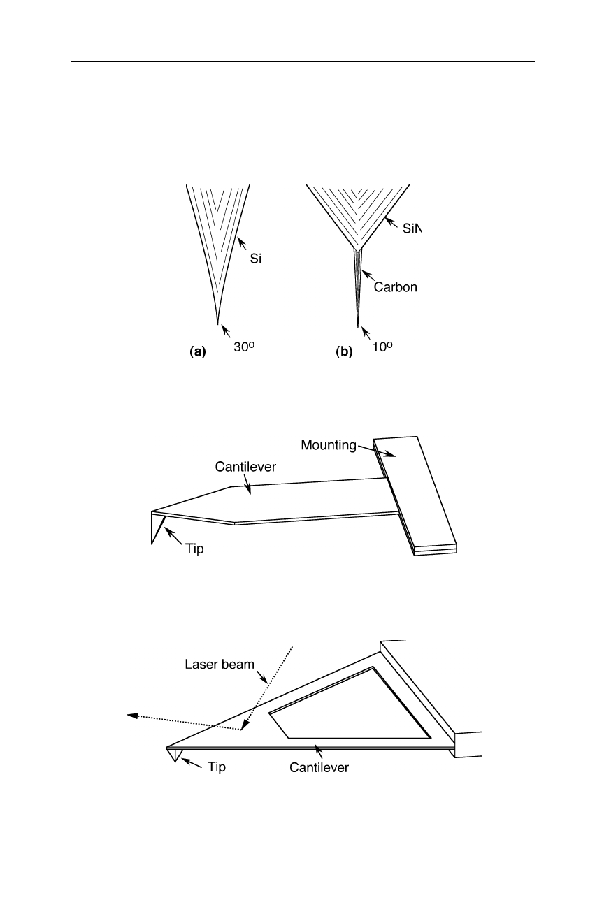
7.1 Atomic force microscope (AFM) 127
density carbon fiber to a silicon tip (Fig. 7.9b). Sharp cantilevers for the
examination of very rough surfaces (Fig. 7.10) and cantilevers with trigonal design
for the purpose of high resistance against torsion (Fig. 7.11) are supplied, e.g., by
Olympus Optical Co. (Tokyo).
Fig. 7.9
(
a
) AFM tip made from silicon. (
b
) Silicon tip with a high density carbon fiber
attached to it
Fig. 7.10
Sharp cantilever geometry for very rough samples (e.g., Olympus Optical Co.,
Tokyo)
Fig. 7.11
A trigonal design of the cantilever (e.g., Olympus Optical Co., Tokyo) causes a
better stability against torsion, compared with rod-shaped cantilevers
128 7 Scanning probe microscopy
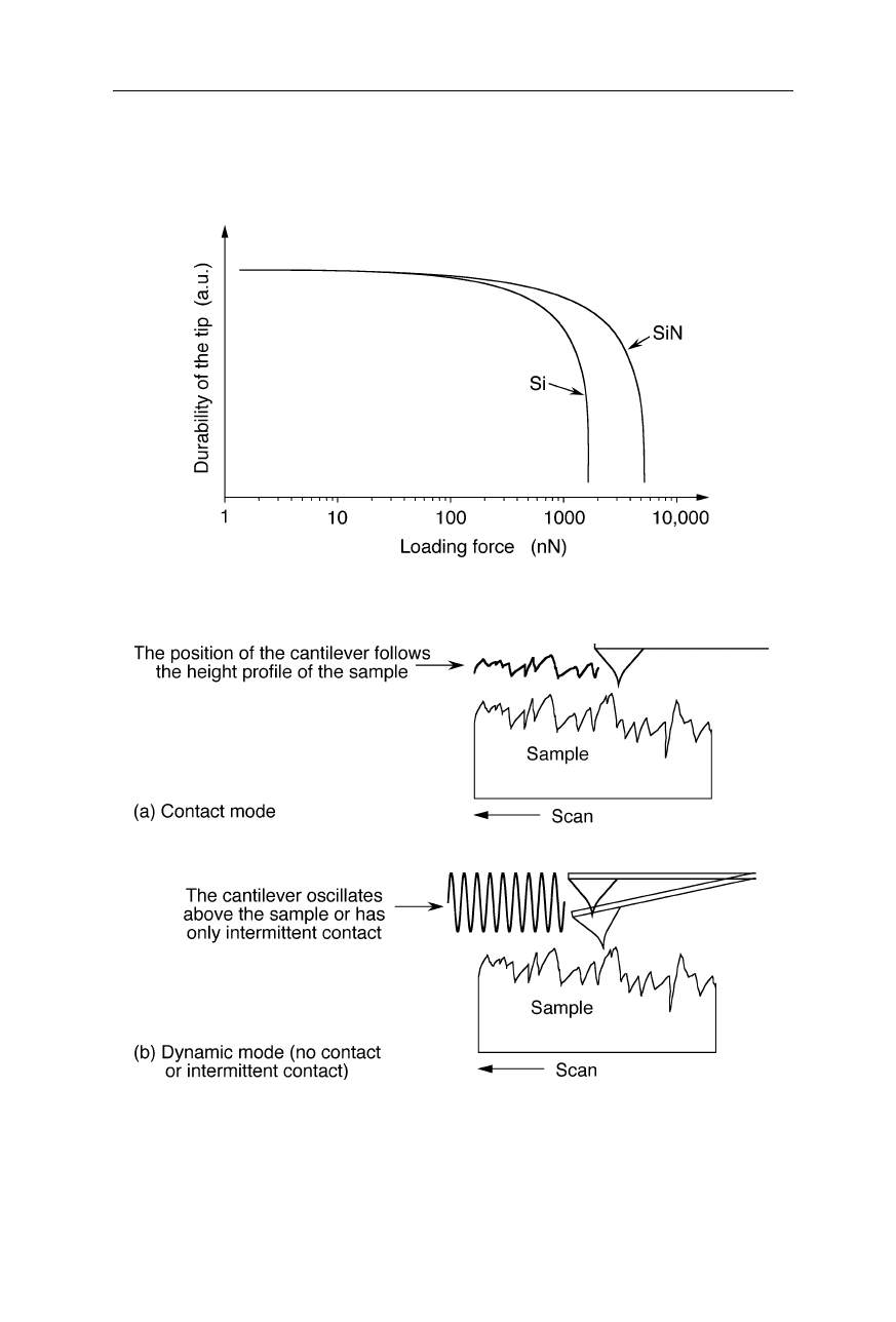
The life of the AFM tip decreases very rapidly with applied force (Fig. 7.12).
High aspect-ratio tips made from silicon or carbon fiber are generally less durable
than low aspect-ratio tips made from silicon nitride.
Fig. 7.12
Example for the wear of two AFM tips due to surface load
Fig. 7.13
(
a
) Contact mode: the cantilever follows the height profile of the sample.
(
b
) Dynamic mode: the cantilever has only intermittent contact or oscillates above the
sample. In the latter mode, oscillations are excited by a piezo crystal and the forces
between tip and sample are very small. This mode permits truly atomic resolution
(Giessibl, 2000)
7.1 Atomic force microscope (AFM) 129
There are two common modes of operation of AFMs (Fig. 7.13): the contact
mode and the dynamic force mode. In the contact mode, the probe tip is in
continuous contact with the sample surface. The force the cantilever exerts on the
substrate in contact mode may perturb the surface of soft biological materials. In
the gentler dynamic mode, the probe tip only oscillates up and down as it is
scanned over the sample surface. Two sub-modes may be distinguished for the
dynamic force mode, the non-contact sub-mode in which the distance between tip
apex and sample surface is always larger than the van-der-Waals distance, and the
tapping sub-mode in which the tip has intermittent contact.
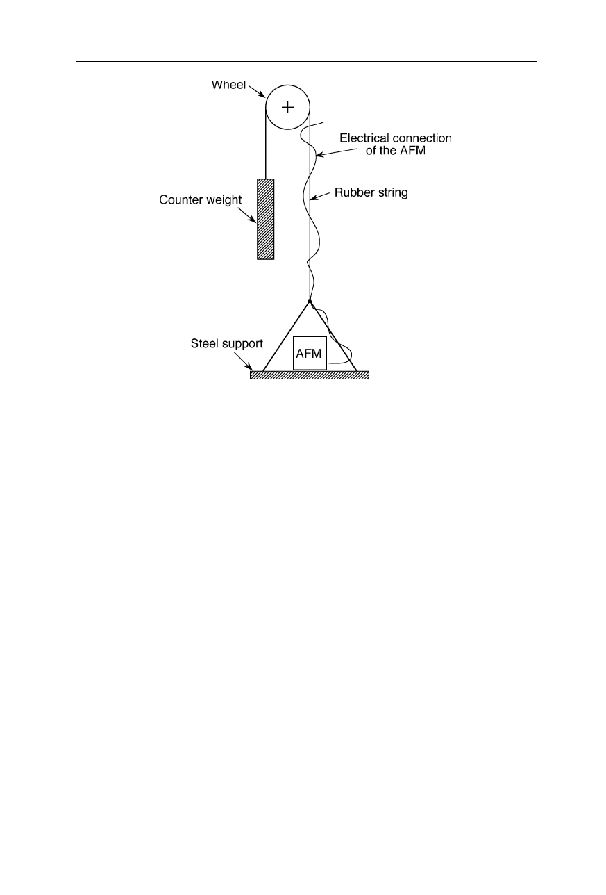
As pointed out, a high degree of protection against external high-frequency
vibrations is obviously crucial for the operation scanning probe microscopes with
atomic resolution. Fig. 7.14 shows a further solution to this problem. Here the
AFM is made from very thick and short plates of steel and the AFM is placed on
three rubber balls that do not transmit fast vibrations. Another technique of
efficient vibrational damping is to hang the AFM on a rubber string (Fig. 7.15).
Fig. 7.14
Robust design of an AFM with atomic resolution. The vibrational damping is
attained by a very rigid construction and an elastic support in form of three rubber balls
130 7 Scanning probe microscopy
Fig. 7.15
“Hanging AFM”: vibrational isolation of an AFM by hanging it on a rubber
string
Important biological applications of AFMs were the direct observation of the
structure of DNA (Lindsay et al., 1989) and the monitoring of actin filament
dynamics in living cells (Henderson et al., 1992). The direct visualization of a
DNA glycosylase searching for damage shows that the glycosylase interrogates
DNA at undamaged sites by introducing drastic kinks (Chen et al., 2002b).
Intramolecular triplex DNA formation results in a kink in the double helix path
(Tiner et al., 2001). A sharp DNA bend is induced by binding of integration host
factor (IHF) to the region between the upstream regulatory sequence and the
promoter sequence (Seong et al., 2002). Single DNA molecule force spectroscopy
can discriminate between different interaction modes of small drug molecules with
DNA by measuring the mechanical properties of DNA and their modulation upon
binding of small drug molecules (Krautbauer et al., 2002) and dye molecules (Kaji
et al., 2001). A decrease of the ionic strength from 50 mM to 1 mM resulted in a
change of the number of nodes (crossings of double helical segments) of a
supercoiled 3000-bp piece of DNA from a 15 to one or two nodes (Cherny and
Jovin, 2001). High resolution fluorescence imaging of
λ
-phage DNA molecules,
intercalated with the dye YOYO-1, by a SNOM/AFM (SNOM, scanning near-
field optical microscope; see Sect. 7.3) resolved the distribution of the dye (Kim et
al., 2001).
AFM proved to be a very useful tool for the study of proteins, yielding some
unique insights into structure and physical properties:
β
-Lactoglobulin forms fine-
stranded aggregates at pH 2 with the diameter of strands being ca. 4 nm (Ikeda and
7.1 Atomic force microscope (AFM) 131
Morris, 2002). AFM technology was used to map out the electrostatic potential of
the transmembrane channel OmpF porin (Fig. 7.16; Philippsen et al., 2002).
AFMs gave crucial topological information of blood cell adhesion on different
sensor materials (Hildebrand et al., 2001). Ac-GWWL(AL)nWWA-Etn peptides
induce the formation of extremely ordered domains in some biologically relevant
membranes (Rinia et al., 2002). The heads of bacteriophage
Φ
KZ and T4 have
different compressibilities (Matsko et al., 2001). Atomic force microscopy
resolved fusion pores in the apical plasma membrane in live pancreatic cells (Cho
et al., 2002) and visualized the growth of Alzheimer's
β
-amyloid-like fibrils
(Goldsbury et al., 2001). Cardiac muscle and skeletal muscle exhibit different
viscous and elastic properties as determined by atomic force microscopy. Cardiac
cells are stiffer (elastic modulus = 100
±
11 kPa) than skeletal muscle cells (elastic
modulus = 25
±
4 kPa; see Mathur et al., 2001). Atomic force microscopy
allowed to visualize the structure of biomolecules, e.g., the native chaperone
complex from
Sulfolobus solfataricus
, in solution under physiological conditions
providing a nanometer resolution topographic image of the sample (Valle et al.,
2001). It is also an excellent technique to study the initial events of mutual cell
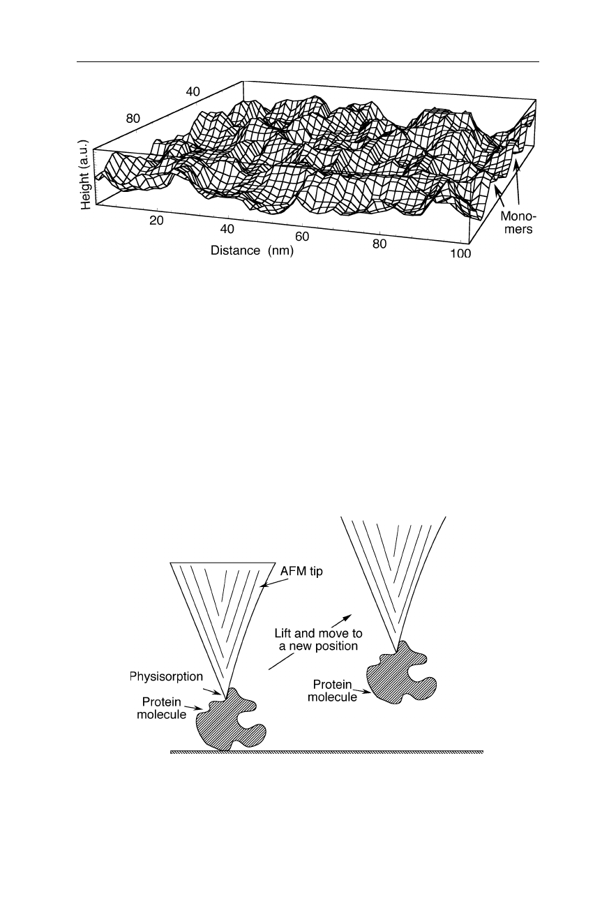
adhesion (Razatos, 2001). An AFM image of a monomolecular film of bovine
serum albumin shows individual monomers and dimers (Fig. 7.17; Gunning et al.,
1996; Morris et al., 1999).
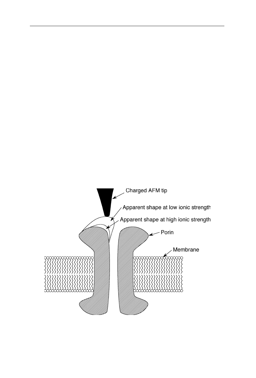
Fig. 7.16
Imaging the electrostatic potential of the transmembrane channel OmpF porin
(Philippsen et al., 2002). Different apparent shapes of the porin are observed at different
ionic strengths. These differences reflect changes of the electrostatic potential which is
experienced by the charged tip of the AFM
132 7 Scanning probe microscopy
Fig. 7.17
AFM image of a monomolecular film of the protein bovine serum albumin
(BSA, M
w,monomer
= 66 kDa) adsorbed at an oil/water interface (Gunning et al., 1996;
Morris et al., 1999). Individual monomers and dimers of BSA can be seen
AFMs are also very useful for the manipulation of macromolecules: proteins
may physisorb to the AFM tip and can then be lifted and manipulated (Fig. 7.18).
The sensitivity of the AFM cantilever, to forces in the pN range, was exploited to
measure folding-unfolding forces within single protein molecules and breakaway
forces between different biomolecules (Jiao et al., 2001; Allison et al., 2002).
Atomic force microscopy has yielded tantalizing insights into the dynamics of pro-
tein self-assembly and the mechanisms of protein unfolding (Furuike et al., 2001;
Yip, 2001). For further, similar applications of AFM technology see Chap. 8.
Fig. 7.18
Manipulation of a protein molecule with an AFM: The tip is lowered till it
touches the macromolecule. Due to the attractive action of the van-der-Waals interaction,
the macromolecule sticks to the tip and can be lifted and moved to a different place