N?lting B. Methods in Modern Biophysics
Подождите немного. Документ загружается.

102 5 Protein infrared spectroscopy
Because of the significantly lower scattering of IR light relative to light of
shorter wavelength, IR microscopes (Fig. 5.14) enable the inspection of most
strongly scattering samples. Computer aided image processing allows two- or
three-dimensional resolution. More complicated microscopes may utilize step-
scan interferometry for photoacoustic depth profiling, monochromators for
spectral analysis and polarizers/analyzers for linear dichroism (LD) analysis.
5.2 Applications
One of the biophysical main applications of FTIR is the characterization of the
structure and conformational changes of proteins (Barrera et al., 2002; Butler et
Fig. 5.15
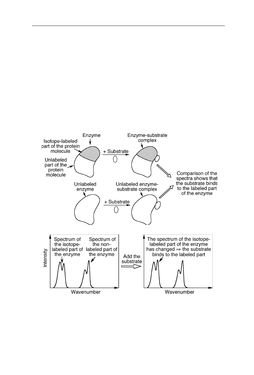
Isotope-edited FTIR spectroscopy (see, e.g., Li et al., 2002; Barth, 2002). Since
the spectrum of the isotope-labeled part of the protein molecule is significantly shifted, it
can be distinguished from the spectrum of the non-labeled part. A change of the protein IR
spectrum upon binding of the substrate to the protein shows which part of the molecule the
substrate binds to. In this example, the magnitude of a peak in the spectrum of the isotope-
labeled part of the protein has changed upon binding of the substrate. This shows that the
substrate binds to the labeled part of the enzyme
5.2 Applications 103
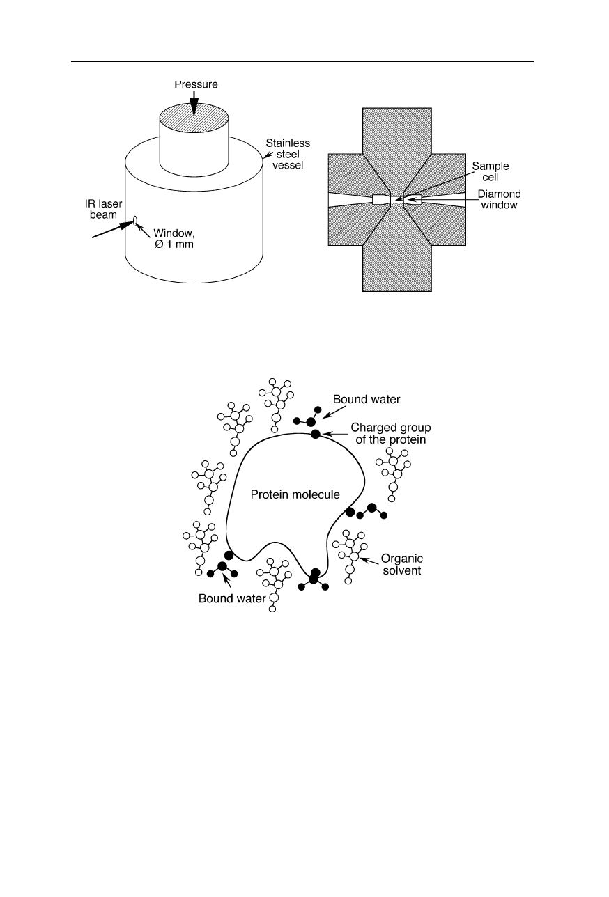
Fig. 5.16
Apparatus to monitor protein unfolding under high pressure with IR. Since the
volume of unfolded protein is less than that of folded protein, high pressure favors
transition to the unfolded state
Fig. 5.17
Protein molecule in organic solvent: only a few strongly bound water molecules
remain attached to the protein molecule
al., 2002; Castellanos et al., 2002; Dong et al., 2002; Hilario et al., 2002; Moritz
et al., 2002; Mui et al., 2002; Noinville et al., 2002), of peptides (Bianco et al.,
2002; Gordon et al., 2002; Huang et al., 2002; Torres et al., 2002), and of DNA
(Lindqvist and Graslund, 2001; Malins et al., 2002). In some cases, interactions
were resolved at the level of individual amino acid residues (Kandori et al., 2002;
Mezzetti et al., 2002; Zhang et al., 2002a).
Isotope-edited FTIR is particularly useful for the structural characterization of
specific macromolecular regions (Fig. 5.15): e.g., the three phosphate stretching
104 5 Protein infrared spectroscopy
vibrations of the phosphate calcium ATPase complex were detected at a
background of 50,000 protein vibrations in an isotope exchange experiment
(Barth, 2002).
Time-resolved step-scan FTIR spectroscopy enables the monitoring of confor-
mational changes of proteins in the microsecond time scale (Bailey et al., 2002).
FTIR spectroscopy allowed to map out the nucleotide binding site of calcium
ATPase (Liu and Barth, 2002). IR and FTIR spectroscopy are two of the only few
methods suitable to monitor conformational changes of proteins under high
pressure (Fig. 5.16; Dzwolak et al., 2002). FTIR spectroscopy on bacterio-
rhodopsin revealed a pre-melting conformational transition at 80
o
C (Heyes et al.,
2002). FTIR is also suitable to investigate the structure and hydration shell of
protein molecules in organic solvents (Fig. 5.17; Costantino et al., 1995). Further,
IR and FTIR spectroscopy was used for the characterization of irradiated starches
(Kizil et al., 2002), and the determination of dihedral angles of tripeptides
(Schweitzer-Stenner, 2002). Molecular changes of preclinical scrapie can be
detected by IR spectroscopy (Kneipp et al., 2002). FTIR spectroscopy can serve
as an optical nose for predicting odor sensation (van Kempen et al., 2002) and for
chemical analysis of drinks (Coimbra et al., 2002; Duarte et al., 2002).
FTIR microscopy at a spatial resolution of 18
µ
m resolved single cells (Lasch
et al., 2002). IR spectroscopy is also a tool for discrimination between different
strains or types of cells (Gaigneaux et al., 2002).
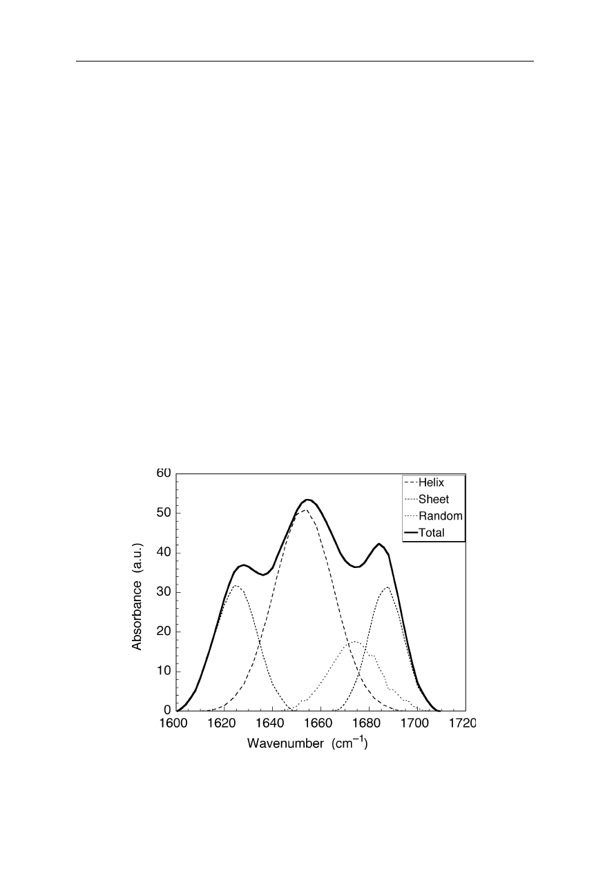
Fig. 5.18
Decomposition of a FTIR spectrum into three components corresponding to
helical structure, sheets and non-regular structure, respectively. Percentages of structure
content and structural changes, e.g., due to protein denaturation, are quantifiable
5.2 Applications 105
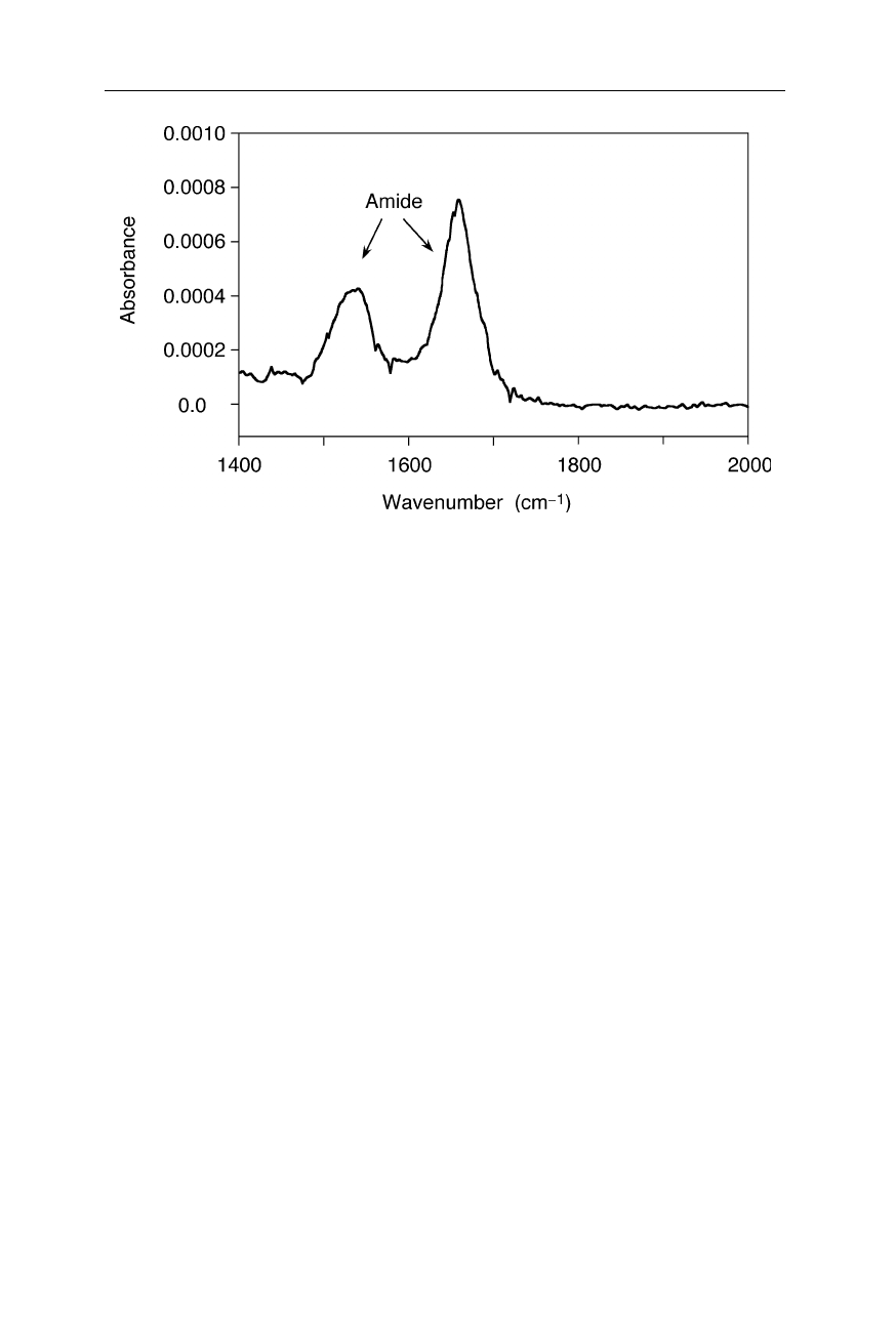
Fig. 5.19
FTIR spectrum of a single molecular monolayer of A126C sperm whale
myoglobin (Jiang et al., 1996). The peaks around 1660 cm
–1
and 1530 cm
–1
correspond to
the amide I and amide II bands, respectively. The spectrum was acquired with a BioRad
FTIR spectrophotometer equipped with a TGS detector
Fig. 5.18 shows an example for the decomposition of a FTIR spectrum of a
protein into the components corresponding to helical, sheet-like and random coil-
like (non-regular) structures, respectively. Such decompositions can be calcu-
lated, e.g., by fitting a linear combination of the base spectra for the secondary
structure components to the measured spectrum.
Fig. 5.19 illustrates the amazing sensitivity of FTIR spectroscopy. The sample
was only two monolayers of a protein. Since at very low sample absorbances it is
quite difficult to avoid the sharp lines of water-vapor absorption, these measure-
ments were taken in a nitrogen-filled chamber at two different, very low concen-
trations of water, and later the water spectrum was subtracted. With this proce-
dure, average artifact and noise levels were reduced to less than 0.00003 ab-
sorbance units.
6 Electron microscopy
6.1 Transmission electron microscope (TEM)
Transmission electron microscopy utilizes the wave properties of moving elec-
trons to generate highly resolved images of specimens.
6.1.1 General design
In 1986 the Nobel prize in physics was awarded by one half to Ernst Ruska for his
fundamental work in electron optics, and for the design of the first electron
microscope (EM), and by one half to Gerd Binnig and Heinrich Rohrer for their
design of the scanning tunneling microscope (see Chap. 7). In some aspects, the
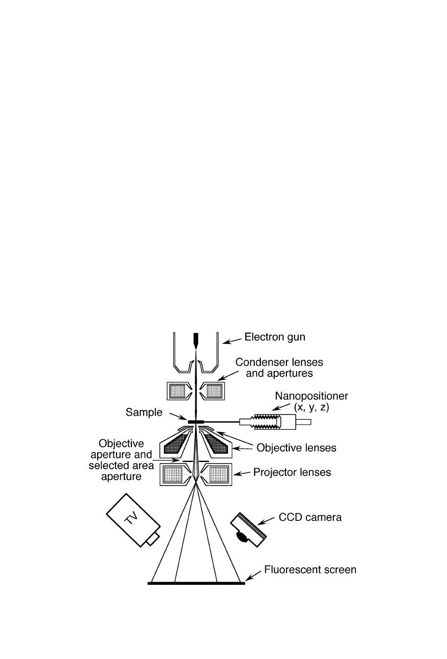
Fig. 6.1
Transmission electron microscope (see text on pp. 107 and 109)
108 6 Electron microscopy
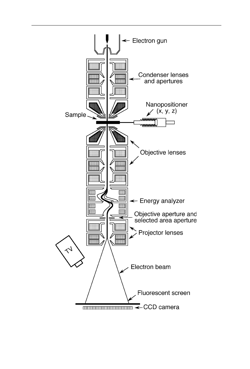
Fig. 6.2
A more complicated design of a transmission electron microscope with an
analyzer which can remove inelastically scattered electrons (see, e.g., LEO Elektronen-
mikroskopie GmbH, Oberkochen, Germany)
6.1 Transmission electron microscope (TEM) 109
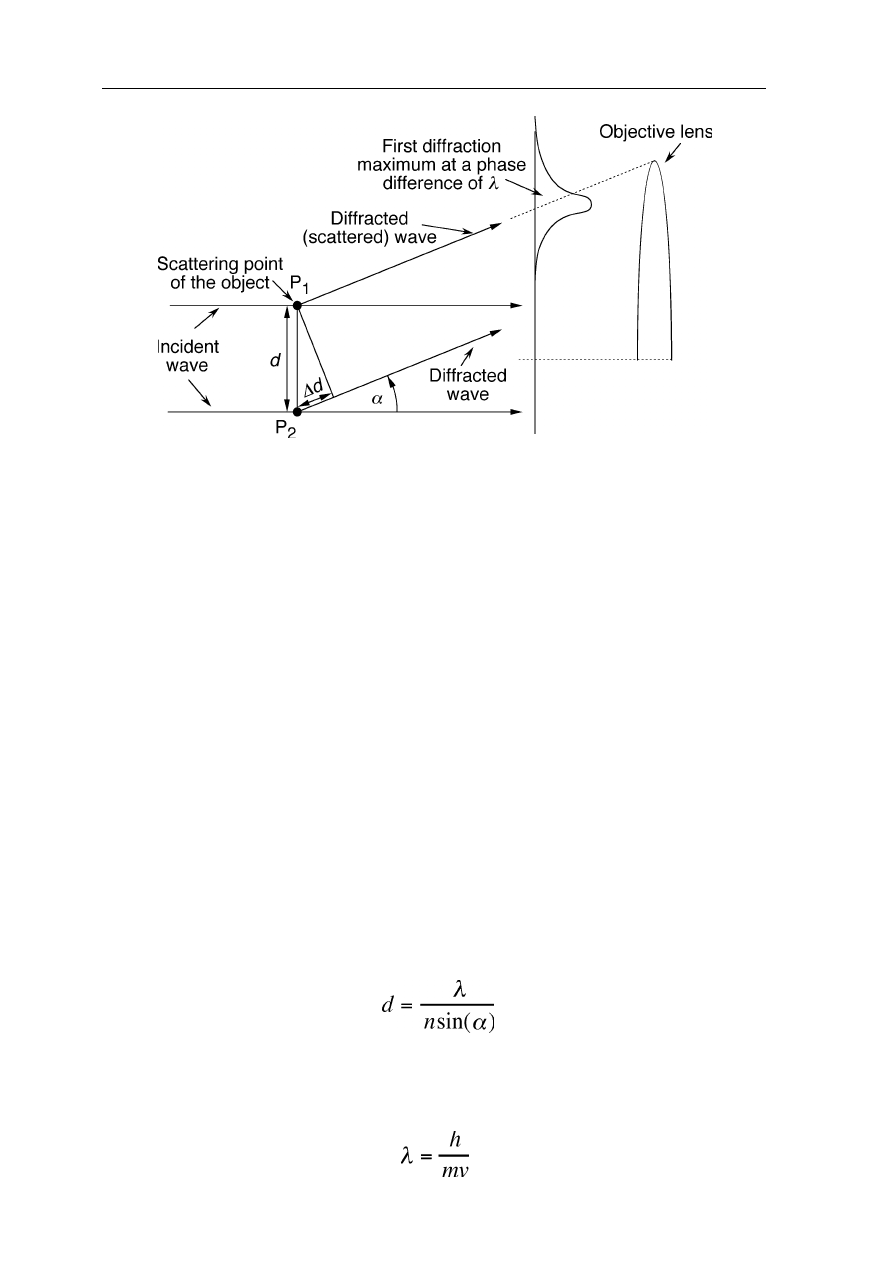
Fig. 6.3
In order to resolve the two points P
1
and P
2
of an object, the objective lens has to
catch the first diffraction maximum of the two points. It appears in the direction where the
diffracted (scattered) waves from the two diffracting (scattering) points have a phase
difference,
∆
d, of one wavelength. Eq. (6.1) was derived from this condition. A typical
objective lens has a bore of 2 mm and a focal length of about 1–2 mm
operation of a transmission electron microscope (TEM) is comparable with that of
a slide projector (Figs. 6.1 and 6.2): Electrons from the electron gun pass through
condenser lenses that focus the electrons onto the sample. The electron beam
shines through the specimen. Objective lenses and projector lenses magnify the
transmitted beam and project it onto the fluorescent viewing screen. Impact of
electrons excites the screen and produces a visible magnified image of the sample.
This image is recorded with various detectors, such as a CCD camera.
6.1.2 Resolution
Electron microscopes enable significantly greater magnification and greater depth
of focus than conventional optical microscopes. High-resolution TEMs permit
spatial resolutions around 0.1 nm (1 Å) at acceleration voltages of 50–600 kV.
Because of the wave nature of the electrons, the resolution limit, d, is given by the
diffraction theory of coherent imaging:
, (6.1)
where
λ
, n, and
α
are the vacuum wavelength, index of refraction of the medium
(=1 in TEMs), and aperture half angle of the objective lens, respectively (Fig.
6.3). The de Broglie relation provides the wavelength,
λ
, of the electrons:
, (6.2)
110 6 Electron microscopy
where
h
= 6.6261
×
10
–34
J s,
m
, and
v
, are the Planck constant, electron mass, and
electron velocity, respectively. For the relativistically high velocities of the
electron beam we have to use Einstein's equations:
,
E
=
mc
2
, (6.3)
and obtain:
, (6.4)
where
e
= 1.6022
×
10
–19
C is the elementary charge,
m
e
= 9.1094
×
10
–31
kg the
electron rest mass,
c
= 2.99792
×
10
8
m s
–1
the speed of light in vacuum,
E
=
E
0
+
∆
E
,
E
0
=
m
e
c
2
,
∆
E
=
V
.
e
the kinetic energy of the electrons, and
V
is the applied
acceleration voltage, typically 200 V – 200 kV. For a voltage of, e.g., 100 kV we
find
λ
= 0.0037 nm. In contrast to most light microscopes, TEMs have small
objective lens apertures of typically
α
= 1–2
o
, and thus according to Eq. 6.1 the
limit of spatial resolution is 0.1–0.2 nm in this example.
6.1.3 Electron sources
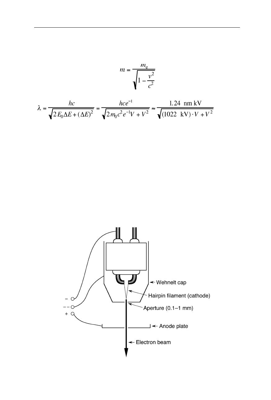
Thermionic electron guns (Fig. 6.4) and cold field emission guns (Fig. 6.5) are
Fig. 6.4
Thermionic electron gun (e.g., Structure Probe, Inc., West Chester, PA)
6.1 Transmission electron microscope (TEM) 111
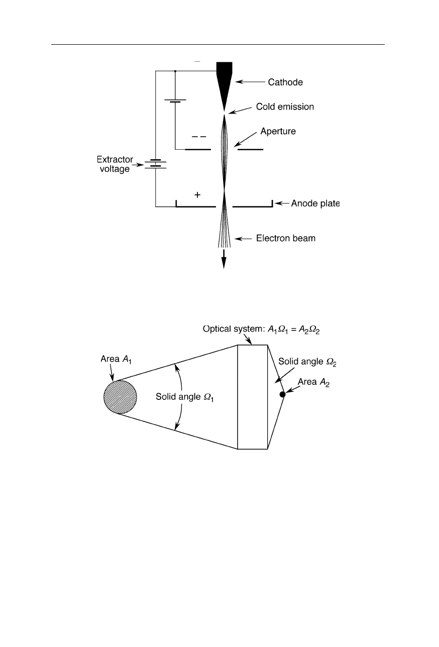
Fig. 6.5
Cold field emission gun focusable with an electrostatic lens comprised of two
apertures with different electrostatic potentials
Fig. 6.6
In the production of an image of an object by an optical system, the product of
area, A, and aperture solid angle,
Ω
, remains about constant
common electron sources. A considerable concern is the brightness and size of
electron sources. Fig. 6.6 illustrates why this is important: For a light beam
passing through an optical system, the product of area and aperture solid angle of
radiation remains constant. Thus, a large source can be focussed on a small spot
only by using a large aperture angle of the optical system. Considering the limited
aperture angles of electron lenses, a source of small size and high brightness is
required to obtain a sufficiently bright picture of the sample.
112 6 Electron microscopy
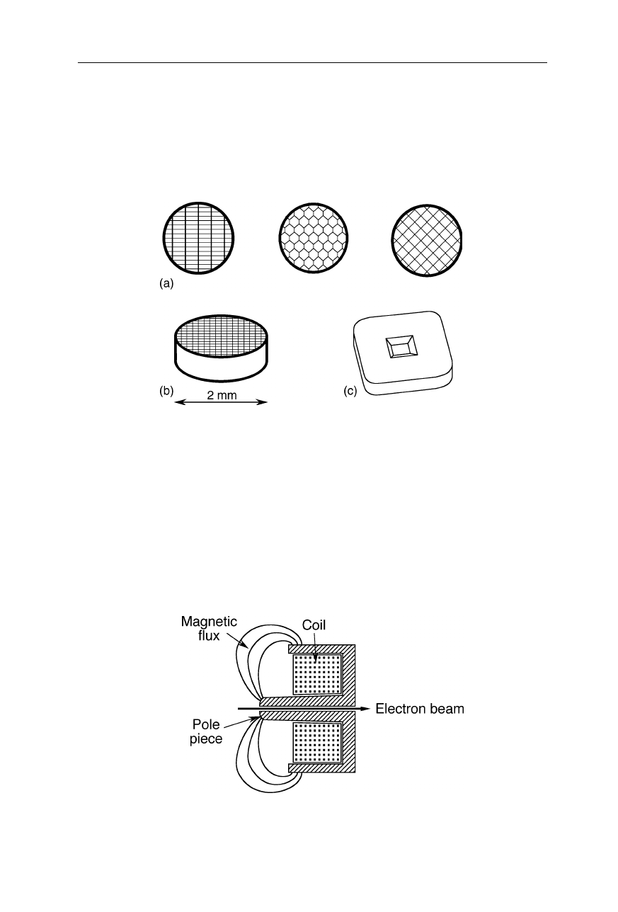
6.1.4 TEM grids
TEM grids (Fig. 6.7) should not get charged during measurement which would
distort the electron path. Usually they are made from conductive chemically inert
non-gassing materials suitable for high vacuum, such as platinum and platinum-
iridium alloys.
Fig. 6.7
TEM grids. (
a
), (
b
) Some common patterns of TEM grids made from a variety of
materials, e.g., platinum, silver, tungsten, molybdenum, stainless steel, or titanium. Among
these materials, platinum is the chemically most inert, but expensive. (
c
) Silicon nitride
“grid” with a single window (from SPI Supplies, West Chester, PA)
6.1.5 Electron lenses
There are magnetic (Figs. 6.8 and 6.9), electrostatic (Fig. 6.5) and compound
lenses (Figs. 6.10 and 6.11). Electron lenses have some similar characteristics like
optical lenses, such as focal length, spherical aberration, and chromatic aberration.
Fig. 6.8
Ernst Ruska's pohlschuh lens: the circular electromagnet is capable of projecting a
precisely circular magnetic field in the region of the electron beam