N?lting B. Methods in Modern Biophysics
Подождите немного. Документ загружается.

4.1 Fourier transform and X-ray crystallography 71
One generally starts with a protein concentration of about 2–50 mg ml
–1
. Usu-
ally, the protein or virus must not contain a significant amount of contaminants,
such as other proteins or viruses, protein or virus fragments, unfolded or misfolded
protein, particulate matter, chemical additions unnecessary for stability or solu-
bility. In most cases compact proteins that do not contain floppy ends, such as
histidine tags or native unstructured peptides, crystallize better. Suitable crystals
have sizes of a few 0.1 mm.
4.1.2.3 Acquisition of the diffraction pattern
For the acquisition of the crystal diffraction pattern (Figs. 4.17–4.19), multi wire
area detectors or CCD area detectors (Fig. 4.20) are commonly used. With the
example of a linear CCD, Fig. 4.21 illustrates the basic principle of operation of
CCDs.
The most common X-ray sources for protein and virus crystallographic analysis
are rotating anode generators (Fig. 4.22) with typically 5–25 kW electrical power
and synchrotrons (Figs. 4.23 and 4.24). Synchrotrons are comparably expensive,
but have a higher brightness enabling shorter measuring times. Reduction of the
exposition time often results in a better quality of the diffraction pattern since
decomposition of the crystal due to radiation damage is reduced.
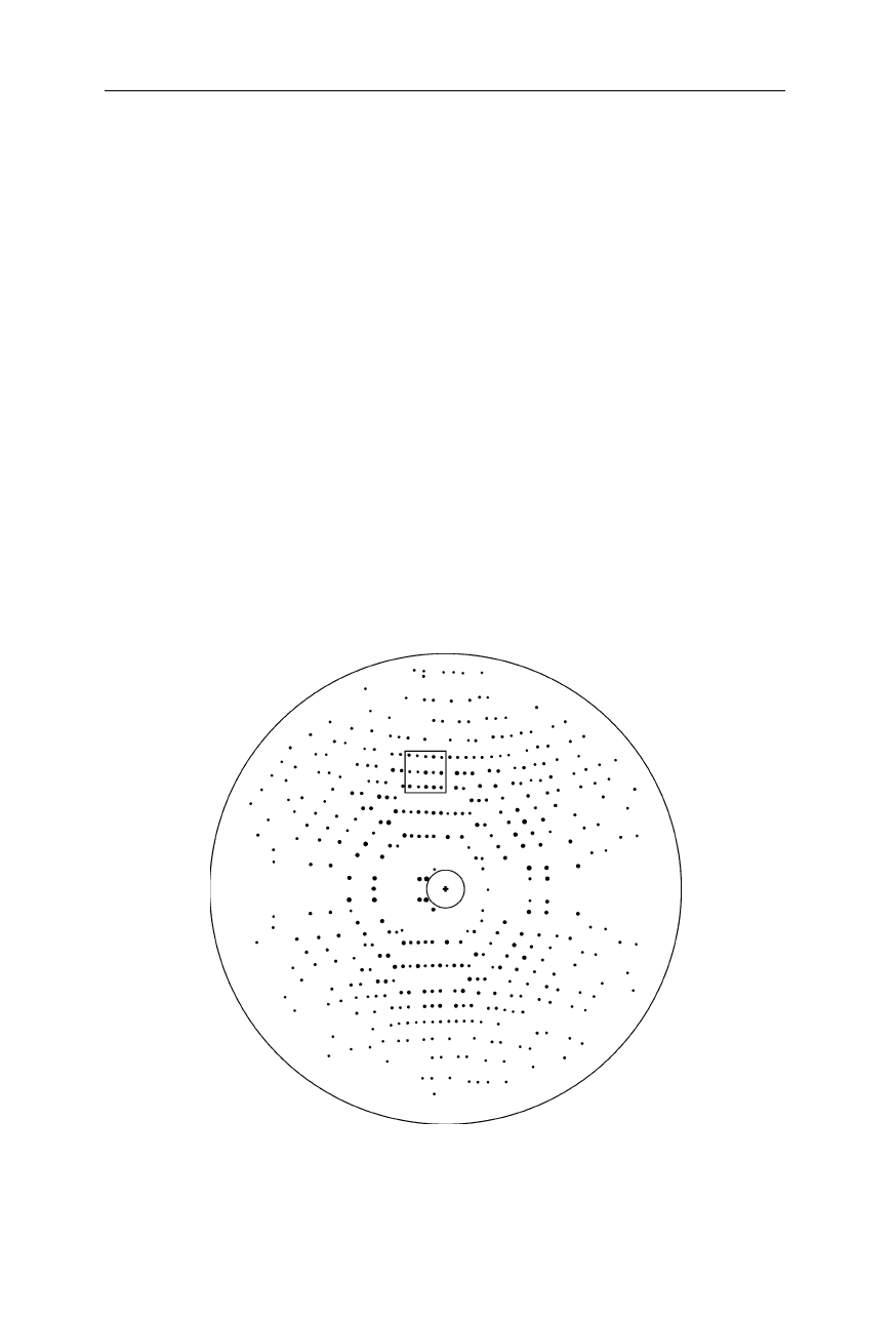
Fig. 4.17
X-ray diffraction pattern of a protein crystal (Norledge et al., 1996). The
highlighted section is referred to in Fig. 4.26
72 4 X-ray structural analysis
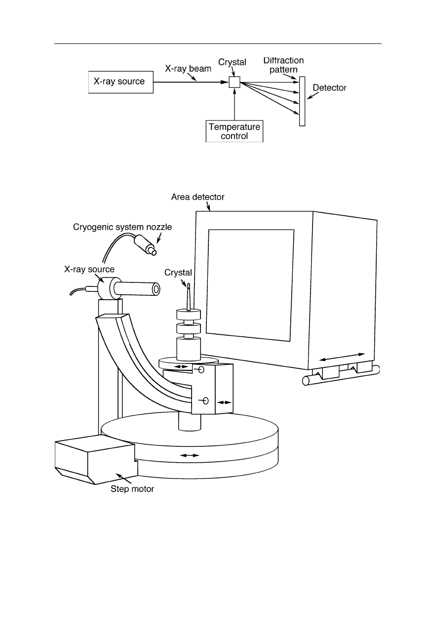
Fig. 4.18
General setup for the acquisition of the diffraction pattern
Fig. 4.19
Setup for acquiring the diffraction pattern with an area detector (see, e.g., area
detectors from Rigaku, The Woodlands, TX). The crystal is cooled with nitrogen from the
cryogenic system nozzle. Cooling the crystal reduces radiation damage, but somewhat
changes the intermolecular distances. Diffraction of the X-rays from the X-ray source by
the crystal are recorded with the area detector with typically 2048
×
2048 or 4096
×
4096
pixels (see next figure)
4.1 Fourier transform and X-ray crystallography 73
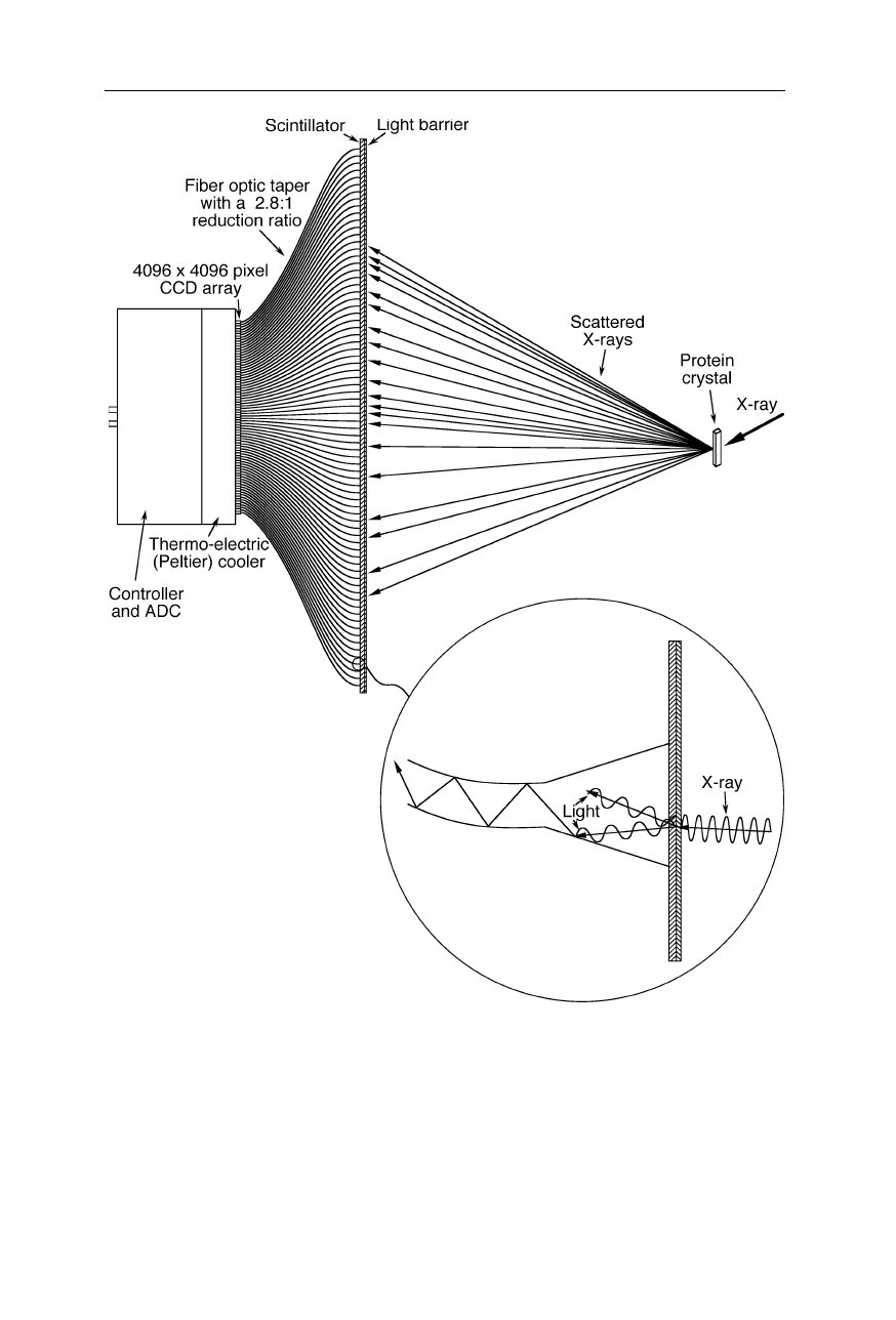
Fig. 4.20
A CCD area detector used for recording of X-ray diffraction patterns. For
reduction of the dark current, this CCD is operated at –40
o
C, allowing it to detect single
photons. The fiber optic taper serves also for blockage of X-rays and thereby prevention of
radiation damage to the sensitive CCD array. At a pixel size of 20
µ
m
×
20
µ
m, the full
well capacity is typically several 100,000 electrons per pixel, enabling the necessary high
dynamic range
74 4 X-ray structural analysis
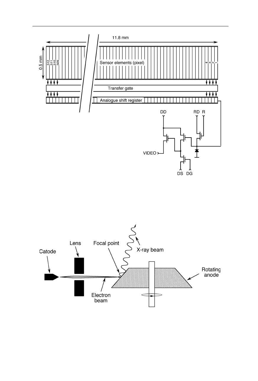
Fig. 4.21
Linear charge coupled device (CCD). The sensor elements generate electrons by
absorption of photons and store the electrons in potential wells. After a certain period of
time, the collected electrons are transferred to the analogue shift register and read out. The
symbols are: DD, drain of the output amplifier; DS, source of the output amplifier; DG,
gate of the output amplifier; RD, drain of the reset transistor; R, clock gate of the reset
transistor (Nölting, 1991)
Fig. 4.22
Rotating anode generator. An electron beam is focussed onto the rotating anode.
It knocks out electrons from the inner electron shells of the anode metal. Reoccupation of
the vacant shells by electrons from higher level shells involves the emission of X-ray
radiation. The interaction of the electron beam with the anode metal generates also a large
amount of heat which is quickly dissipated by rotating the anode below the spot of
incidence of electrons
4.1 Fourier transform and X-ray crystallography 75
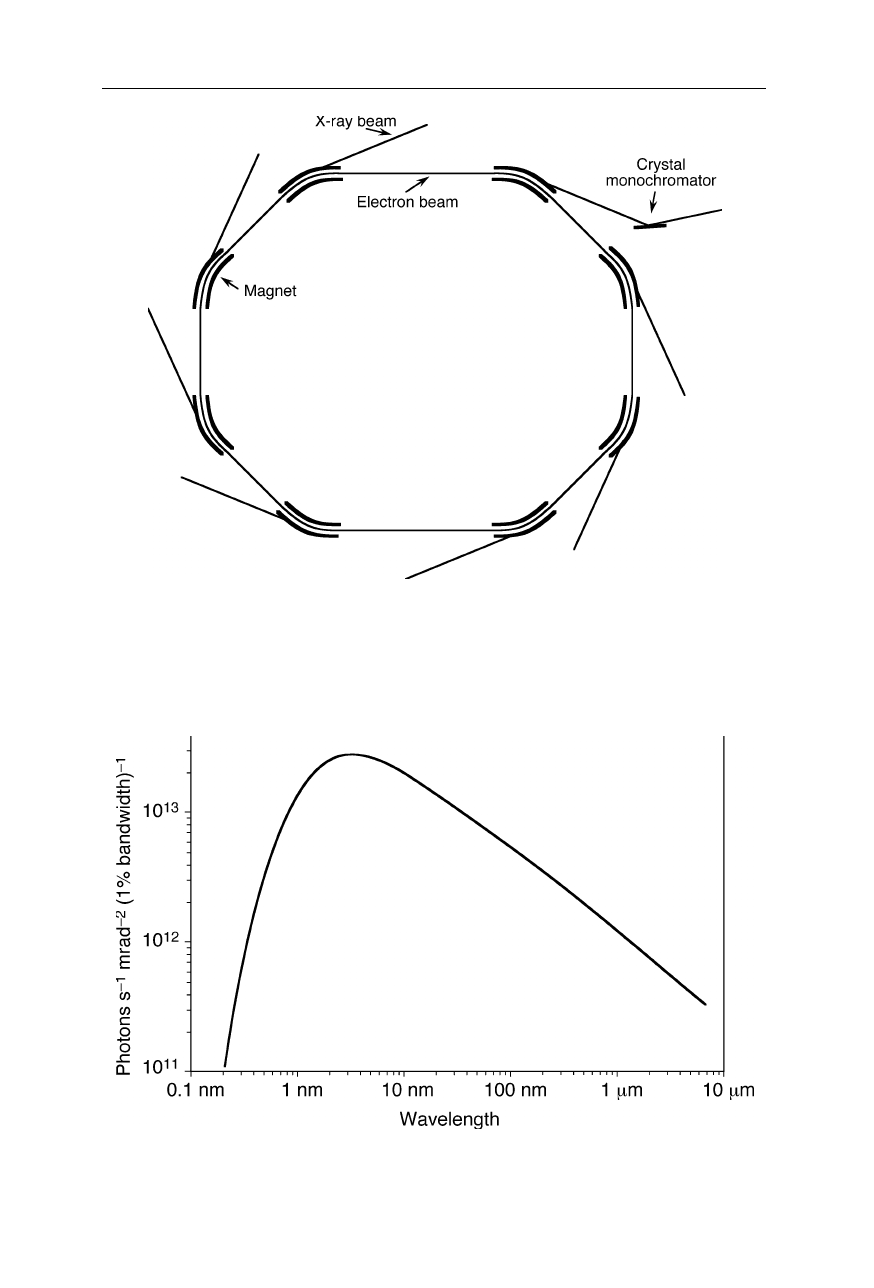
Fig. 4.23
Design of a synchrotron. Ions or electrons are accelerated to a speed close to
the speed of light and forced on a curved trajectory. A broad spectrum of radiation is
produced along the curved sections of the beam. For protein crystallography, a certain
wavelength, e.g., 1 Å, is selected by a monochromator
Fig. 4.24
Emission of the Berlin Electron Synchrotron Storage Ring (BESSY I)
76 4 X-ray structural analysis
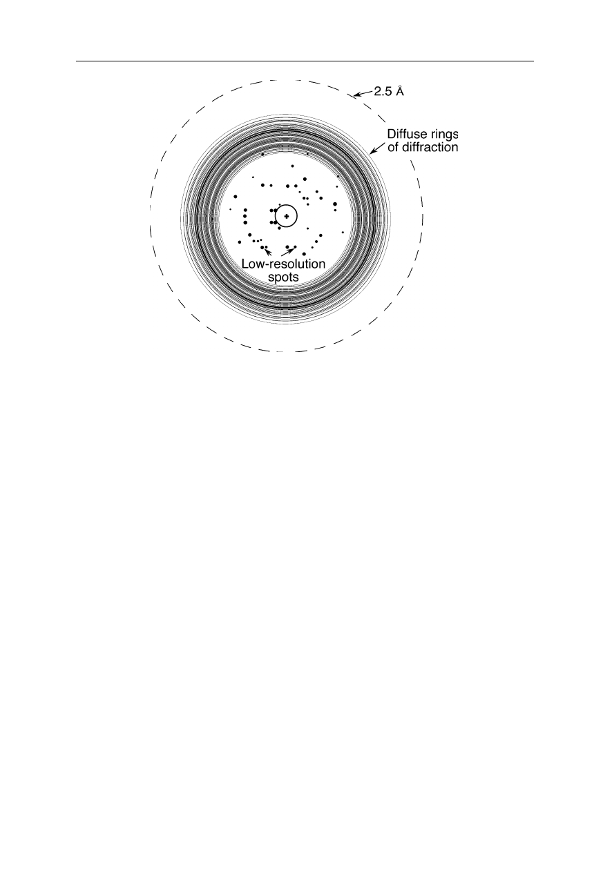
Fig. 4.25
Diffraction pattern of a poorly scattering crystal: only a few spots near the center
are observed, any high resolution information is absent. The dashed circle indicates the
area corresponding to a resolution of 2.5 Å. If diffraction spots would be visible up to this
circle, the resolution of the obtained structure would be 2.5 Å. One can see that the
resolution is much lower in this example
Already superficial inspection of the diffraction pattern provides a lot of
information about the quality of the crystals: since the information about fine
details of the protein structure is found at large diffraction angles, the absence of
spots far outside the center of the diffraction pattern shows that only a low
resolution will be obtained (Fig. 4.25).
4.1.2.4 Determination of the phases: heavy atom replacement
As mentioned earlier, after measurement of the diffraction pattern, determination
of the phase information is required. If we do not have information from mole-
cules with a similar structure, or anomalously scattering atoms in the molecule, the
method of choice may be the heavy atom replacement: the diffraction pattern of
the original (native) crystal is compared with crystals that contain a single or a few
heavy atoms at fixed positions. Those crystals can be prepared, e.g., by diffusing
a solution of a heavy atom salt into the protein crystal.
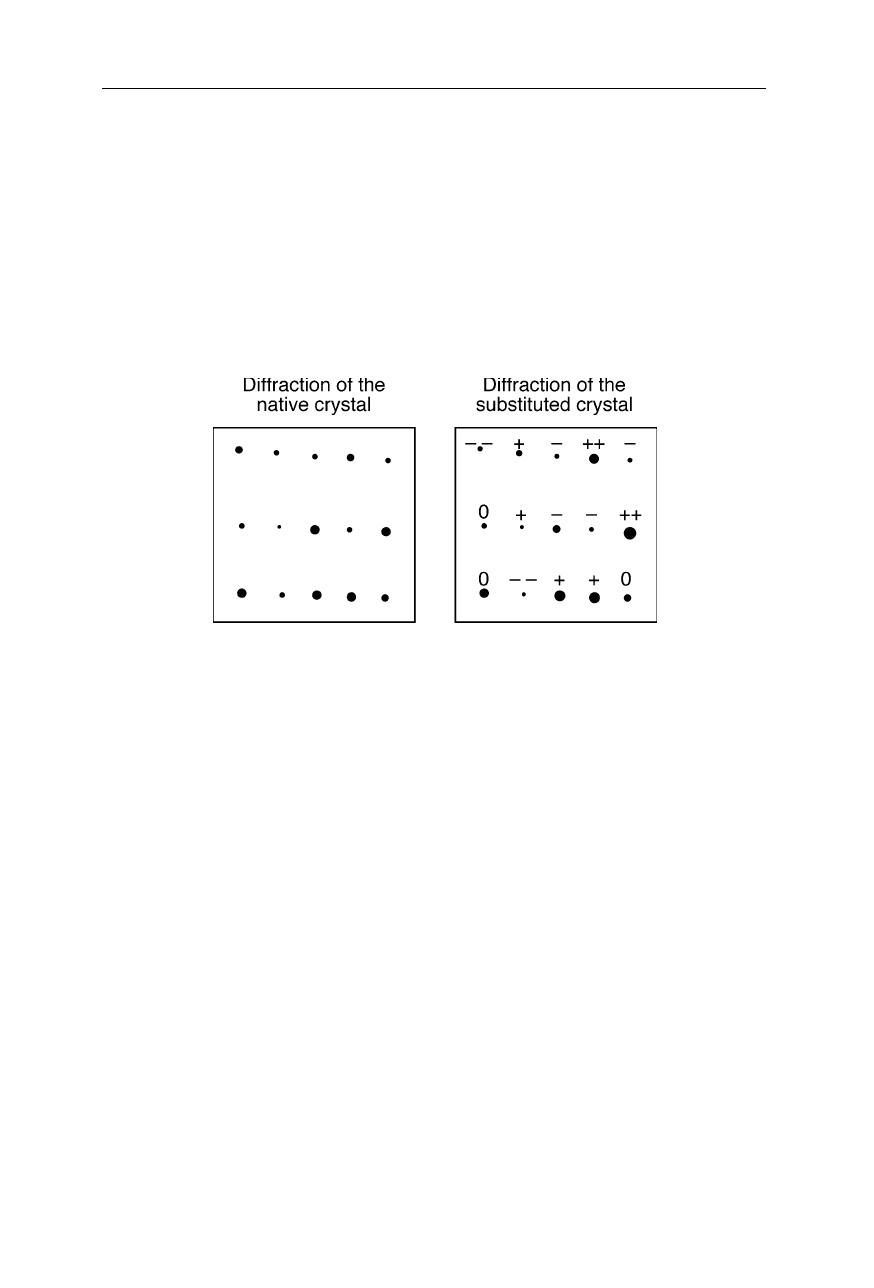
Fig. 4.26 depicts sections of the diffraction pattern of the native protein crystal
and heavy atom derivatized crystal, respectively. The diffraction spots labeled
with “++” are significantly increased in intensity for the heavy atom derivative.
This shows that they belong to phases with a large magnitude. How can we make
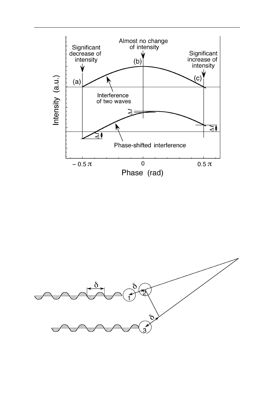
this conclusion? See Fig. 4.27 which, in the upper part, shows the intensity of an
interferogram of two waves as function of phase: when we introduce a small shift
4.1 Fourier transform and X-ray crystallography 77
to one of the waves (lower part), a large increase of intensity of the interferogram
is found for large positive phases. Essentially no change of intensity occurs at
phases around zero. Thus, analogously we can conclude that diffraction spots
which increase in intensity only slightly between native crystal and heavy atom
derivative belong to phases around zero or
π
. So, by comparing the intensities of
the spots between native crystal and the heavy atom derivative we can estimate the
phases of the individual diffraction spots. With only one heavy atom derivative,
an uncertainty of two possibilities remains for each spot, but this can easily be
removed with a further, different heavy atom derivative of the protein crystal.
Fig. 4.26
Section of the diffraction pattern of a protein crystal. Left: “native” crystal.
Right: heavy atom derivative
Another way of showing the importance and meaning of phases in crys-
tallography is illustrated in Fig. 4.28: Atoms with different phases and relative
positions may cause a diffraction spot at the same position. Thus, without
information from heavy atom replacement, or from diffraction patterns of proteins
with similar structure or other information, we cannot deduce the protein structure
from the diffraction pattern. Theoretically one could also try out all possible
phases and see if it leads to a meaningful structure, but currently for macro-
molecules the computational effort would be much too high.
It should be noted that the problem of loss of phase information occurs only in
the common methods of recording the crystal diffraction, such as with a
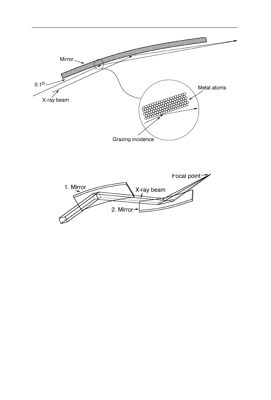
photographic film or a semiconductor detector. The use of lenses or mirrors to
produce an image like in an microscope would prevent this loss of information
(see p.65). Unfortunately, currently we cannot build a lens which is sufficiently
suitable for focussing X-rays of less than a few Å wavelength: the surface of a
conventional lens would not be smooth enough and the bulk of the lens would act
like a non-regular grating. Further, it is also very difficult to build highly precise
X-ray mirrors (Figs.
4.29 and 4.30). X-ray mirror microscopes using soft radiation
currently reach only a few 10 nm resolution. More importantly, the radiation
damage would prevent atomic resolution of a single protein molecule or virus.
78 4 X-ray structural analysis
Fig. 4.27
Interferogram of two waves: within the phase interval [–
π
/2,
π
/2], a small phase
shift causes a large negative amplitude change,
∆
I, for large negative phases (
a
), and a
large positive amplitude change for large positive phases (
c
), but almost no amplitude
change for zero phases (
b
). Thus, e.g., from a large amplitude increase of a diffraction spot
upon application of a small phase shift by an additional heavy atom, we can conclude that
the phase of the spot has a large magnitude. Analogously one can estimate the phases from
the observation of various intensity changes of diffraction spots upon derivatization of the
crystal with a heavy atom. For the complete phase interval, (–π
,
π
], there are still two
phases for each amplitude change (not shown). This uncertainty is removed by using data
from a second heavy atom derivative
Fig. 4.28
Different phases and relative positions may cause a diffraction spot at the same
position: the atom pairs (1,2) and (2,3) with completely different relative locations cause
positive interference at the same position
4.1 Fourier transform and X-ray crystallography 79
Fig. 4.29
Common X-ray mirrors are only suitable for low angles of incidence
Fig. 4.30
A pair of X-ray mirrors with grazing incidence focuses an X-ray beam to a spot
The mathematics behind the method of heavy atom replacement is exemplary
illustrated in Figs. 4.31–4.34. Fig. 4.31a represents an array of atoms. Fig. 4.31b
is the absolute of the Fourier transform of this array. From the imaginary and real
parts (Fig. 4.32) of this Fourier transform, the phase was calculated (Fig. 4.33).
Fig. 4.34a represents the same array as above, but with one additional heavy atom
causing a small change of the Fourier transform. The difference of the absolutes
of Fourier transforms between native array and heavy atom derivatized array is
shown in Fig. 4.34b. Comparing this difference of the absolutes of Fourier trans-
forms with the absolutes of the phases of the native array (Fig. 4.33b), we find a
connection between phases (Fig. 4.33b) and amplitude differences (Fig. 4.34b).
This connection allows the magnitude of the phase angles to be determined. As
mentioned, the remaining ambiguity of sign is removed by including the data from
a second isomorphous heavy atom derivative.
80 4 X-ray structural analysis
(a) f1(x,y)
FT
(b) |F1(h,k)|
Fig. 4.31
(
a
) Representation of an array of atoms. (
b
) Absolute of the Fourier transform
of (
a
)