N?lting B. Methods in Modern Biophysics
Подождите немного. Документ загружается.

92 5 Protein infrared spectroscopy
5.1 Spectrometers and devices
5.1.1 Scanning infrared spectrometers
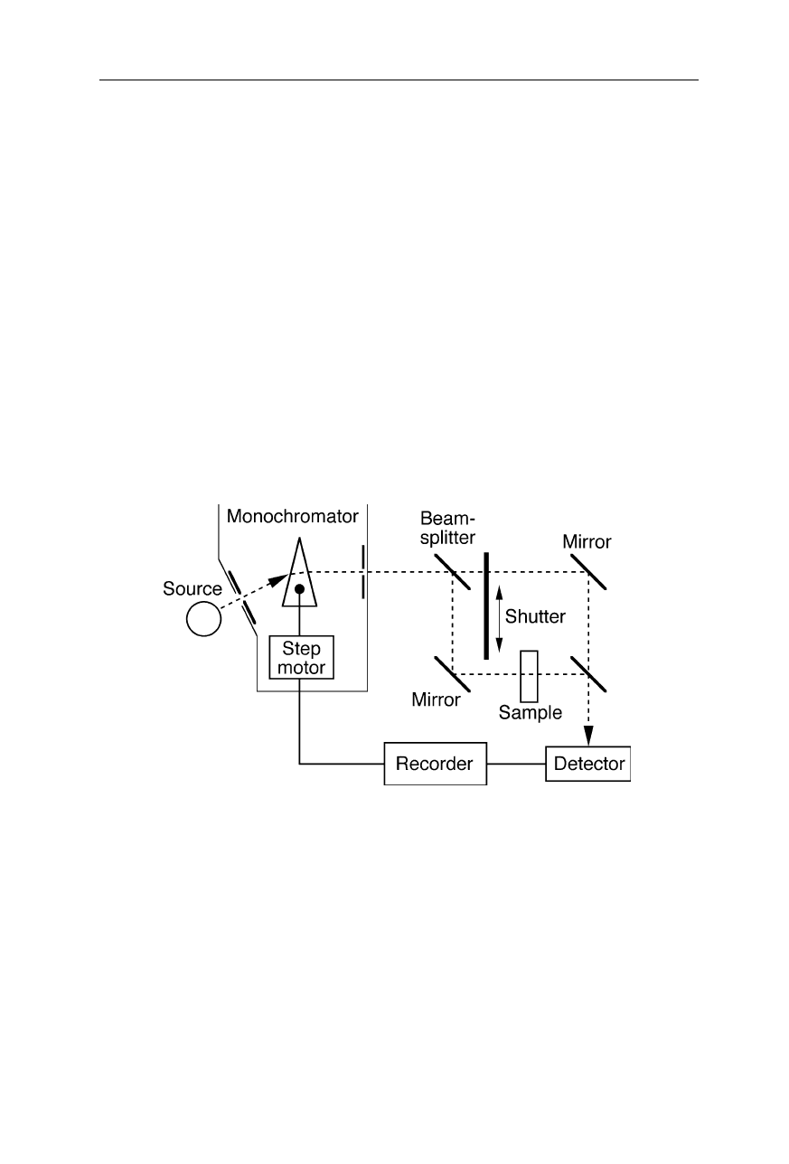
Early IR spectrometers (Fig. 5.2) were constructed similarly to scanning UV/VIS
absorption spectrometers. The emission of the source, e.g., a thermal source
operated at 1000
o
C, is passed through a monochromator selecting a single
wavelength. The monochromatic beam is split into two beams – one having the
sample in the path. A shutter passes through only one of the two beams at a time.
Both beams are alternatingly detected by an IR detector, e.g., a pyroelectric
detector, and compared which each other. The optical density of the sample is
calculated from the logarithm of the intensity quotient. The use of light modula-
tion is quite indispensable since the problem of background radiation is much
more severe than in UV/VIS spectrometers. Spectra are recorded by scanning the
wavelength region of interest. This scanning principle of operation is still widely
used in IR spectrometers with time resolutions in the femtosecond to nanosecond
region, where infrared lasers serve as IR source (see Nölting, 2005).
Fig. 5.2
Example of a scanning infrared (IR) spectrometer. The monochromator separates
the radiation of the IR source into its different wavelengths and selects one wavelength at a
time. A beam splitter separates the monochromatic beam into sample beam and reference
beam. The absorption coefficient, according to the chemical and structural properties of
the sample molecules, is calculated using the detected intensity quotient between both
beams, the pathlength, and the sample concentration
5.1.2 Fourier transform infrared (FTIR) spectrometers
FTIR spectrometers (Figs. 5.3–5.7) use the technique of Michelson interferometry
and have the advantage of using a larger part of the emission of the IR source
during the measurement of a spectrum, compared with scanning IR spectrometers
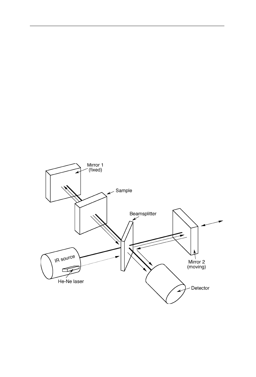
5.1 Spectrometers and devices 93
that are based on monochromators which select only one wavelength at a time.
The better usage of radiation improves the inherent signal-to-noise ratio,
especially for strongly absorbing samples for which the measurement may be
photon shot noise limited. Also the spectral resolution of FTIR spectrometers,
which is limited by the path length of the moving mirror, is often better than that
of scanning IR spectrometers.
In FTIR spectrometers (Fig. 5.3) the beam of radiation from the IR source is
focused on a beam splitter constructed such that half the beam is transmitted to a
moving mirror and the other half is reflected to a fixed mirror. Both the moving
mirror and the fixed mirror reflect the beam back to the beam splitter which
reflects the half of both beams to the detector where they interfere according to
their phase difference. The light intensity variation with optical path difference,
called interferogram, is the Fourier transform of the incident light spectrum (light
intensity as a function of the wavenumber). Absorption spectra are obtained by
measuring interferograms with a sample and with an empty sample cell in the
beam and inverse Fourier transforming the interferograms into spectra (Figs.
5.4
–5.6).
Fig. 5.3
Typical design of FTIR spectrometers. The lamp, e.g., a thermal source, emits a
beam of infrared radiation. A Michelson interferometer, consisting of a beamsplitter, a
fixed mirror and a moving mirror, splits the beam into two beams and generates an
interference of them. The sample inserted in one of the beam paths changes the inter-
ference. Interferograms with and without sample are recorded and the absorption of the
sample is calculated by inverse Fourier transform (see Fig. 5.6)
94 5 Protein infrared spectroscopy
Fig. 5.4 shows three examples of interference of the two monochromatic light
beams of the interferometer resulting in different intensities of the interferogram.
Eq. 5.1 describes the intensity of the interferogram,
I
interferogram
, for the interference
of two polychromatic beams of equal intensity in the FTIR spectrometer:
, (5.1)
where
δ
is the phase difference of the two beams,
const
a constant,
I
beam
the
intensity of the beams, and
v
the wavenumber. From the interferogram, the inten-
sity of the beams can be calculated by inverse Fourier transform:
(5.2)
Analogously, the intensity of the beam with the sample in the path is calculated
from the corresponding interferogram. The absorption is given by the logarithm
of the intensity quotient of blank to sample.
Fig. 5.4
Interference of two monochromatic light waves with equal intensity.
Top:
both
beams have the same phase; their interference yields the maximum of the interferogram,
i.e., the sum of both intensities.
Middle:
at a phase difference of
λ
/4, the intensity of the
interferogram equals the intensity of the interfering beams.
Bottom:
at a phase difference
of
λ
/2, both beams extinguish each other
5.1 Spectrometers and devices 95
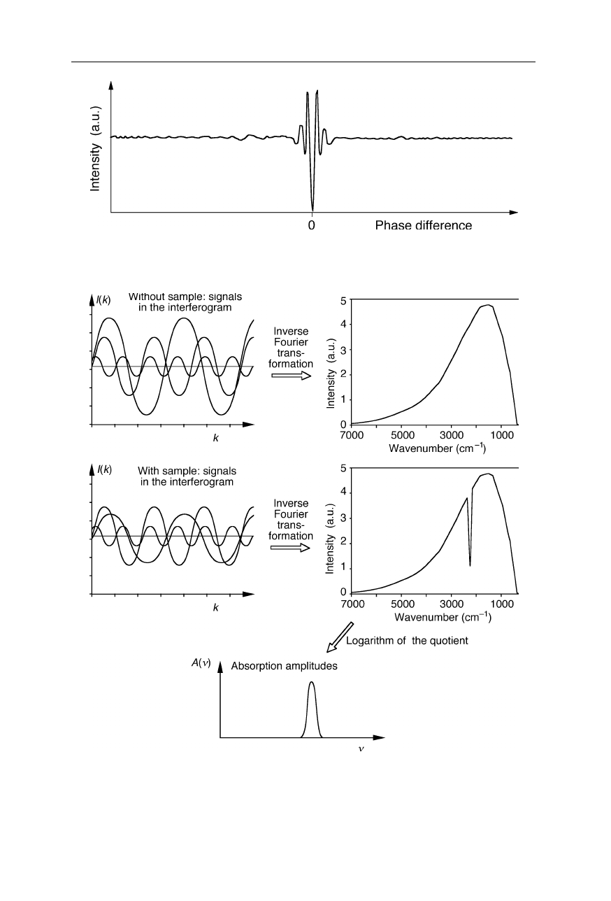
Fig. 5.5
Example of an interferogram of two polychromatic light beams
Fig. 5.6
Principle of operation of a FTIR spectrometer. IR intensities at the detector are
recorded both for the sample cell filled with solvent and for the sample cell filled with
sample. Inverse Fourier transform of the two interferograms yields the IR intensities. The
IR absorption spectrum is calculated using the logarithm of the intensity quotient
96 5 Protein infrared spectroscopy
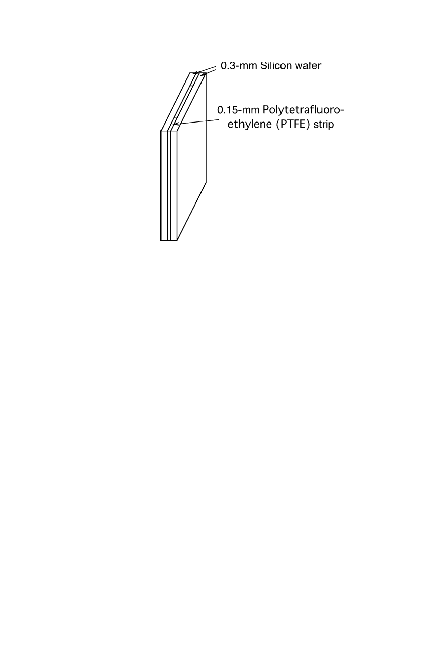
Fig. 5.7
Sample cell for FTIR experiments. The transparent walls of the cell are made
from silicon wafers supplied by a manufacturer of electronic chips
A very suitable material for the manufacture of sample cells, sample holders,
and windows is silicon (Fig. 5.7). Polished silicon wafers of 0.5 – 1 mm thickness
are sufficiently transparent from 400 to 4000 cm
–1
(25 – 2.5
µ
m wavelength)
(Jiang et al., 1996). Only the fragility and the high refractive index of this mate-
rial might be problematical in some experimental set-ups. Used infrared sources
are often thermal sources operated at about 1000
o
C. Beam splitters made from a
thin germanium film evaporated on a potassium bromide (KBr) or cesium iodide
(CsI) slide are transparent down to about 400 cm
–1
(25
µ
m wavelength) and 200
cm
–1
(50
µ
m wavelength), respectively. Liquid nitrogen cooled mercury cadmium
telluride (MCT) detectors and deuterated triglycine sulfate (DTGS) pyroelectric
detectors are frequently applied for infrared detection. For an excellent intro-
duction into the instrumentation of FTIR spectroscopy see Perkins, 1986.
5.1.3 LIDAR, optical coherence tomography, attenuated total reflec-
tion and IR microscopes
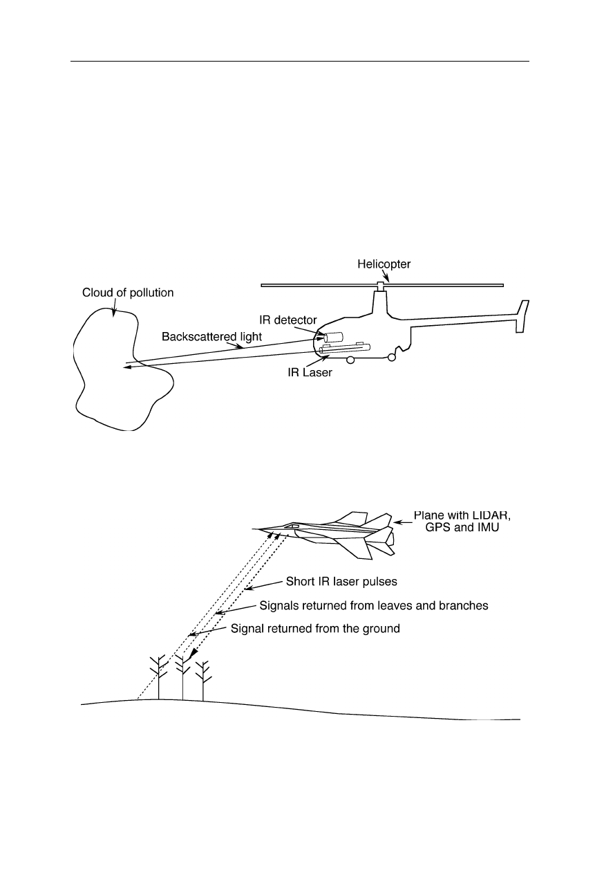
IR spectroscopy is exquisitely suitable for remote sensing of clouds of biological
agents (Fig. 5.8). The IR LIDAR set-up consists of a pulsed IR laser and an IR
detector which senses the backscattered light from the laser. Since the light
travels extremely fast, the detector senses the return echo before the next pulse is
sent. The time it takes for the laser pulse to travel down and back is a measure of
the distance. Mobile commercial LIDAR systems quite often employ an
integrated global positioning system (GPS) to determine the own position.
5.1 Spectrometers and devices 97
Equipped, e.g., with an optical modulator which rapidly changes the direction
of the beam, and mounted on top of a roof, the IR LIDAR can scan the 360
o
-
environment at distances of 0 to several 10 km. This method has importance, for
example, for early warning systems of smog in large cities and for three-
dimensional analysis of forest structure and terrain (Fig. 5.9). Remote sensing of
changes in forest structure utilizes the information of time and intensity of multiple
reflections from leaves and branches. Effects of environmental pollutants and
pests are quickly detectable in vast areas and economic damage is largely
reducible.
Fig. 5.8
Remote sensing of environmental changes, e.g., a cloud of biological material,
with an IR LIDAR (light detection and ranging; measurement of light backscatter)
Fig. 5.9
Remote survey of forest structure and terrain with IR LIDAR technology. The
plane is equipped with a GPS and an inertial measurement unit (IMU). The latter contains
several gyroscopes and an accelerometer and can determine the position and angle of tilt
with some accuracy during periods of failure of the GPS
98 5 Protein infrared spectroscopy
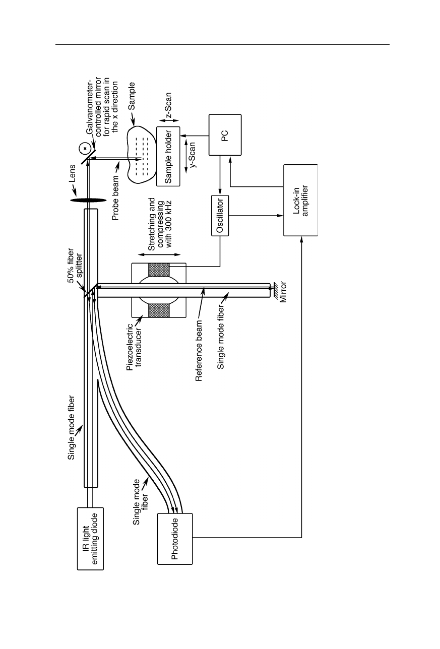
Fig. 5.10 Optical coherence tomography (OCT). The IR light from
a light emitting diode is split into reference and probe beams. Ligh
t
of the probe beam reflected from the sample is interfered with l
ight of the reference beam, and the interference is detected by
the
photodiode. The pathlength of the reference beam is modulated by stretching an optical fiber with a piezoelectric transducer.
Light fro
m
the sample which has traveled the same distance as the referen
ce beam interferes constructively. Its signal is extracted from
the
interference intensity by a lock-in amplifier (Duncan et al., 1998)
5.1 Spectrometers and devices 99
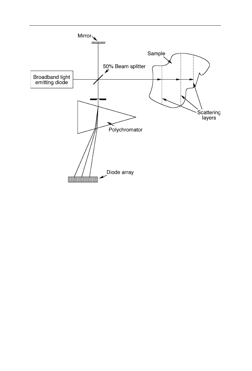
Fig. 5.11
Spectral domain optical coherence tomography (SDOCT) (Andretzky et al.,
1998; Häusler and Lindner, 1998). Polychromatic backscattered light from different
depths interferes with polychromatic light of a reference beam. The interference of the
beams is analyzed with a polychromator and a multichannel detector. From the spectral
changes due to interference, information about the depth of the scattering layer is obtained
Another important variant of IR spectroscopy on biological samples is optical
coherence tomography (OCT). OCT (Figs. 5.10 and 5.11) utilizes echoes of
infrared light waves backscattered off the internal microstructures within
biological objects to obtain images on a
µ
m scale. In the design of Fig. 5.10, IR
radiation backscattered from the sample is interfered with a reference beam. Light
from a scattering layer in the sample with a certain depth has the same phase as the
reference beam and thus interferes constructively, i.e., produces a high inter-
ference intensity. Light from slightly deeper or shallower scattering layers cause a
lower interference intensity. By modulating the phase of the reference beam and
detecting the interference intensity with a lock-in amplifier, the signals from layers
with different depths are extracted from the interference intensity (Duncan et al.,
1998). Fig. 5.11 depicts a second design variant of optical coherence tomography
(Andretzky et al., 1998; Häusler and Lindner, 1998). Here the information on
depth is gained by analyzing the spectrum of the backscattered light.
100 5 Protein infrared spectroscopy
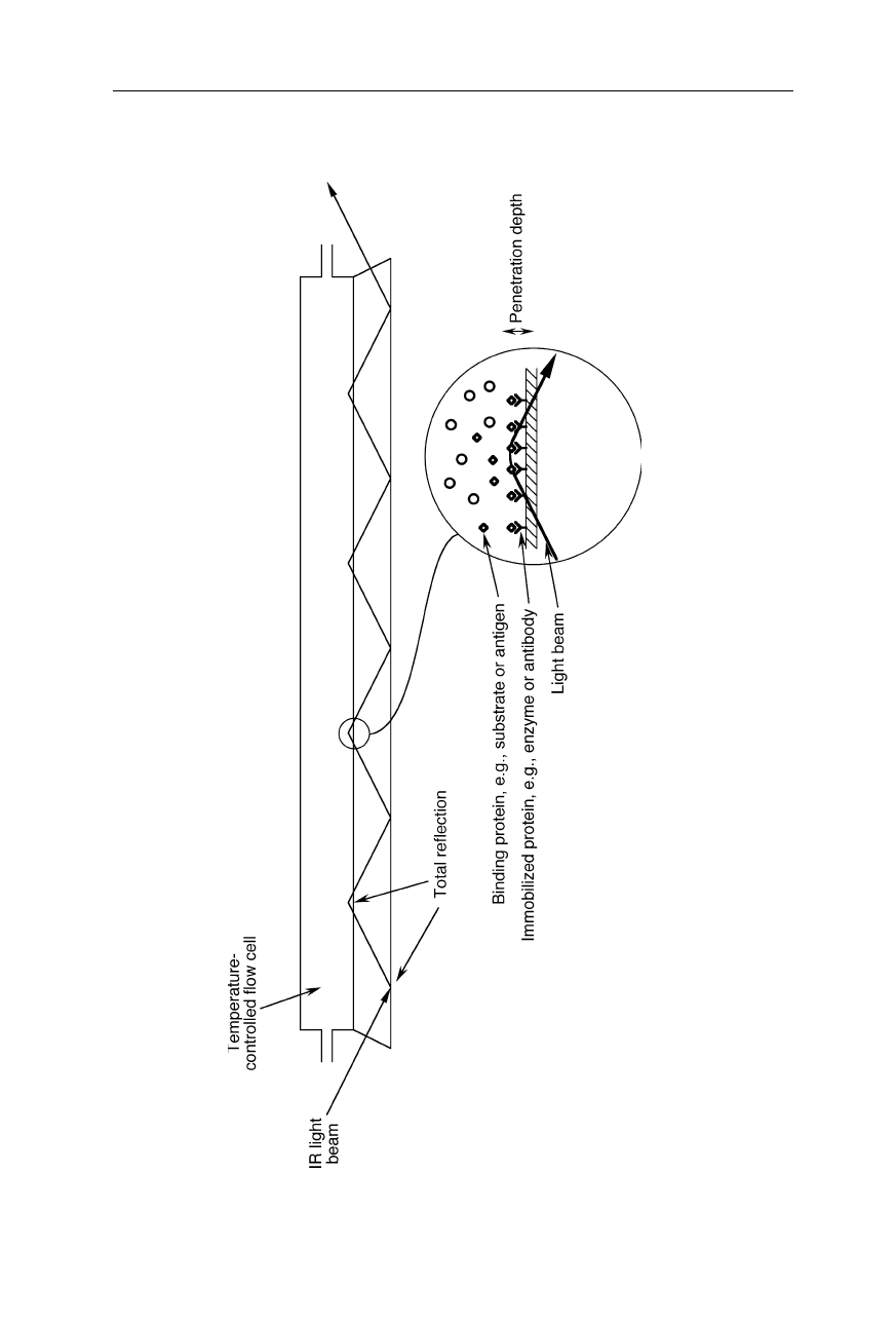
Fig. 5.12 Flow cell for attenuated total reflection (ATR) infrare
d spectroscopy (Fringeli et al., 1998; Feughelman et al., 2002; Snabe
and
Petersen, 2002). The internally total-reflected light slightly leaves the waveguide and so can probe the sample molecules on t
he outside of the
waveguide. The part of the light wave which leaves the wavegu
ide at the total reflection points is called evanescent wave. On
ly very little
sample is needed. Using a large number of reflections can lead to a more than 100-fold amplification of the measured signal
5.1 Spectrometers and devices 101
The next IR spectroscopic technique to be mentioned is attenuated total
reflection (ATR) infrared spectroscopy (see, e.g., Fringeli et al., 1998; Ding et al.,
2002; Feughelman et al., 2002; Snabe and Petersen, 2002; Figs. 5.12 and 5.13).
Here the coefficient of internal total reflection of an IR beam in a waveguide is
changed by a sample deposited on the surface of the waveguide. An advantage of
ATR on thin layered samples is the dramatic increase of the effective optical path-
length and sensitivity through multiple reflections compared with conventional
transmission spectroscopy on such a sample.
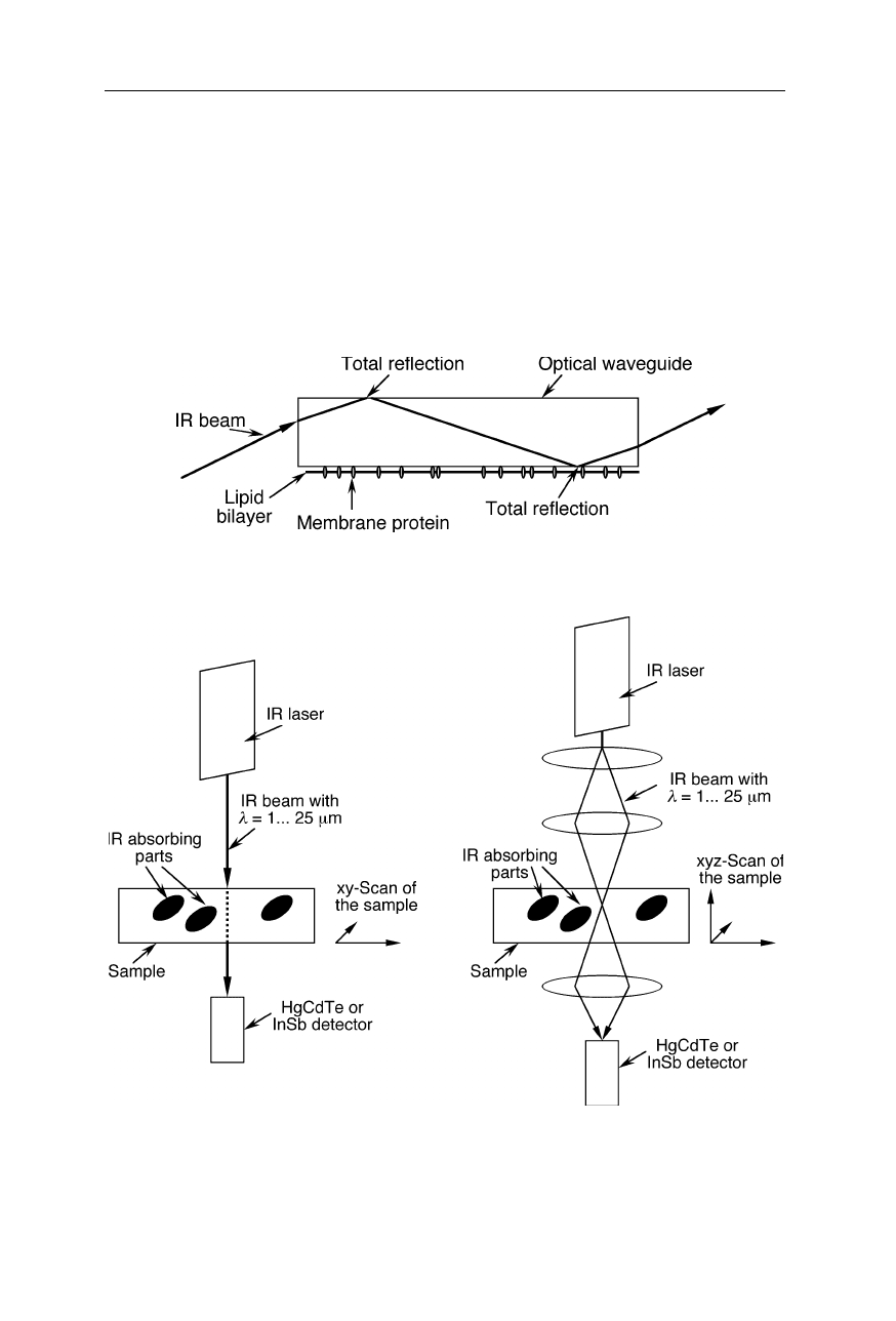
Fig. 5.13
Attenuated total reflection (ATR) infrared spectroscopy on membrane proteins
(see, e.g., Ding et al., 2002)
Fig. 5.14
Scanning IR microscope. The focussed beam from an IR laser is passed through
the sample and detected. Left: simple microscope with planar resolution, especially suit-
able for thin layer samples. Right: microscope with three-dimensional resolution: for ac-
quisition of the image, the sample is moved in xyz-directions