N?lting B. Methods in Modern Biophysics
Подождите немного. Документ загружается.

7.2 Scanning tunneling microscope (STM) 133
7.2 Scanning tunneling microscope (STM)
In 1986 Gerd Binnig and Heinrich Rohrer were awarded the Nobel Prize for
Physics for the groundbreaking invention of the STM. It was the first member of
the family of scanning probe microscopes (SPM) that can characterize surface
morphology with atomic resolution. In contrast to the AFM, its principle of
operation (Fig. 7.19) requires electrically conductive samples. A sharp tip located
on a flexible cantilever is used to probe the distance between the tip and sample
surface, as judged by the tunneling current (Fig. 7.20). Since the tunneling current
also depends on the chemical nature of sample and tip, the STM also serves for
characterization of electronic properties of solid samples.
Significant complications on the way towards higher resolution of proteins are
the undesired excitation of the soft biological material by the high current of
STMs, typically pA–nA, and the distribution of conductivity within the sample
distorting the image (Fig. 7.21). Low currents and stable attachment of the sample
to the support are required for high resolution images of biological macro-
molecules.
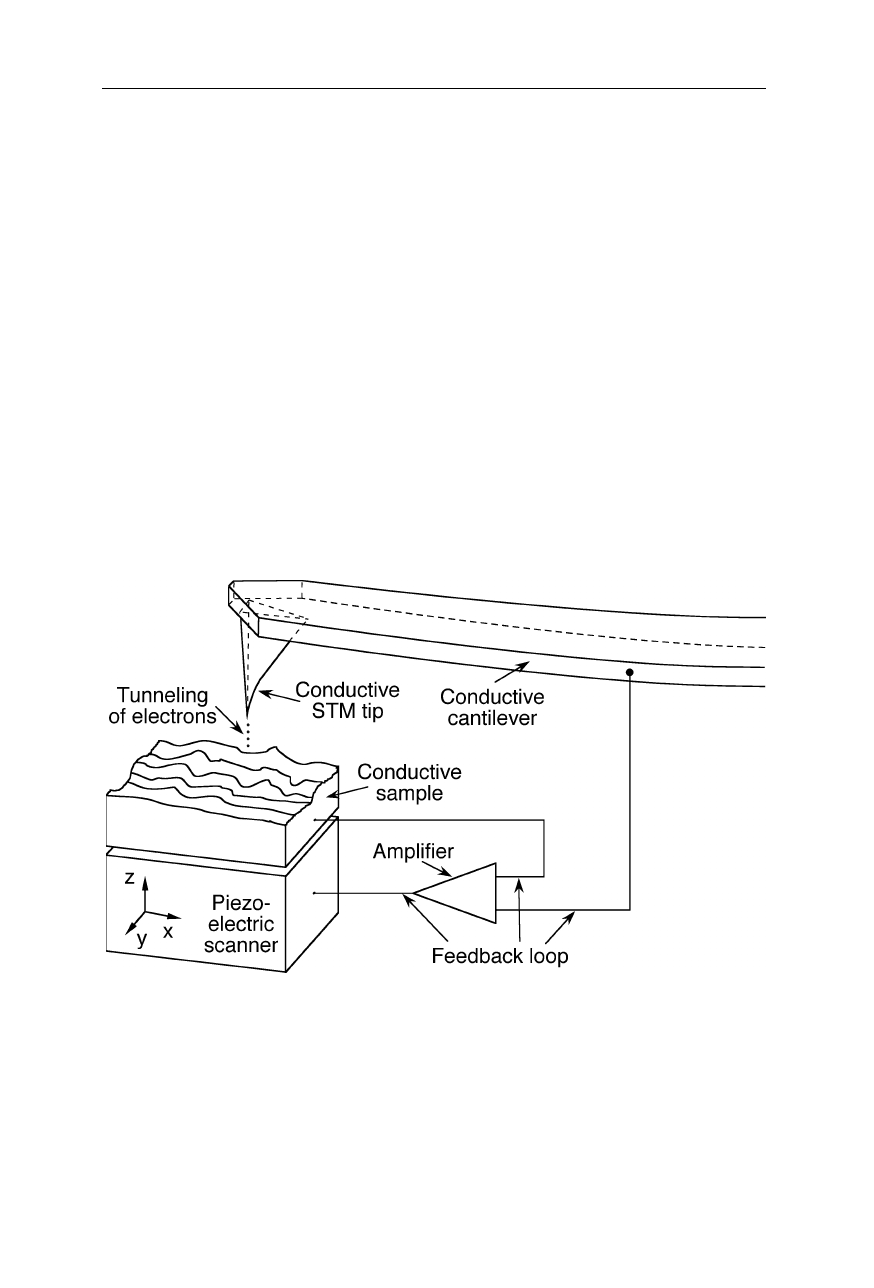
Fig. 7.19
Principle of operation of STMs. A finely sharpened electrically conductive tip is
first positioned within about 1 nm of the sample by mechanical translation stages (not
shown) and the piezoelectric scanner. At this small separation, electrons tunnel through
the gap between tip and sample (Fig. 7.20). The tunneling current depends on the applied
bias voltage between tip and sample, the distance, the tip shape, and the chemical
compositions of sample and tip. The feedback loop ensures constant height or constant
current. Tunneling current and feedback voltage are a measure of surface morphology and
composition (Binnig et al., 1982a, 1982b, 1983; Binnig and Rohrer, 1987)
134 7 Scanning probe microscopy
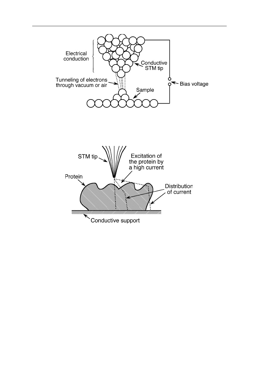
Fig. 7.20
When the distance between conductive tip and conductive sample is lowered to a
few Å, electrons can traverse the gap with some transmission probability. The STM meas-
ures not purely distance like the AFM, but the local density of electronic states
Fig. 7.21
Different possible current paths in STM measurements of extended biological
structures lower the resolution and complicate the interpretation of data
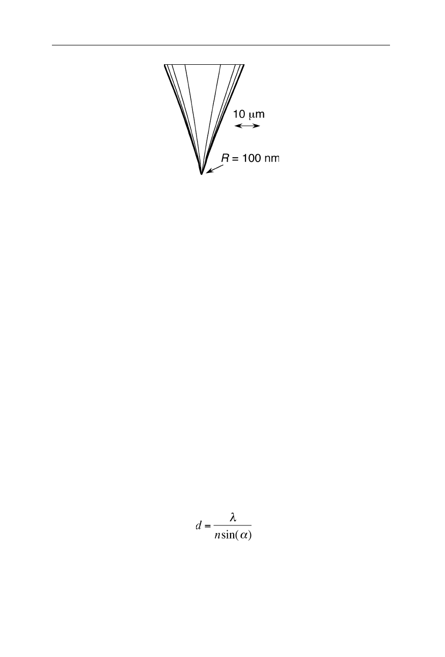
Similar as in atomic force microscopy, the quality of the tip is crucial for a high
resolution (Figs. 7.7–7.11, 7.22). Commonly tips are micromachined and/or
electrochemically etched and have apex radii of 5–100 nm.
STM images of a 1:1 mixture of K344C cytochrome P450
cam
/ putidaredoxin
adsorbed on gold (111) showed a regular array of pairs of the two proteins
(Djuricic et al., 2002). I21C/E25C plastocyanin essentially maintains its native
redox properties upon immobilization onto a gold substrate as shown by the full
potentiostatic control of the electron transfer reaction during STM imaging
(Andolfi et al., 2002). Scanning tunneling microscopy demonstrated that the
otherwise nearly linear mammalian metallothionein-2 molecule bends by about 20
degrees at its central hinge region between the domains in the presence of ATP
(Maret et al., 2002). Electrochemical scanning tunneling microscopy on thiol-
derivatized DNA immobilized on a gold (111) single crystal surface showed
7.3 Scanning nearfield optical microscope (SNOM) 135
Fig. 7.22
An etched tungsten tip for STM (see also Figs. 7.7–7.11)
potential-dependent orientation changes of the DNA in the potential range from
200 to 600 mV (Zhang et al., 2002b). A STM study of morphology and electron
transport features in cytochrome c offers evidence for sequential discrete electron-
tunneling effects (Khomutov et al., 2002). Tunneling in proteins adsorbed onto a
conductive substrate may depend on the applied potential (Facci et al., 2001). The
resistance of a single octanedithiol molecule is 900 M
Ω
(Cui et al., 2001). The
ability to site-specifically introduce cysteine residues and to engineer tags, such as
histidine tags and biotin-acceptor peptides, allow the creation of ordered
immobilized protein structures that can be characterized both electrochemically
and topographically by using scanning probe microscopy and cyclic voltammetry
(Gilardi et al., 2001).
7.3 Scanning nearfield optical microscope (SNOM)
7.3.1 Overcoming the classical limits of optics
SNOMs (Figs. 7.23–7.25), also known as NSOMs, utilize a light source with a
diameter smaller than the wavelength of the light (Synge, 1928; Ash and Nicholls,
1972; Pohl et al., 1984; Betzig et al., 1986, 1991, 1992; Toledo-Crow et al., 1992;
Williamson et al., 1998; Egawa et al., 1999; Heimel et al., 2001). By means of
this technological innovation they achieve a resolution which may be well beyond
the resolution limit,
d
, of classical Abbe-Fourier optics (see also Sect. 6.1.2):
, (7.1)
where
λ
,
n
, and
α
are the vacuum wavelength, refractive index of the medium
between sample and objective lens, and half angle of aperture, respectively. For
visible light with
λ
= 500 nm,
n
= 1.6, and
α
near 90
o
, we obtain a resolution limit
of classical optics of about 300 nm. Using UV light and image processing can
136 7 Scanning probe microscopy
yield improvement beyond this, but it is clear from Eq. 7.1, that classical optics
can hardly penetrate the 100-nm resolution barrier. SNOMs have been the first
optical microscopes that significantly overcame the limit of Eq. 7.1.
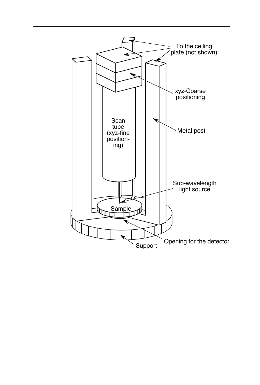
Fig. 7.23
A design of a SNOM. An optical probe emits light from points above the 2D
sample. The subwavelength probe tip takes advantage of the physical effect of optical
tunneling. In this SNOM it is made using a metal coated tapered glass fiber: a first taper
for the probe was manufactured by melt-drawing, and a second taper at the very end of the
probe was etched. The damping of the horizontally vibrating SNOM probe caused by
shear forces is taken as a measure for the distance from the sample. A shear force feedback
loop which involves an interferometric measurement of the horizontal position of the probe
tip keeps it at constant height (Betzig et al., 1992). Near-field optical properties of the
sample surface are mapped out by scanning each point within a certain area
7.3 Scanning nearfield optical microscope (SNOM) 137
Fig. 7.24
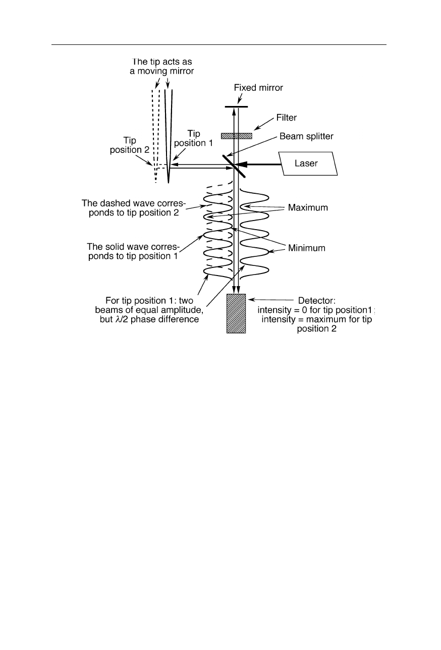
Design of an interferometer used for the detection of shear force of the SNOM
tip. The principle of operation is based on the extinction of light at the position of the
detector due to the interference of the two incident light beams of equal amplitude when
the probe tip (movable mirror) is in position 1. A small perturbation of the position of the
tip causes a small deviation from
λ
/2 of the phase shift of the two beams inciding on the
detector which leads to a non-negligible detector signal. The light filter in the fixed path
ensures an equal amplitudes of the two interfering beams
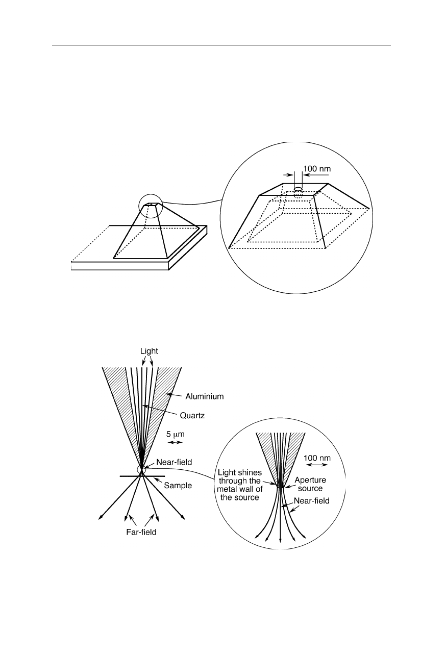
Generally the working principle of SNOMs is as follows (Figs. 7.23–7.25):
The subwavelength light source is positioned a few
µ
m above the specimen
surface with the help of mechanical coarse translation stages and a piezoelectric
fine translation device, e.g., a piezoelectric scan tube. Transmission is measured
below the specimen. An image of the specimen surface is obtained by moving the
light source (or the sample in some designs) with the help of the piezoelectric fine
translation device in horizontal direction. To avoid crashes with the sample, in
many SNOMs the light source is oscillated over the specimen surface and damp-
ing of oscillations due to source-sample interactions detected. The piezoelectric
fine translation device somewhat lifts the subwavelength light source (or lowers
the sample) when damping increases. Near-field optical excitation of the sample
can be seen as a dipole-dipole energy transfer (Sekatskii and Dietler, 1999).
138 7 Scanning probe microscopy
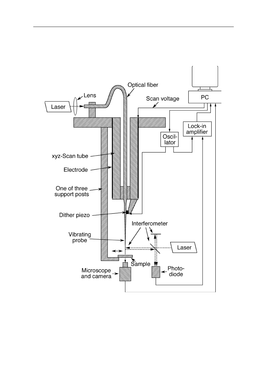
Fig. 7.25
A design of a SNOM with a xyz-coarse positioning and a xyz-fine positioning
There is virtually no resolution limit of SNOMs as long as one can manufacture
light sources of sufficient small size (see next section) and detect very small
intensity differences of light passing through thin sample layers.
7.3.2 Design of the subwavelength aperture
The most common methods to manufacture subwavelength apertures are (a)
adiabatic pulling of an optical fiber during heating (Betzig et al., 1991;
Williamson and Miles, 1996; Figs. 7.26a and 7.27), (b) etching (Muramatsu et al.,
1999), in particular tube etching (Turner, 1983; Stöckle et al., 1999a; Figs. 7.26b
7.3 Scanning nearfield optical microscope (SNOM) 139
and 7.28), and (c) microfabrication (Schurmann et al., 2000; Mitsuoka et al.,
2001), e.g., by ion beam milling (e.g., Veerman et al., 1998).
Fig. 7.26
(
a
) Melt-drawn probe: in the upper part of the probe, the light is reflected from
the walls by total internal reflection (TIR). TIR is the phenomenon involving the reflection
of all the incident light off a boundary when both (i) the light is traveling in the more
optically dense medium and approaching the less optically dense medium and (ii) the angle
of incidence is greater than the so-called critical angle. (
b
) Etched probe. A desirable high
brightness is obtained by a large cone angle. An optical aperture is formed by rotational
evaporation of the etched fiber with an opaque metal, e.g., aluminum
Fig. 7.27
Fabrication of a probe by adiabatic pulling of an optical fiber during heating
(melt-drawing; see, e.g., Williamson and Miles, 1996)
140 7 Scanning probe microscopy
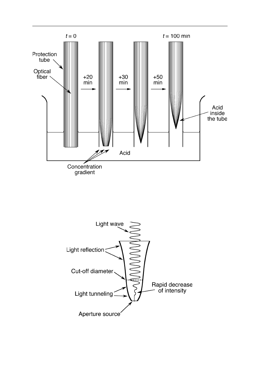
Fig. 7.28
Tube-etching of a quartz glass fiber with hydrofluoric acid (Turner, 1983;
Stöckle et al., 1999a, 1999b): the tip forms due to concentration gradients of acid and
desolved fiber. The tube serves also for suppression of convection of the acid. These tube-
etched tips are inexpensive and have large cone angles permitting high light throughputs
Fig. 7.29
Propagation of a light wave in a SNOM probe tip. Up to the cut-off diameter
(about equal to the wavelength), the light travels with only little attenuation. Beyond this
critical diameter, the light intensity very rapidly falls off
7.3 Scanning nearfield optical microscope (SNOM) 141
Usually the aforementioned probe tips are coated with a metal. Since the light
transmission of the tip dramatically depends on the distance from the cut-off
region to the aperture (Fig. 7.29), the optical throughput of etched tips with cone
angles around 30
o
is, in general, 2–3 orders of magnitude better than that of heat-
pulled fiber probes. Even brighter sources can be microfabricated (Fig. 7.30).
Fig. 7.30
SNOM tip made from silicon nitride. It was manufactured by using photo-
lithography, potassium hydroxide etching, and electron beam nanolithography (Zhou et
al., 1998, 1999)
Fig. 7.31
Optical far-field and near-field in the vicinity of a small optical tip for a SNOM.
Since the light can transmit through thin layers of metal, the diameter of the light beam in
this design cannot be less than a few 10 nm
142 7 Scanning probe microscopy
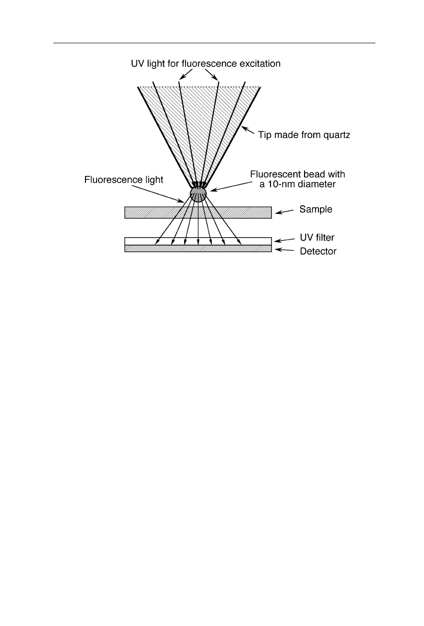
Fig. 7.32
A 10-nm sized light source for SNOMs made from fluorescent material. UV
light excites fluorescence of the 10-nm sized bead at the end of the probe. The
transmission of the fluorescence light through the sample is sensed by a detector which is
covered with an UV-absorbent layer. Antioxidants may be added to the fluorescent
material to enhance stability and life. The resolution for thin-layered samples enabled by
this type of light sources depends on the size of the fluorescent bead and the detectability
of small absorbance differences of the fluorescent light
In the aforementioned methods of source fabrication, a limitation for the source
size is the transmission of light through thin layers of metal (Fig. 7.31). Sources
of 10 nm diameter could hardly be made just by machining an opening in a metal
plate or in an optical fiber since light would significantly shine through the walls
of the opening. Smaller light sources involve the excitation of small fluorescent
particles (Fig. 7.32).
7.3.3 Examples of SNOM applications
An important applicability is seen in cell biology, microbiology (Meixner and
Kneppe, 1998), and proteomics (Gao et al., 2001). SNOM imaging visualized
domains of photosystem II core complex bound to lipid monolayers (Trudel et al.,
2001). Topographic, friction, fluorescence, and surface potential distributions for
a Langmuier-Blodgett film can simultaneously be observed using a SNOM-AFM
with a thin step-etched optical fiber probe (Horiuchi et al., 1999). Fig. 7.33
demonstrates a 50-nm resolution.