N?lting B. Methods in Modern Biophysics
Подождите немного. Документ загружается.

7.4 SICM, SThM and further scanning probe microscopes 143
Fig. 7.33
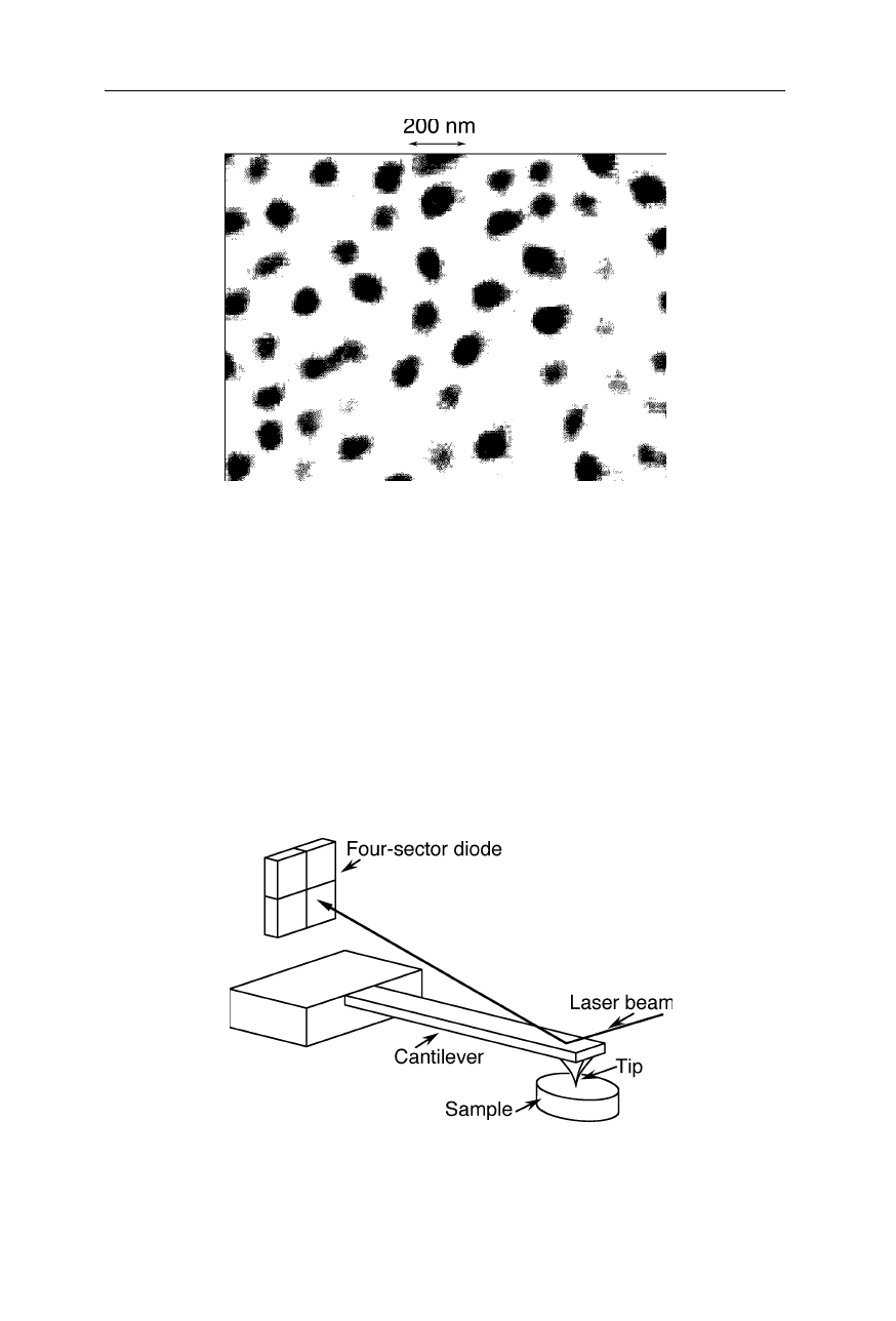
Absorption of latex beads with approximately 100 nm diameter taken with a
SNOM at a resolution of about 50 nm (OMICRON, Taunusstein, Germany)
7.4 Scanning ion conductance microscope, scanning
thermal microscope and further scanning probe
microscopes
Important scanning probe microscopes are also the magnetic force microscope,
scanning Hall probe microscope (Chang et al., 1992a, 1992b), friction force
microscope (Fig. 7.34; Howald et al., 1995), scanning ion conductance micro-
Fig. 7.34
Example of a friction force microscope: the detector has sectors in both vertical
and horizontal direction so that the torsion of the cantilever can be estimated and a
frictional (lateral) force be calculated (see, e.g., Howald et al., 1995)
144 7 Scanning probe microscopy
scope (Fig. 7.35; Hansma et al., 1989; Korchev et al., 1997, 2000a, 2000b;
Stachelberger, 2001; Bruckbauer et al., 2002a), and scanning thermal microscope
(Fig. 7.36; Mills et al., 1998, 1999). There are two common types of thermal
probes for scanning thermal microscopes: thermocouples and thermal resistors.
The thermocouple probe in Fig. 7.36 involves two dissimilar metal wires which
bisect over the top of a blunt silicon nitride pyramid (Mills et al., 1998). A
potential biophysical application is the elucidation of local heating effects in
biological tissue due to cell metabolism.
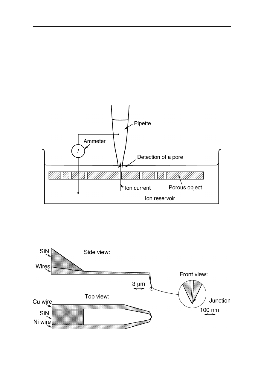
Fig. 7.35
Detection of membrane pores by a scanning ion conductance microscope. The
probe is a nanopipette filled with electrolyte solution. The current between pipette and ion
reservoir starts increasing when the pipette tip approaches a pore. The technique permits
100-nm resolution characterization of distribution and sizes of pores
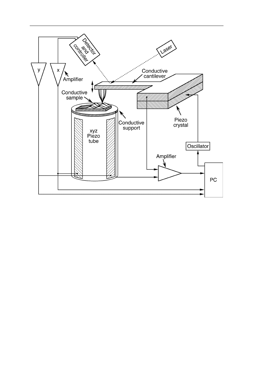
Fig. 7.36
Design of the tip of a scanning thermal microscope (Mills et al., 1998, 1999). A
submicrometer-sized Cu/Ni thermocouple at the end of the cantilever detects the thermal
microenvironment
7.4 SICM, SThM and further scanning probe microscopes 145
Fig. 7.37
STM with simultaneous AFM capability: AFM and STM sense different
physical properties. The combined information provides greater insight into the chemical
nature of the sample ant its physico-chemical properties
The STM with simultaneous AFM capability (Fig. 7.37) provides simultane-
ously information about surface relief and chemical composition of the specimen.
8 Biophysical nanotechnology
8.1 Force measurements in single protein molecules
Atomic force microscope (AFM)-related techniques can induce and monitor the
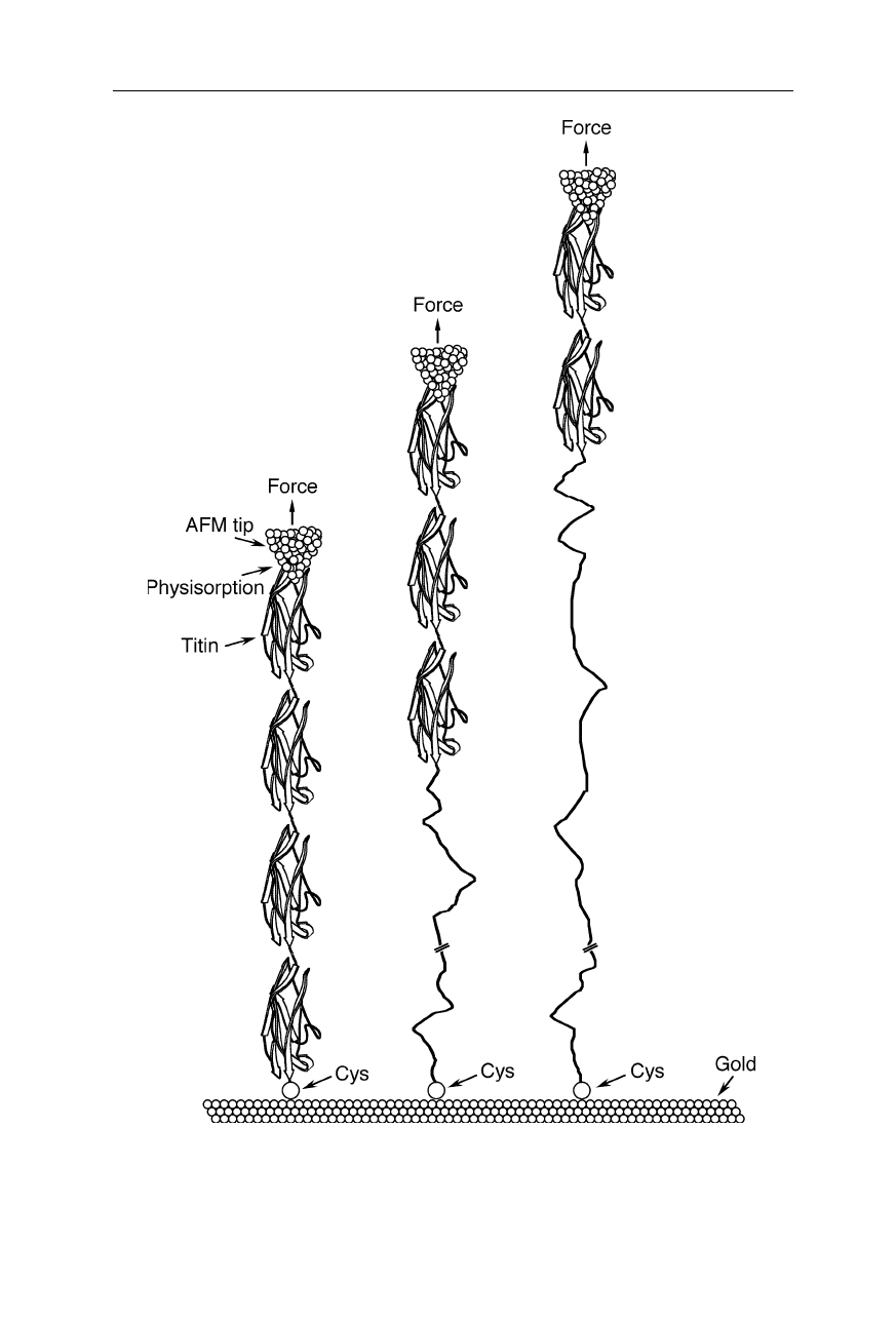
unfolding of single protein molecules. Experiments on the protein titin, which is a
main component of skeletal muscles (Figs. 8.1–8.3), revealed that the force for
unfolding of its individual domains with cross sections of less than 5 nm
2
is of the
order of 100–300 pN and dependent on the pulling speed (Rief et al., 1997; Gaub
and Fernandez, 1998; Carrion-Vasquez et al., 1999). A similar investigation on
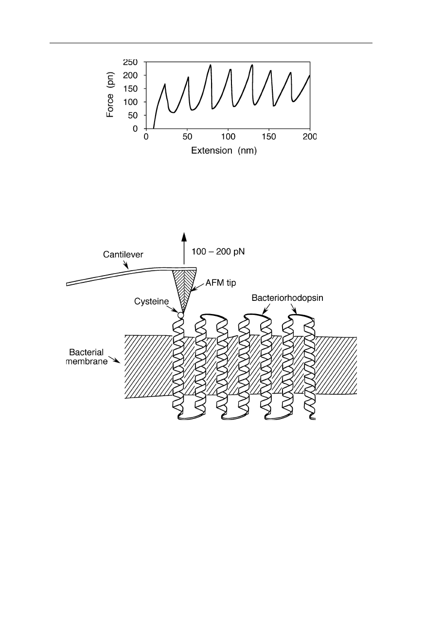
bacteriorhodopsin showed that its helices are anchored to the bacterial membrane
with 100–200 pN (Fig. 8.4; Oesterhelt et al., 2000). Similarly, single-molecule
force spectroscopy on spider dragline silk protein molecules revealed that the
molecule unfolds through a number of rupture events, indicating a modular
structure within single silk protein molecules (Oroudjev et al., 2002). The mini-
mal unfolding module size of 14 nm indicates that the modules are composed of
38 amino acid residues (Oroudjev et al., 2002). Adhesion between two adjacent
cell surfaces of the eukaryote Dictyostelium discoideum involves discrete
interactions characterized by an unbinding force of about 23 pN. This force
probably originates from interactions of individual “contact site A” (csA)
glycoprotein molecules (Fig. 8.5; Benoit et al., 2000).
Fig. 8.1
Molecular architecture of skeletal muscle fibers. AFM-related techniques contrib-
uted to the understanding of the role of individual titin molecules in such fibers: some
skeletal muscle proteins can withstand drags of 600 kp cm
–2
(see Figs. 8.2 and 8.3; Rief et
al., 1997; Gaub and Fernandez, 1998; Carrion-Vasquez et al., 1999)
148 8 Biophysical nanotechnology
Fig. 8.2
Unfolding of a titin fragment with the help of an AFM (Gaub and Fernandez,
1998; Carrion-Vasquez et al., 1999). The unfolding force for the protein, anchored with a
cysteine (Cys) to a gold surface, ranged from about 100 to 300 pN (see Fig. 8.3)
8.1 Force measurements in single protein molecules 149
Fig. 8.3
Sketch of a force-distance curve for the unfolding of a titin fragment with the help
of an AFM (see Fig. 8.2). The saw tooth-shaped force-extension curve reflects the un-
folding of individual titin domains according to an all-or-non mechanism (Gaub and
Fernandez, 1998; Carrion-Vasquez et al., 1999)
Fig. 8.4
Unfolding of individual bacteriorhodopsins. A force of 100–200 pN is required
to remove a bacteriorhodopsin helix from the bacterial membrane (Oesterhelt et al., 2000)
Recoverin, a calcium-myristoyl switch protein, binds to a phospholipid bilayer
in the presence of Ca
2+
with an adhesion force of 48 ± 5 pN (Desmeules et al.,
2002). Single molecules of
holo
-calmodulin (i.e., the calcium-loaded form)
require a significantly larger force of unfolding by an AFM tip than single
molecules of the
apo
-form (Hertadi and Ikai, 2002). Single molecules of the giant
filamentous protein titin exhibit mechanical fatigue when exposed to repeated
stretch and release cycles (Kellermayer et al., 2001). For further AFM studies on
single protein molecules see also Sects. 7.1, 8.2, and 8.3.
150 8 Biophysical nanotechnology
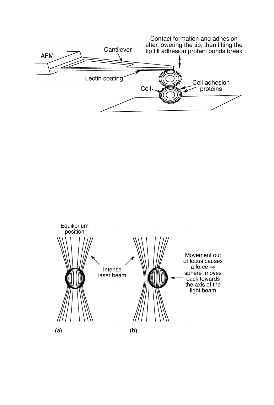
Fig. 8.5
Measurement of discrete interactions in cell adhesion (Benoit et al., 2000)
8.2 Force measurements in a single polymerase-DNA
complex
DNA polymerases catalyze DNA replication. The replication reaction requires
single-stranded DNA (ssDNA) as a template. In the course of the reaction, a
complementary strand of ssDNA is synthesized to the original ssDNA. Already
during the polymerization reaction, both strands coil around each other, leading to
a shortening of the end-to-end distance of the DNA. Exerting strain on the DNA
strand during polymerization can stop and even revert the extension reaction
Fig. 8.6
Optical tweezers for the measurement of the effect of template tension on T7
polymerase activity (Fig. 8.7; Smith et al., 1996; Wuite et al., 2000). Regarding the
method of optical tweezers see also the groundbreaking studies of single-molecule
mechanics by Florin et al. (Florin et al., 1997; Jeney et al., 2001; Pralle and Florin, 2002)
and Smith et al. (Bustamante et al., 2000; Liphardt et al., 2001; Smith et al., 2001)
8.2 Force measurements in a single polymerase-DNA complex 151
Fig. 8.7
Measurement of the effect of template tension on T7 polymerase activity (Smith et
al., 1996; Wuite et al., 2000): the polymerase which catalyses DNA replication can work
against a maximum force of about 34 pN. Exonuclease activity increases about 100-fold
above 40 pN template tension.
(Figs. 8.6 and 8.7; Smith et al., 1996; Wuite et al., 2000). For the measurement of
the small forces in the single DNA-protein complex, an optical trap was used: a
small bead with DNA attached to it is held into position and moved by an intense
laser beam. The main mechanism of the action of such optical tweezers is com-
monly as follows: The bending of light rays through the refractive sphere is
connected with a change of momentum of the light which exerts a force back on
the sphere. When the sphere is out of focus of the light beam, these light deflec-
tion forces pull the sphere back into focus. Another mechanism is as follows:
When an isotropically scattering bead moves out of focus, the momentum of the
photons scattered in the direction of the movement increases due to the Doppler
effect which decelerates the bead. The larger the light intensity the larger is the
deceleration of the movement. Thus, the Brownian motion out of focus is ener-
getically unfavorable relative to the motion into focus. Choosing a wavelength
just below an absorption maximum of the bead increases the trapping force since
then movement causes increased absorption due to the Doppler-shift of the
wavelength and thus an additional momentum slowing down the bead.
152 8 Biophysical nanotechnology
8.3 Molecular recognition
AFM-related techniques allow the direct measurement of individual intermolecu-
lar interactions (Figs. 8.8–8.11; Florin et al., 1994; Dammer et al., 1995, 1996;
Merkel et al., 1999; Strunz et al., 1999; De Paris et al., 2000; Fritz et al., 2000;
Schwesinger et al., 2000; Zocchi, 2001; Prechtel et al., 2002; see also Sect. 7.1).
Virtually any intermolecular interaction forces, e.g., of antibody-antigen interac-
tions, are measurable with the technique illustrated in Fig. 8.8.
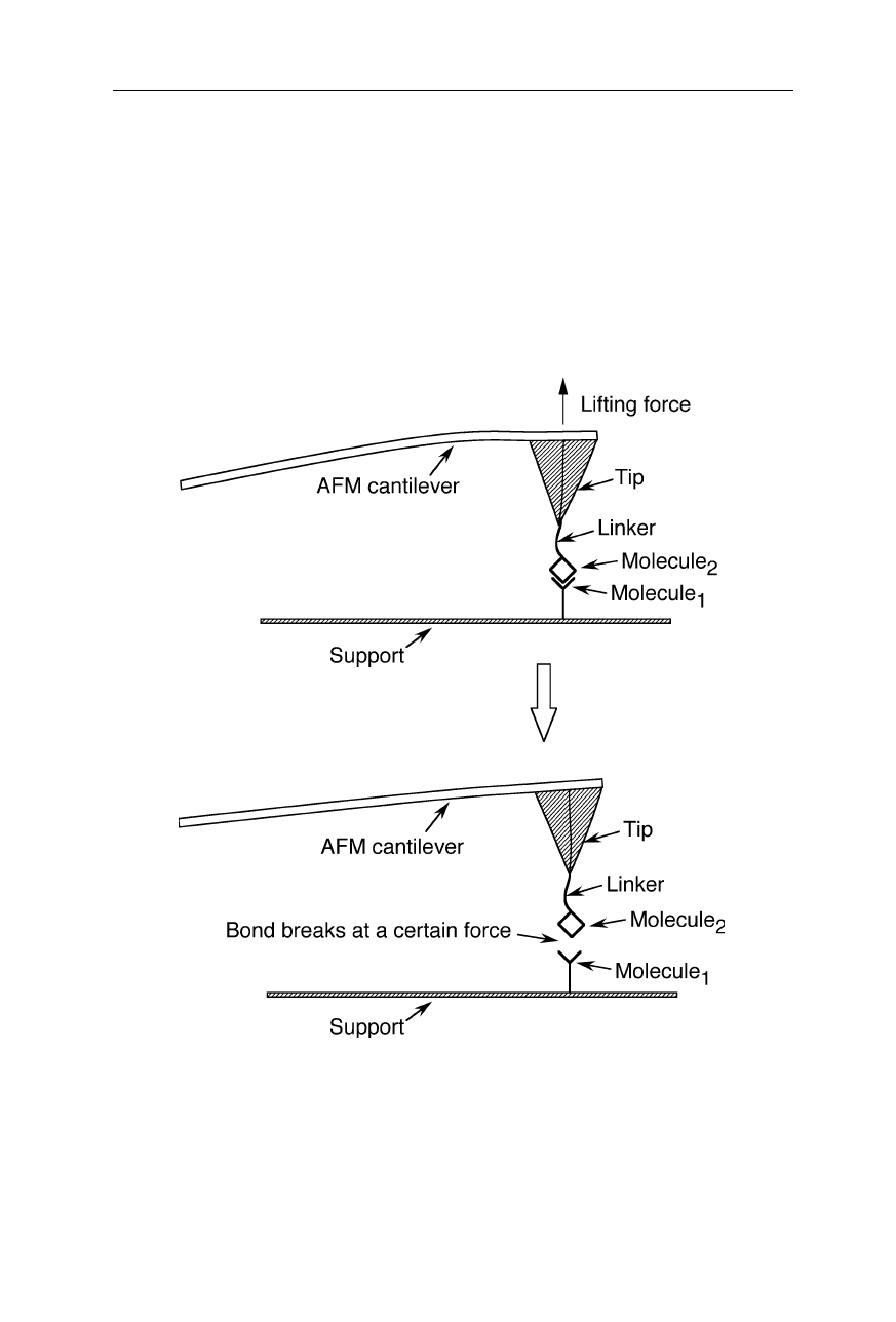
Fig. 8.8
Direct measurement of intermolecular interactions by an AFM-related technique.
One of the interacting molecules is immobilized on the surface of the support, the other is
connected to the AFM tip by a linker. The tip is approached to the surface so that a spe-
cific interaction can take place. Retracting the cantilever ruptures the biophysical interac-
tion. The strength of the interaction is determined from the retract force distance curves
(De Paris et al., 2000; Schwesinger et al., 2000; see also Sect. 7.1)
8.3 Molecular recognition 153
Fig. 8.9
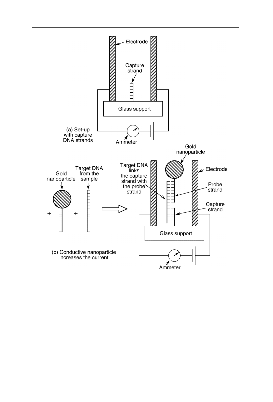
Sensor of biological agents using recognition between single DNA molecules
(Park et al., 2002; Service, 2002; see also Demers et al., 2002). (
a
) Two electrodes and
single-stranded capture DNA strands are attached to a glass substrate. The capture DNA is
complementary to one end of the target DNA of the agent. (
b
) Target DNA and probe
strand DNA was added. The probe strand DNA has a gold nanoparticle attached and is
complementary to the other end of the target DNA. When all three strands of DNA
hybridize together, the gold nanoparticle gets held between the two electrodes. This is
detected by an increase of current
Fig. 8.9 illustrates a new type of DNA sensor with potential application for
detection of biological contaminants (Park et al., 2002; Service, 2002). It is based
on the change of electrical conductivity when gold particles attached to DNA bind