Messerschmidt U. Dislocation Dynamics During Plastic Deformation
Подождите немного. Документ загружается.

402 10 Quasicrystals
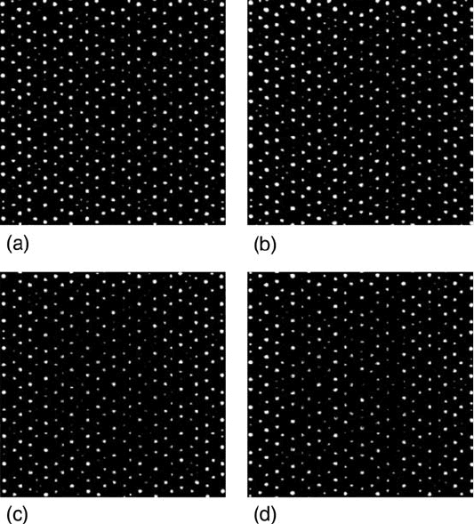
Fig. 10.8. Two-dimensional quasiperiodic structures generated by the density-wave
method. (a) Perfect pattern. (b) Pattern of (a) distorted by a spatial variation of
the phonon degree of freedom. (c) Pattern distorted by varying the phason degree
of freedom. (d) Dislocation. Reprinted with permission from Socolar et al. [679].
Copyright (1986) by American Physical Society (http://link.aps.org/abstract/PRB/
v34/p3345/y1986)
of the cut and projection procedure. The undisturbed arrangement of lat-
tice points is demonstrated in Fig. 10.8a, showing different kinds of clusters
forming the structure. Looking at this figure at a glazing angle reveals the
different atom rows. In Fig. 10.8b, the same structure is distorted by spatially
varying displacements of only phononic character. The originally straight rows
of atoms are now curved, corresponding to elastic strains. The effect of varying
phasonic displacements is demonstrated in Fig. 10.8c. The rows of atoms are
straight again but they are jagged, i.e., they show lateral displacements. Sim-
ilar images were revealed by high-resolution electron microscopy (e.g., [681]).
Relaxation of the phason faults requires local diffusion of atoms. Therefore,
these processes occur far more slowly than phonon relaxations. The dynamics
of long-wavelength phason fluctuations was studied by changes in the speckle
contrast in a coherent X-ray-scattering experiment [682]. According to that,
phason fluctuations in i-Al–Pd–Mn are frozen-in below 500
◦
C. At 650
◦
C, the
10.2 Defects in Quasicrystals 403
corresponding diffusion coefficient is 2.2 × 10
−18
m
2
s
−1
with an activation
energy of 2.3(±1)eV. This diffusion coefficient is in the range of those of
Pd and Mn. Phason flips were observed in situ at a high temperature in a
decagonal Al–Cu–Co quasicrystal by high-resolution TEM [683].
10.2.3 Dislocations
As in crystals, plastic deformation is realized by the motion of dislocations.
A recent review has been given by Bonneville, Caillard and Guyot [684].
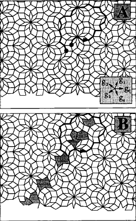
Figure 10.9 shows the result of a computer simulation of the shearing and
introduction of an edge dislocation into a two-dimensional decagonal qua-
sicrystal from [685]. A Burgers circuit (see Sect. 3.1.1) is carried out around
the dislocation in B and the corresponding reference circuit is shown in A. It
has to be closed by the Burgers vector of the dislocation. Since the shearing
was carried out in physical space, there remain a number of places with vio-
lations of the matching rules marked by grey shading, i.e., of phason defects.
Therefore, the Volterra process to generate a dislocation should not be per-
formed in the physical space but in the higher-dimensional hyperspace so
that in icosahedral quasicrystals the Burgers vector is a six-dimensional vec-
tor [686]. The cutting procedure similar to Figs. 3.7 and 3.8 is carried out along
a five-dimensional half-plane comprising the whole perpendicular space. The
Fig. 10.9. Creation of phason strain by the introduction of a dislocation into a
two-dimensional decagonal quasicrystal. From [685]. Copyright Taylor & Francis
Ltd. (1995) (http://www.informaworld.com)
404 10 Quasicrystals
dislocation line is a four-dimensional manifold, a one-dimensional line in phys-
ical space plus the whole orthogonal space, since after (10.5), the strain field
does not depend on the coordinates of the perpendicular space. The geomet-
ric properties of dislocations and their motion in the quasiperiodic lattice are
described in detail in [687].
In analogy to (3.1), the Burgers vector B in quasicrystals can be defined
by a line integral over the displacements U along a closed curve C around the
dislocation in the physical space E
B =
C
dU =
C
du
+du
⊥
= b
+ b
⊥
. (10.6)
Thus, the integration over the phonon strain field yields the parallel part b
of the Burgers vector. It corresponds to the usual Burgers vector in crystals
describing the continuous elastic strains around the dislocation line. For the
edge dislocation, b
is perpendicular to the dislocation line, whereas for the
screw dislocation it is parallel. In many processes, b
replaces the Burgers
vector known from crystals. The perpendicular part of the Burgers vector b
⊥
represents the phason strain field around the dislocation line. Both phenom-
ena can be observed in the two-dimensional density wave image of an edge
dislocation in Fig. 10.8d. The lattice rows are bent around the inserted half-
row characterizing the elastic strain field of the dislocation. In addition, there
appears a number of jags in the atom rows.
The full six-dimensional Burgers vector of (three-dimensional) quasicrys-
tals can be determined by several methods. As shown by Wollgarten et
al. [688], the contrast extinction criterion (2.9) of diffraction contrast TEM
can be extended to yield
G · U = g
· u
+ g
⊥
· u
⊥
=0, (10.7)
with G = g
+ g
⊥
. Since the cut in six-dimensional space has an irrational
orientation, each g
belongs to a single G in the six-dimensional reciprocal
lattice, and accordingly also to a single g
⊥
in reciprocal space [654,689]. The
criterion (10.7) can be fulfilled in two ways. The first one is by making both
terms zero, i.e.,
g
· u
= g
⊥
· u
⊥
=0. (10.8)
This is called a “strong” contrast extinction condition. Then, the dislocation
contrast disappears for a whole systematic row of reflections through the origin
of the diffraction pattern. Two such conditions have to be found to determine
the direction of the Burgers vector in both the physical and orthogonal space.
In the second case,
g
· u
= −g
⊥
· u
⊥
. (10.9)
This is the “weak” contrast extinction condition, which is fulfilled only for
periodically spaced reflections within a systematic row but not for other reflec-
tions of the same row. The determination of the length of the Burgers vector
requires a quantitative analysis of the dislocation contrasts as in crystals.

10.2 Defects in Quasicrystals 405
Wang and Dai [690] were the first to use convergent beam electron diffrac-
tion (CBED) to determine the six components of the Burgers vector in
icosahedral quasicrystals. In this technique, the dislocation image is super-
imposed with the defocus CBED pattern where zero-order Laue zone (ZOLZ)
lines split n times when a dislocation line with |G · B| = n is crossed. B
is determined completely by six independent equations G · B = n.Afurther
method employs high-resolution TEM by observing lattice fringe images taken
in two different orientations of the specimen [691].
The elastic properties of dislocations in quasicrystals are treated in the
framework of an extended elasticity theory (e.g., [692], for an introduction
see, e.g., [693, 694]). Considering only short-time elastic responses, icosahe-
dral quasicrystals behave isotropically so that their elastic properties can be
described by two elastic constants (e.g., the Lam´e constants λ and μ). They
can be measured in the usual way by ultrasound transmission. In the gen-
eralized Hooke’s law, there appear also two phason elastic constants K
1
and
K
2
, which are determined indirectly by diffuse X-ray or neutron scattering
(e.g., [695]), and a phonon–phason coupling term R. Like for strains, there
occur stress components in physical space and orthogonal space. The stresses
in real space depend on the elastic strains via the Lam´e constants, and on
the phason strains via the coupling constant. On the other hand, the stresses
in perpendicular space depend on the phason strain via the phason elastic
constants, and on the phonon strains via the coupling constant. A set of elas-
tic constants for i-Al–Pd–Mn used in another terminology in [692] comprises
λ = 85, μ = 65, K
1
=0.084, K
2
=0.036, and R =0.41 (all in GPa). The elas-
tic energy of a quasicrystal is composed of three terms, one from the phonon
part, one from the phason part, and a coupling term.
Not only the phononic strains of dislocations but also the phasonic ones
decrease with 1/r,wherer is the distance from the dislocation line. Only pure
screw and edge dislocations along a twofold orientation have simple displace-
ment fields with displacements only parallel to the line direction for the screws,
and perpendicular to it for edges. In all the other cases, the properties are not
so simple. The equilibrium positions of parallel dislocations with respect to
glide were calculated in [696]. This is based on a generalized Peach–Koehler
force, in analogy to (3.22),
F
PK
=
b
σ
+ b
⊥
σ
⊥
× ξ, (10.10)
where σ
⊥
is the stress tensor in perpendicular space. In [697], the formula is
quoted for one-dimensional hexagonal quasicrystals.
In terms of the extended elasticity theory, the energy of a screw dislocation
in the same quasicrystal is given by [697]
E
d
=
c
44
b
2
3
+ K
2
b
2
⊥3
+2R
3
b
3
b
⊥3
ln (R/r
0
)
4π
,
with R and r
0
being the outer and inner cut-off radii as introduced in
Sect. 3.2.2. Thus, the dislocation energy depends on the squares of both
406 10 Quasicrystals
Burgers vector components b
and b
⊥
. Assuming that the phason part of the
dislocation energy is proportional to the number of matching rule violations,
there should arise a linear dependence on b
⊥
[679,698]. Though the necessity
of the extension of the elasticity theory for quasicrystals is quite obvious, the
consequences have widely been ignored so far in interpreting the experimental
results on dislocation motion and plastic deformation.
The microprocesses of dislocation motion depend on the temperature. If
the temperature is high enough for a rapid phason rearrangement, the equilib-
rium state of the phason stress field around the dislocation can move together
with the phonon field. The phason flips accompanied with the motion occur
also far from the dislocation itself as illustrated by a simulation of the dislo-
cation motion in a decagonal quasicrystal [699]. However, if the temperature
is not so high, the dislocation may decompose [700] and the phononic part
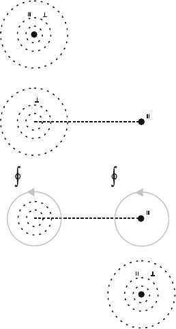
of it will move alone as it is outlined highly schematically in Fig. 10.10. In
Fig. 10.10a, the dislocation of Burgers vector b
+ b
⊥
sketched as a full cir-
cle is surrounded by its phason cloud marked by dashed circles. Caillard and
coworkers called such a dislocation a perfect dislocation (e.g., in [684,701]). In
Fig. 10.10b, a dislocation with only the parallel part of the Burgers vector b
has moved from A to B owing to an external stress. It may be called an imper-
fect dislocation. In its wake, the imperfect dislocation created a layer of high
b
b
b+b
b+b
A
B
b
A
B
b
A
B
b
A
B
du
⊥
=0 du
⊥
= b
⊥
(a)
(b)
(d)
(c)
Fig. 10.10. Schematic of the motion of a perfect dislocation. (a) Starting dislocation
with phonon and phason stress fields. (b) Intermediate state with separation of
phonon and phason stress fields. (c) Burgers circuits around dislocations A and B.
(d) Final state
10.2 Defects in Quasicrystals 407
phason disorder called a phason wall and indicated by the thick dashed line.
At the original site A, there remains a dislocation with the Burgers vector b
⊥
representing the phasonic strain field of the original dislocation. The situation
is illuminated by the two grey Burgers circuits around A and B in Fig. 10.10c.
While the dislocation in real space at B has only the Burgers vector b
,the
Burgers circuit in orthogonal space around B yields the Burgers vector b
⊥
when crossing the concentrated phason strain of the phason wall. The Burg-
ers circuit around A crosses the phason wall in opposite direction resulting in
a contribution of −b
⊥
from the phason wall, which cancels the contribution
of the phason strain from the original dislocation. The whole system is not in
thermal equilibrium. The equilibrium configuration with a perfect dislocation
at B as in Fig. 10.10d can only be attained by diffusional retiling, where the
phason field at A and the phason wall dissolve. Computer simulations on a
two-dimensional decagonal quasicrystal estimate the stress necessary to cre-
ate the phason wall by glide at zero temperature to amount to τ
ph
≈ (1/27) μ
[685]. This would be a very high stress. Therefore, quasicrystals were consid-
ered brittle at low temperatures. On the other hand, measurements from the
width of pairs of dislocations moving together by climb yield very low values
[702].
Since the imperfect dislocations have only a phononic Burgers vector b
,
they fulfill only the strong extinction condition (10.8) with g
· b
=0.In
the orientation where the corresponding perfect dislocations obey the weak
extinction condition, a residual contrast remains. The phason walls have a
displacement vector which can be defined either by r
= b
or by r
⊥
= b
⊥
.
The shift vectors of phason walls are determined in a way similar to that of
stacking faults in crystals. Since g
·r
is irrational in quasicrystals, extinction
occurs only for g
· r
= 0, but not for g
· r
= integer. A TEM diffraction
contrast analysis of phason faults is published in [703].
If the temperature is not too low, the distribution of the phason defects
in the solid can be rearranged by spreading of the phasons of the wall and by
annealing to form the equilibrium phason distribution around A and the new
cloud around the dislocation at B. The process is called phason dispersion
or retiling. All the existing phasons certainly do not diffuse to the dislo-
cation at B. Instead, the retiling occurs by local diffusion processes which,
however, include chemical diffusion over distances larger than the atomic dis-
tances. Thus, the atomic mobility has to be high enough. At the end, the
new equilibrium configuration of phonon and phason strain fields may be re-
established (Fig. 10.10d). During rapid deformation at lower temperatures,
dislocations are certainly generated as imperfect dislocations. If the Burgers
vector in hyperspace is a fraction of a lattice vector, the dislocation is a partial
dislocation.
An important issue of dislocation motion in quasicrystals was the ques-
tion whether the dislocations move by glide or climb. Although the first in
situ straining experiments in a TEM on i-Al–Pd–Mn single quasicrystals were
performed at a relatively high temperature (at 0.88 T
m
,withT
m
being the
408 10 Quasicrystals
melting temperature) [647], they immediately showed that dislocation motion
in this material is of crystallographic character, i.e., many dislocations showed
straight crystallographically oriented segments, and they moved on crystal-
lographic high-symmetry planes. Therefore, for several years it had been
assumed that the dislocations move by glide without questioning this hypothe-
sis. To prove the mode of dislocation motion, it is necessary to determine both
the direction of the parallel component of the Burgers vector and the plane of
motion of always the same dislocations. This is only possible by in situ strain-
ing experiments in a TEM. Only recently, Caillard and coworkers succeeded in
performing such experiments, at first during in situ heating where the disloca-
tions moved under internal stresses [704], and then also during in situ straining
[705]. Both experiments showed dislocations moving by pure climb. Thus,
climb is certainly a main mode of dislocation motion in quasicrystals. The
following microstructural results have to be considered keeping this in mind.
10.3 Microscopic Observations of Dislocations
While information on dislocation mechanisms of quasicrystal deformation
has been gained by TEM on a number of materials of either icosahedral
or decagonal symmetry, for other materials like Al–Cu–Fe the evidence of
dislocation motion is still missing, in spite of a wealth of macroscopic defor-
mation data available. The following sections are restricted to two single
quasicrystalline materials, icosahedral Al-21at%Pd-8.5at%Mn and decago-
nal Al-15at%Ni-15at%Co. These materials may be considered prototypes of
quasicrystals. Their dislocation microstructures have most frequently been
studied, also by the author of this book and his coworkers.
10.3.1 i-Al–Pd–Mn
As it was mentioned earlier, the dislocation properties strongly depend on the
temperature. This influences also the macroscopic deformation parameters,
which will be described in detail in Sect. 10.4. At high temperatures where
diffusion is rapid, quasicrystals can be deformed up to high strains. After a
yield drop effect, there develops a range of almost steady state deformation.
Most deformation experiments and microstructure studies were carried out
in this temperature range. Only later, experiments succeeded at lower tem-
peratures where deformation at low rates is accompanied with very strong
work-hardening. The following results consider both ranges. In situ strain-
ing experiments, however, were successful only in the high-temperature range
where the flow stresses are not so high.
Transmission Electron Microscopy of Deformed Specimens
The first experiment which proved a dislocation mechanism of the plastic
deformation of quasicrystals was the observation by Wollgarten et al. [646] of
10.3 Microscopic Observations of Dislocations 409
an increase of the dislocation density after deformation. Later on, the Burgers
vectors of deformation-induced dislocations were analyzed by the CBED tech-
nique [706]. 60 dislocations were chosen from specimens deformed at 800
◦
C
up to different plastic strains, and at different temperatures down to 732
◦
C
at a fixed strain, all in the high-temperature range. Fifty-two of the 60 dis-
locations had parallel parts b
of the Burgers vectors in twofold directions,
32 of which with a length of |b
| =0.183 nm. Five dislocations had parallel
Burgers vectors in the fivefold direction. They are super-partial dislocations.
To characterize the contribution of phason strain, the so-called strain accom-
modation parameter ζ = |b
⊥
|/|b
|, i.e., the ratio between the lengths of the
perpendicular and parallel parts of the Burgers vectors, was calculated. In
deformed samples, the values varied between τ
7
≈ 29.0andτ
3
≈ 4.2witha
maximum of τ
5
≈ 11.1. Higher values of the ratio occurred more frequently
with increasing plastic strain. In the in situ and post-mortem TEM studies
of the group of Caillard [702,704, 705] performed at lower temperatures, only
dislocations with longer parallel parts of the Burgers vectors b
were analyzed,
i.e., of 0.257, 0.296, 0.348, 0.456, 0.513, and 0.563 nm.
In addition to the Burgers vectors, also the line directions were deter-
mined of 25 dislocations with twofold Burgers vectors [706]. Forty percent
of the lines were oriented along twofold directions. The character β of the
dislocations ranged between 7 and 90
◦
. Thus, pure screw dislocations have
not been observed. The parallel parts of the Burgers vectors and the line
directions span the slip planes of the dislocations, and the dislocations were
believed to glide on these planes, although there were already hints that this
may be wrong.
The dislocation microstructure of deformed samples was also imaged by
diffraction contrast in the HVEM allowing thick specimens to be investigated
so that the three-dimensional character of the dislocation structures could be
studied [707]. After deformation, some of the samples were cooled under load
to better preserve the microstructure. Some micrographs reveal dislocations
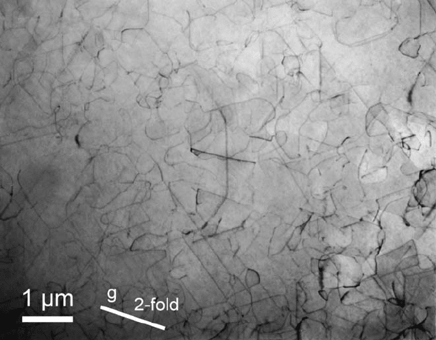
being arranged in bands, but inside the bands, they form a homogeneous three-
dimensional network at high temperatures, as shown in Fig. 10.11. Diffraction
contrast analysis using strong contrast extinctions at two different g vectors
indicates that dislocations with different Burgers vectors form the nodes of
the network. There exist groups of dislocations with their parallel component
of the Burgers vector being parallel to the compression axis, i.e., these dis-
locations experience a climb force but no glide force. At 800
◦
C, the average
link length in the network is of the order of magnitude of L =0.5 μm. Many
dislocation segments are quite straight and oriented along crystallographic
directions. This is remarkable for the high temperature.
At the lowest temperature with steady state deformation, many disloca-
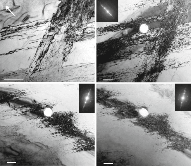
tions form narrow bands. In Fig. 10.12, taken near a twofold pole parallel to the
compression axis, two bands cut each other. Dislocations are arranged on two
steeply inclined planes, most probably planes perpendicular to threefold direc-
tions. The bands include elongated dislocations as well as strongly bowed-out
410 10 Quasicrystals
Fig. 10.11. Dislocation structure in an i-Al–Pd–Mn single quasicrystal deformed
at 770, 800, and 818
◦
C along a fivefold axis. Image taken near this pole. From [707].
Copyright Elsevier (2004)
dislocation segments and many small dislocation loops, as shown in Fig. 10.13.
The radii of curvature of the bowings and the loops are as small as 70 nm.
The dislocation density inside the bands is approximately 1.2 × 10
14
m
−2
.
Figures 10.12b to d exemplify the analysis of the parallel components of the
Burgers vector of the band running downwards from the upper right corner
of the figure. The hole in the specimen may serve as a marker. Because of
the bending of the specimen, a field-limiting aperture of 2 μmdiameterwas
placed near the center for taking the diffraction patterns shown in the insets.
Thus, the respective diffraction conditions are fulfilled only near the centers
of the images. The discussed dislocation band is extinguished in Figs. 10.12c
and d. Accordingly, the parallel component of the Burgers vector is perpen-
dicular to the extension of the band and to the compression axis. Thus, these
dislocations are near the edge orientation and experience neither a glide force
nor a climb force from the applied stress.
At a lower temperature of 532
◦
C, where plastic deformation is achieved in
stress relaxation tests in the strong work-hardening range, many dislocation
segments are straight and oriented crystallographically but other segments
very strongly bow out between cusps indicating pinning agents, as marked by
an arrow in Fig. 10.14. The distances between the cusps (obstacle distances)
10.3 Microscopic Observations of Dislocations 411
1µm
(a)
1µm
(d)
1µm
(c)
1µm
(b)
g
Fig. 10.12. Slip bands after steady state deformation by ε = 2% at 580
◦
Cat
10
−6
s
−1
along a twofold axis with analysis of the parallel component of the Burgers
vector of the band running approximately vertically. The deformation curve is shown
in Fig. 10.32b. Compression axis and image normal [0/00/00/2] in (a, b), g vectors
(
¯
1/00/10/0) (a, b), (1/00/10/0) (c) and (
¯
1/0
¯
1/
¯
10/1) (d). The insets show the
respective diffraction patterns. From [708]
are of the order of magnitude of 100 nm and the radii of curvature of the
bowed-out segments are about δ = 50 nm. At the lowest temperatures of 487
and 482
◦
C, where plastic deformation was obtained in conventional compres-
sion tests, different dislocation structures are observed. Some specimen regions
again exhibit the straight crystallographically oriented dislocation segments.
However, Fig. 10.15a presents a totally different dislocation structure. The
dislocations are arranged in strongly curved loops, but the cusps indicating
pinning agents at 532
◦
C are missing now. Frequently, the area inside the loops
exhibits a homogeneous area contrast, depending on the diffraction conditions
applied. Stereo pairs show that the loops belong to different families of planes
but that the individual loops have segments which bow out onto planes that
differ from those of the main parts of the loops. In another specimen shown
in Fig. 10.15b, two ordinary dislocation bands are crossing. In addition to the
