Martin P.M. Handbook of Deposition Technologies for Films and Coatings, Third Edition: Science, Applications and Technology
Подождите немного. Документ загружается.


Characterization of Thin Films and Coatings 861
[81] E.P.T.M. Suurmeijer, A.L. Boers, Low-energy ion reflection from metal surfaces, Surf. Sci. 43 (1974)
309–352.
[82] A.G.J. Dewit, R.P.N. Bronckers, J.M. Fluit, Oxygen-adsorption on Cu (110) – determination of atom
positions with low-energy ion scattering, Surf. Sci. 82 (1979) 177–194.
[83] M. Aono, Y. Hou, C. Oshima, Y. Ishizawa, Low-energy ion scattering from the Si (001) Surface, Phys. Rev.
Lett. 49 (1982) 567.
[84] M. Aono, R. Souda, Quantitative surface atomic-structure analysis by low-energy ion-scattering
spectroscopy (ISS), Jpn. J. Appl. Phys. Part 1 24 (1985) 1249–1262.
[85] H. Niehus, G. Comsa, Determination of surface reconstruction with impact-collision alkali ion-scattering,
Surf. Sci. 140 (1984) 18–30.
[86] H. Niehus, Analysis of the PT (110) – (1X2) surface reconstruction, Surf. Sci. 145 (1984) 407–418.
[87] H. Niehus, W. Heiland, E. Taglauer, Low-energy ion-scattering at surfaces, Surf. Sci. Rep. 17 (1993)
213–303.
[88] J.W. Rabalais, Low-energy ion-scattering and recoiling, Surf. Sci. 299 (1994) 219–232.
[89] K. Eiperssmith, K. Waters, J.A. Schultz, Atomic-beam modifications of insulator surfaces, J. Am. Ceram.
Soc. 76 (1993) 284–291.
[90] G.S. Herman, Surface structure determination of CeO
2
(001) By angle-resolved mass spectroscopy of
recoiled ions, Phys. Rev. B 59 (1999) 14899.
[91] L.V. Goncharova, D.G. Starodub, E. Garfunkel, T. Gustafsson, V. Vaithyanathan, J. Lettieri, D.G. Schlom,
Interface structure and thermal stability of epitaxial SrTiO
3
thin films on Si (001), J. Appl. Phys. 100 (2006)
044103.
[92] B.W. Busch, J. Kwo, M. Hong, J.P. Mannaerts, B.J. Sapjeta, W.H. Schulte et al., Interface reactions of
high-kappa Y
2
O
3
gate oxides with Si, Appl. Phys. Lett. 79 (2001) 2447–2449.
[93] E.P. Gusev, H.C. Lu, T. Gustafsson, E. Garfunkel, Growth-mechanism of thin silicon-oxide films on Si (100)
studied by medium-energy ion-scattering, Phys. Rev. B 52 (1995) 1759–1775.
[94] V. Shutthanandan, S. Thevuthasan, Y. Liang, E.M. Adams, Z. Yu, R. Droopad, Direct observation of
atomic disordering at the SrTiO
3
/Si interface due to oxygen diffusion, Appl. Phys. Lett. 80 (2002)
1803–1805.
[95] J.C. Vickerman, D. Briggs, TOF-SIMS: Surface Analysis by Mass Spectrometry, SurfaceSpectra/IM,
Manchester/Chichester (2001).
[96] J.C. Vickerman, A.A. Brown, N.M. Reed, Secondary Ion Mass Spectrometry: Principles and Applications,
Oxford University Press, Oxford (1989).
[97] A. Brunelle, O. Laprevote, Recent advances in biological tissue imaging with time-of-flight secondary ion
mass spectrometry: polyatomic ion sources, sample preparation, and applications, Curr. Pharm. Des. 13
(2007) 3335–3343.
[98] Glow discharge plasmas in analytical spectroscopy.
<http://www.loc.gov/catdir/description/wiley034/2002072636.html> (2003).
[99] M. Betti, L.A. de las Heras, Glow discharge mass spectrometry in nuclear research, Spectrosc. Eur. 15
(2003) 15–24.
[100] M. Betti, L.A. de las Heras, Glow discharge spectrometry for the characterization of nuclear and
radioactively contaminated environmental samples, Spectrochim. Acta B Atom. Spectrosc. 59 (2004)
1359–1376.
[101] P. Konarski, K. Kaczorek, M. Cwil, J. Marks, SIMS and GDMS depth profile analysis of hard coatings,
Vacuum 82 (2008) 1133–1136.
[102] V.E. Krohn, G.R. Ringo, Ion-source of high brightness using liquid-metal, Appl. Phys. Lett. 27 (1975)
479–481.
[103] R.L. Seliger, J.W. Ward, V. Wang, R.L. Kubena, High-Intensity scanning ion probe with submicrometer spot
size, Appl. Phys. Lett. 34 (1979) 310–312.
[104] L.W. Swanson, Liquid-metal ion sources – mechanism and applications, Nucl. Instrum. Meth. Phys. Res.
218 (1983) 347–353.

862 Chapter 16
[105] L.A. Giannuzzi, F.A. Stevie, Introduction to Focused Ion Beams Instrumentation, Theory, Techniques, in:
and Practice, Springer, New York (2005).
[106] P. Sigmund, Theory of sputtering. I. Sputtering yield of amorphous and polycrystalline targets, Phys. Rev.
184 (1969) 383.
[107] M.D. Uchic, L. Holzer, B.J. Inkson, E.L. Principe, P. Munroe, Three-dimensional microstructural
characterization using focused ion beam tomography, MRS Bull. 32 (2007) 408–416.
[108] E.L. Principe, Focused Ion Beam Systems: Basics and Applications, Cambridge University Press,
Cambridge (2007).
[109] C.A. Volkert, A.M. Minor, Focused ion beam microscopy and micromachining, MRS Bull. 32 (2007)
389–395.
[110] S. Hofmann, Sputter depth profile analysis of interfaces, Rep. Prog. Phys. 61 (1998) 827–888.
[111] S. Hofmann, Sputter-depth profiling for thin-film analysis, Phil. Trans. R. Soc. Lond. Ser. A 362 (2004)
55–75.
[112] B.R. Chakraborty, Sputter depth profiling of nanoscale interfaces by optimizing depth resolution in
secondary ion mass spectrometry, in: 12th ISMAS Symposium cum Workshop on Mass Spectrometry,
Indian Society for Mass Spectrometry, Dona Paula, Goa (2007).
[113] S. Hofmann, Characterization of nanolayers by sputter depth profiling, Appl. Surf. Sci. 241 (2005)
113–121.
[114] G. Gillen, A. Fahey, M. Wagner, C. Mahoney, 3D molecular imaging SIMS, Appl. Surf. Sci. 252 (2006)
6537–6541.
[115] N. Winograd, The magic of cluster SIMS, Anal. Chem. 77 (2005) 142A–149A.
[116] Y. Yamamoto, S.H. Kiyoshi Yamamoto, XPS-depth analysis using C
60
ion sputtering of buried interface in
plasma-treated ethylene-tetrafluoroethylene-copolymer (ETFE) film, Surf. Interface Anal. 40 (2008)
1631–1634.
[117] J. Cheng, N. Winograd, Depth profiling of peptide films with TOF-SIMS and a C-60 probe, Anal. Chem. 77
(2005) 3651–3659.
[118] A. Wucher, S.X. Sun, C. Szakal, N. Winograd, Molecular depth profiling of histamine in ice using a
buckminsterfullerene probe, Anal. Chem. 76 (2004) 7234–7242.
[119] A.G. Shard, F.M. Green, I.S. Gilmore, C-60 ion sputtering of layered organic materials, Appl. Surf. Sci. 255
(2008) 962–965.
[120] M.P. Seah, Cluster ion sputtering: molecular ion yield relationships for different cluster primary ions in static
SIMS of organic materials, Surf. Interface Anal. 39 (2007) 890–897.
[121] C.M. Mahoney, A.J. Fahey, G. Gillen, C. Xu, J.D. Batteas, Temperature-controlled depth profiling of poly
(methyl methacrylate) using cluster secondary ion mass spectrometry. 2. Investigation of sputter-induced
topography, chemical damage, and depolymerization effects, Anal. Chem. 79 (2007) 837–845.
[122] B.-Y. Yu, Y.-Y. Chen, W.-B. Wang, M.-F. Hsu, S.-P. Tsai, W.-C. Lin et al., Depth Profiling of organic films
with X-ray photoelectron spectroscopy using C60+ and Ar+ co-sputtering, Anal. Chem. 80 (2008)
3412–3415.
[123] M.P. Seah, S.J. Spencer, F. Bensebaa, I. Vickridge, H. Danzebrink, M. Krumrey et al., Critical review of the
current status of thickness measurements for ultrathin SiO
2
on Si. Part V. Results of a CCQM pilot study,
Surf. Interface Anal. 36 (2004) 1269–1303.
[124] M.P. Seah, Intercomparison of silicon dioxide thickness measurements made by multiple techniques: the
route to accuracy, J. Vac. Sci. Technol. A 22 (2004) 1564–1571.
[125] J.T. Grant, in: D. Briggs, J.T. Grant (Eds.), Surface Analysis by Auger and X-ray Photoelectron
Spectroscopy, SurfaceSpectra/IM Publications, Manchester/Chichester (2003).
[126] D. Briggs, J.C. Riviere, in: D. Briggs, M.P. Seah (Eds.), Practical Surface Analysis: By Auger and X-ray
Photo-Electron Spectroscopy, Wiley, Chichester (1983).
[127] J.H. Hubbell, P.N. Trehan, N. Singh, B. Chand, D. Mehta, M.L. Garg et al., A review, bibliography, and
tabulation of K, L, and higher atomic shell X-ray-fluorescence yields, J. Phys. Chem. Ref. Data 23 (1994)
339–364.

Characterization of Thin Films and Coatings 863
[128] M. Kudo, in: D. Briggs, J.T. Grant (Eds.), Surface Analysis by Auger and X-ray Photoelectron
Spectroscopy, IM, Chichester (2003).
[129] M.P. Seah, I.S. Gilmore, Quantitative AES. VIII: Analysis of Auger electron intensities from elemental data
in a digital Auger database, Surf. Interface Anal. 26 (1998) 908–929.
[130] K.D. Childs, C.L. Hedberg, Handbook of Auger Electron Spectroscopy: A Book of Reference Data for
Identification and Interpretation in Auger Electron Spectroscopy, Physical Electronics, Eden Prairie, MN
(1995).
[131] M.P. Seah, A system for the intensity calibration of electron spectrometers, J. Electron Spectrosc. Relat.
Phenom. 71 (1995) 191–204.
[132] J. Cazaux, Mechanisms of charging in electron spectroscopy, J. Electron Spectrosc. Relat. Phenom. 105
(1999) 155–185.
[133] D.R. Baer, A.S. Lea, J. Geller, J. Hammon, L. Kover, M.P. Seah, et al., Approaches to analyzing insulators
with Auger electron spectroscopy: update and overview (2009) d.o.i.: 10.1016/j.elspec.2009.03.02.
[134] S. Thevuthasan, W. Jiang, W.J. Weber, Cleaving oxide films using hydrogen implantation, Mater. Lett. 49
(2001) 313–317.
[135] R.F. Egerton, Electron Energy-Loss Spectroscopy in the Electron Microscope, Plenum Press, New York
(1996).
[136] D.B. Williams, C.B. Carter, Transmission Electron Microscopy: A Textbook for Materials Science, Plenum
Press, New York (1996).
[137] E.J. Kirkland, Advanced Computing in Electron Microscopy, Plenum Press, New York (1998).
[138] M. De Graef, Introduction to Conventional Transmission Electron Microscopy, Cambridge University Press,
Cambridge (2003).
[139] B. Fultz, J.M. Howe, Transmission Electron Microscopy and Diffractometry of Materials, Springer, Berlin
(2002).
[140] J.C.H. Spence, High-Resolution Electron Microscopy, Oxford University Press, Oxford (2003).
[141] D.C. Joy, A.D. Romig, J. Goldstein, Principles of Analytical Electron Microscopy, Plenum Press, New York
(1986).
[142] E. Ruska, Uber Fortschritte im Bau und in der Leistung des magnetischen Elektronenmikroskops, Z. Phys. A
87 (1934) 580–602.
[143] S.J. Pennycook, M. Varela, C.J.D. Hetherington, A.I. Kirkland, Materials advances through
aberration-corrected electron microscopy, MRS Bull. 31 (2006) 36–43.
[144] M. Haider, S. Uhlemann, E. Schwan, H. Rose, B. Kabius, K. Urban, Electron microscopy image enhanced,
Nature 392 (1998) 768–769.
[145] O. Scherzer, Spharische Und Chromatische Korrektur Von Elektronen-Linsen, Optik 2 (1947) 114–132.
[146] D.J. Smith, Development of aberration-corrected electron microscopy, Microsc. Microanal. 14 (2008) 2–15.
[147] D.A. Muller, L.F. Kourkoutis, M. Murfitt, J.H. Song, H.Y. Hwang, J. Silcox et al., Atomic-scale chemical
imaging of composition and bonding by aberration-corrected microscopy, Science 319 (2008) 1073–1076.
[148] S.A. Chambers, C.M. Wang, S. Thevuthasan, T. Droubay, D.E. McCready, A.S. Lea et al., Epitaxial growth
and properties of MBE-grown ferromagnetic Co-doped TiO
2
anatase films on SrTiO
3
(001) and LaAlO
3
(001), Thin Solid Films 418 (2002) 197–210.
[149] C.M. Wang, S. Azad, V. Shutthanandan, D.E. McCready, C.H.F. Peden, L. Saraf, S. Thevuthasan,
Microstructure of ZrO
2
–CeO
2
hetero-multi-layer films grown on YSZ substrate, Acta Mater. 53 (2005)
1921–1929.
[150] C.M. Wang, S. Thevuthasan, F. Gao, D.E. McCready, S.A. Chambers, The characteristics of interface misfit
dislocations for epitaxial alpha-Fe
2
O
3
on alpha-Al
2
O
3
(0001), Thin Solid Films 414 (2002) 31–38.
[151] C.M. Wang, V. Shutthanandan, S. Thevuthasan, G. Duscher, Direct imaging of quantum antidots in MgO
dispersed with Au nanoclusters, Appl. Phys. Lett. 87 (2005) 153115.
[152] Y.J. Kim, Y. Gao, G.S. Herman, S. Thevuthasan, W. Jiang, D.E. McCready, S.A. Chambers, Growth and
structure of epitaxial CeO
2
by oxygen-plasma-assisted molecular beam epitaxy, J. Vac. Sci. Technol. A 17
(1999) 926–935.

864 Chapter 16
[153] J.B. Hudson, Surface Science: An Introduction, John Wiley & Sons, New York (1998).
[154] S.A. Chambers, Epitaxial growth and properties of thin film oxides, Surf. Sci. Rep. 39 (2000) 105–180.
[155] G. Binnig, H. Rohrer, C. Gerber, E. Weibel, Surface studies by scanning tunneling microscopy, Phys. Rev.
Lett. 49 (1982) 57.
[156] J. Tersoff, in: D.A. Bonnell (Ed.), Scanning Tunneling Microscopy and Spectroscopy: Theory, Techniques,
and Applications, VCH, New York (1993).
[157] H.E. Hoster, M.A. Kulakov, B. Bullemer, Morphology and atomic structure of the SiC (000
¯
1) 3 ×3 surface
reconstruction, Surf. Sci. 382 (1997) L658–L665.
[158] H. Jaffe, Piezoelectric ceramics, J. Am. Ceram. Soc. 41 (1958) 494–498.
[159] D.A. Bonnell, in: D.A. Bonnell (Ed.), Scanning Tunneling Microscopy and Spectroscopy: Theory,
Techniques, and Applications, VCH, New York (1993).
[160] M.F. Crommie, C.P. Lutz, D.M. Eigler, Confinement of electrons to quantum corrals on a metal-surface,
Science 262 (1993) 218–220.
[161] R.J. Hamers, in: D.A. Bonnell (Ed.), Scanning Tunneling Microscopy and Spectroscopy: Theory,
Techniques, and Applications, VCH, New York (1993).
[162] G. Binnig, C.F. Quate, C. Gerber, Atomic force microscope, Phys. Rev. Lett. 56 (1986) 930–933.
[163] E. Meyer, Atomic Force Microscopy, Progress in Surface Science 41 (1992) 3–49.
[164] N.A. Burnham, R.J. Colton, in: D.A. Bonnell (Ed.), Scanning Tunneling Microscopy and Spectroscopy:
Theory, Techniques, and Applications, New York (1993).
[165] Y. Liang, A.S. Lea, D.R. Baer, M.H. Engelhard, Structure of the cleaved CaCO
3
(10
¯
14) surface in an
aqueous environment, Surf. Sci. 351 (1996) 172–182.
[166] A.S. Lea, A. Pungor, V. Hlady, J.D. Andrade, J.N. Herron, E.W. Voss, Manipulation of proteins on mica by
atomic force microscopy, Langmuir 8 (1992) 68–73.
[167] F.J. Giessibl, Atomic-resolution of the silicon (111) – (7X7) surface by atomic-force microscopy, Science
267 (1995) 68–71.
[168] R. Garcia, A. San Paulo, Dynamics of a vibrating tip near or in intermittent contact with a surface, Phys.
Rev. B 61 (2000) 13381–13384.
[169] T. Hugel, M. Seitz, The study of molecular interactions by AFM force spectroscopy, Macromol. Rapid
Commun. 22 (2001) 989–1016.
[170] Guide to Scanner and Tip Related Artifacts in Scanning Tunneling Microscopy and Atomic Force
Microscopy, E2380-04, ASTM International, West Conshohocken, PA (2004).
[171] T.F. Kelly, M.K. Miller, Invited Review Article: Atom probe tomography, Rev. Sci. Instrum. 78 (2007).
[172] E.W. Muller, J.A. Panitz, S.B. McLane, Atom-probe field ion microscope, Rev. Sci. Instrum. 39 (1968) 83.
[173] T.F. Kelly, K. Thompson, E.A. Marquis, D.J. Larson, Atom probe tomography defines mainstream
microscopy at the atomic scale, Microsc. Today 14 (2006) 34–40.
[174] <http://www.imago.com/imago/techNote/viewAction.do?tnid=25>
[175] <http://www.cameca.fr/html/atom_probe_technique.html>

CHAPTER 17
Atmospheric Pressure Plasma Sources
and Processing
Hana Bar
´
ankov
´
a and Ladislav B
´
ardos
Uppsala University,
˚
Angstr
¨
om Lab., Plasma Group, Box 534, SE-751 21 Uppsala, Sweden
17.1 Introduction 865
17.2 Generation of Non-Equilibrium Plasma at Low and High Gas Pressures 867
17.2.1 Electrical Breakdown of Gas at Reduced Pressures 867
17.2.2 Electrical Breakdown of Gas at High Pressures 869
17.2.3 Suppression of Streamers in High-Pressure Plasmas 870
17.2.4 Space-Charge Sheaths at Low and High Gas Pressures 871
17.3 Examples of Cold Atmospheric Plasma Sources 871
17.4 Characteristic Features and Typical Applications of Cold Atmospheric Plasma 873
17.4.1 Typical Features and Application Abilities of Atmospheric Plasma 874
17.4.2 Applications of Atmospheric Plasma in Coating and Surface Processing 876
17.5 General Conclusions 878
17.1 Introduction
Plasma processing represents a very broad spectrum of methods and devices utilizing
electrical discharges in selected gases or vapors for interactions with solids, liquids, or gases.
In general, all these interactions are based on charged particles (electrons, ions) generated in
the discharge and on their control by applied electric and magnetic fields. Although the
designation plasma should be used only for those parts of the discharge where the number
density of negative electrons (and negative ions in electronegative gases) is equivalent to the
number density of positive ions, in practice the whole discharge is simply denoted as plasma.
The densities and energies of charged particles depend on many factors, for example on the
power generator (applied field, frequency, pulsing properties, etc.), on the type of gas
(molecular, atomic, chemical composition, etc.), and very strongly also on the gas pressure. In
the processing technology typical plasma systems usually work at reduced or low gas
pressures, where rarefied gas brings about low collision frequency between particles. Under
these conditions it is possible for electrons to attain very high energies but at the same time the
gas particles can remain at low energy and temperature. Such plasmas are called
Copyright © 2010 Peter M. Martin. Published by Elsevier Inc.
All rights reserved.
865

866 Chapter 17
non-equilibrium or non-thermal, or simply cold plasmas. When the gas pressure is higher the
frequency of collisions increases and the particle energies become closer and closer to each
other, to the state when all particle energies are practically equivalent (above approximately
100 torr). This is the case of equilibrium or thermal plasma, composed of high-temperature
gas particles. High temperatures of over 10,000 K can be reached in thermal plasma.
It is important to note that in most cases only a certain portion of gas particles is in an ionized
state. For example, the degree of ionization in low-pressure plasma processing devices such as
magnetrons or arc evaporators is typically less than 1%. At pressures of about 10 mtorr
(1.33 Pa) where the density of neutral gas particles is about 10
14
cm
−3
, the density of ions is
typically less than 10
12
cm
−3
(typically 10
9
–10
10
cm
−1
in magnetrons and 10
10
–10
12
cm
−1
in
arc evaporators).
Atmospheric pressure plasma systems, mainly those based on thermal equilibrium plasma,
have been used for material processing for more than 100 years. An example of everyday
technology based on thermal atmospheric plasma is electric welding. A high current arc is
capable of melting metallic electrodes as well as conductive surfaces to be welded together. In
most thermal plasma sources more than 80% of the applied power is transferred into heat.
Simple arc sources forming jet plasmas are often used for thermal spraying of powders.
Industrial torches operate at direct current (DC) powers roughly from 10 kW up to 1 MW
and at gas flows of the order of 1000 slm. The gas close to the cathode tip can reach
temperatures of about 10,000
◦
C. The temperature then decreases in both the radial and axial
directions [1]. In ‘transferred arcs’ the plasma can be heated to temperatures as high
as about 15,000
◦
C at absorbed power up to about 10 MW. The arc current can reach the
order of 1 kA. Water-cooled electromagnetic coils can cause rotation of the arc to stabilize it
and to decrease thermal load at the anode surface. Very hot equilibrium plasmas can be
produced in inductive (electrodeless) plasma sources. One example is a plasma torch with
radio-frequency (RF) inductive coil (frequency order from 1 to 10 MHz) powered up to
about 100 kW. The plasma temperature of about 10,000
◦
C can be measured in the axis of the
coil [2].
High gas temperatures in thermal plasmas are obviously not desirable in ‘softer’ surface
treatments such as coating, cleaning, or dry etching. For this purpose a cold plasma is required,
where the main energy carriers are electrons while heavier particles (ions, gas atoms, and
molecules) remain cold. So far, large-scale industrial technology is based mostly on cold
plasmas at reduced or low gas pressures where the neutral gas temperature is below 1000
◦
C
and long mean free paths allow large volumes of the plasma. During recent decades the
non-equilibrium (cold) plasmas at reduced gas pressure became a widespread standard
industrial technology for coating and surface processing in microelectronics, machinery,
optics, etc. However, from the end of the twentieth century there is rapidly growing interest in
replacing low-pressure plasma systems with cold atmospheric plasma systems, where
expensive vacuum equipment can be eliminated.

Atmospheric Pressure Plasma Sources and Processing 867
In many cases the low-temperature non-equilibrium atmospheric plasma devices have certain
limitations and cannot compete with low-pressure plasma. At the same time they have a very
promising unique potential for a number of new non-conventional applications, including new
surface treatments and processing of materials. Typical features of the atmospheric plasma are
reviewed below, mainly with respect to surface processing, to give a realistic picture about this
emerging technology.
17.2 Generation of Non-Equilibrium Plasma at Low and High Gas
Pressures
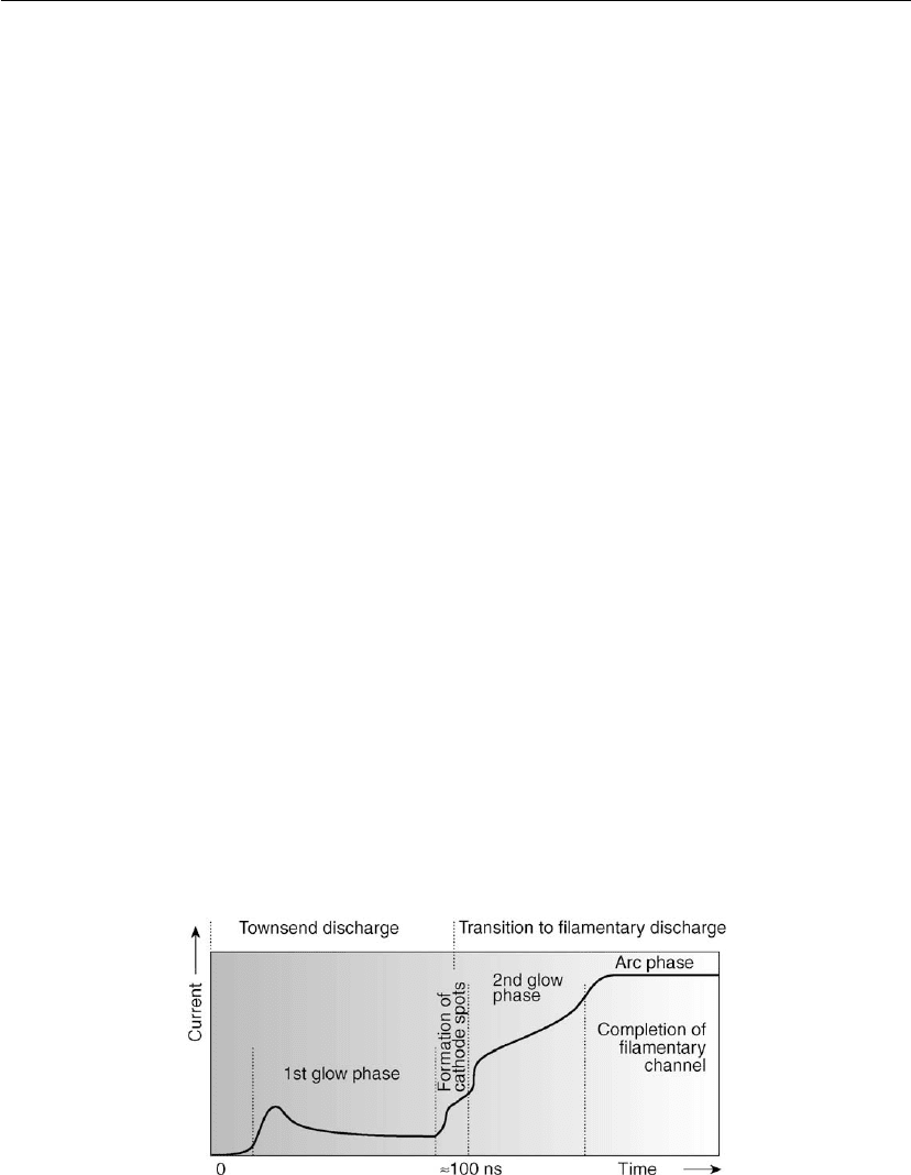
In 1970 Kekez et al. [3] studied gas breakdown processes in hydrogen and krypton in an
adjustable gap (distance d) between two parallel planar electrodes ({3/4} inch in diameter) at
different gas pressures p up to atmospheric pressure. They found that the time function of the
current flowing in the gap after applying the pulsed voltage always has several characteristic
phases. A model approach of their observations describing the individual phases is shown in
Figure 17.1. Providing that the voltage in the pulse exceeds the breakdown value the current
starts at a moderate level resembling a glow discharge where the ionization follows
Townsend’s mechanism with ionization avalanches, and the gas breakdown follows the
well-known Paschen law. During later phases (roughly after 100 ns) there is a growing
probability of forming large current filaments, or streamers, representing local arcs.
17.2.1 Electrical Breakdown of Gas at Reduced Pressures
Townsend avalanches proceed in a space-charge-free field to the point where a secondary
emission from the cathode provides sufficient feedback of electrons to maintain the stable
discharge. In a simplified description of the DC avalanche each electron emitted from the
cathode moves in an electric field toward the anode at a distance d from the cathode and
collides with the neutral gas particles forming exp (αd) new electrons and the same number of
Figure 17.1: Model approach describing individual phases of gas breakdown in hydrogen and
krypton in an adjustable gap (distance d) between two parallel planar electrodes {3/4} inch in
diameter at different gas pressures p up to the atmospheric pressure. (According to [3].)

868 Chapter 17
positive ions. The coefficient α is the so-called 1st Townsend coefficient. To fulfill the
requirement of equal numbers of opposite charged particles in the plasma, one ion must
remain in the space to compensate the initial electron. The rest of [exp (αd) − 1] ions should
return to the cathode where they can, with a probability of γ (3rd Townsend coefficient), form
γ [exp (αd) − 1] secondary electrons. When the number of secondary electrons can replace the
initial electron, we may write a simple balance equation, which actually represents the Paschen
breakdown condition, 1 = γ [exp (αd) − 1]. By multiplying both sides by gas pressure p and
after a simple mathematical operation the equation can be rewritten into the form:
pd = p/α ln (1 + 1/γ).
The DC breakdown always follows the well-known Paschen function V
b
= f (pd) [4]. The
Paschen function (curve) has a concave shape with certain minimum breakdown voltage at a
certain value of pd. For a fixed geometry (d = constant) the breakdown voltages V
b
are higher
at both low and high gas pressures. In a typical low-pressure plasma processing systems with
gas pressures in the interval 0.1 Pa ≤ p ≤ 100 Pa and distances d between electrodes of a few
centimeters the voltages required to start the plasma can be reasonably low (V
b
≤ 1 kV). It
should be noted that similar principles to those in DC breakdown are also valid for RF
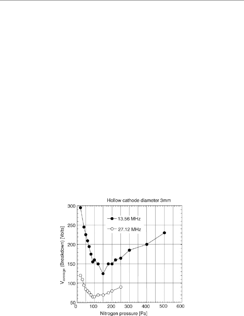
breakdown. An example of an experimental Paschen curve for breakdown in a 3 mm diameter
cylindrical hollow cathode in nitrogen at pressures between 20 and 500 Pa for two different
frequencies is shown in Figure 17.2 [5]. It is seen that the average voltage for the breakdown
can even be below 100 V.
Figure 17.2: An experimental function of the discharge ignition voltage versus the nitrogen
pressure in the radio frequency hollow cathode system at two different frequencies. (According
to [5].)

Atmospheric Pressure Plasma Sources and Processing 869
17.2.2 Electrical Breakdown of Gas at High Pressures
In contrast to low-pressure breakdown, atmospheric and higher pressure breakdown in dry air
requires more than 30 kV with a 1 cm gap between electrodes. Even when the distance is
shortened to d ≈ 1 mm it still requires about 3 kV at 1 atmosphere. The experimental DC
values in a 1 mm gap reported in [6] are 3.2 kV in air, 1.5 kV in Ar, and 0.75 kV in He.
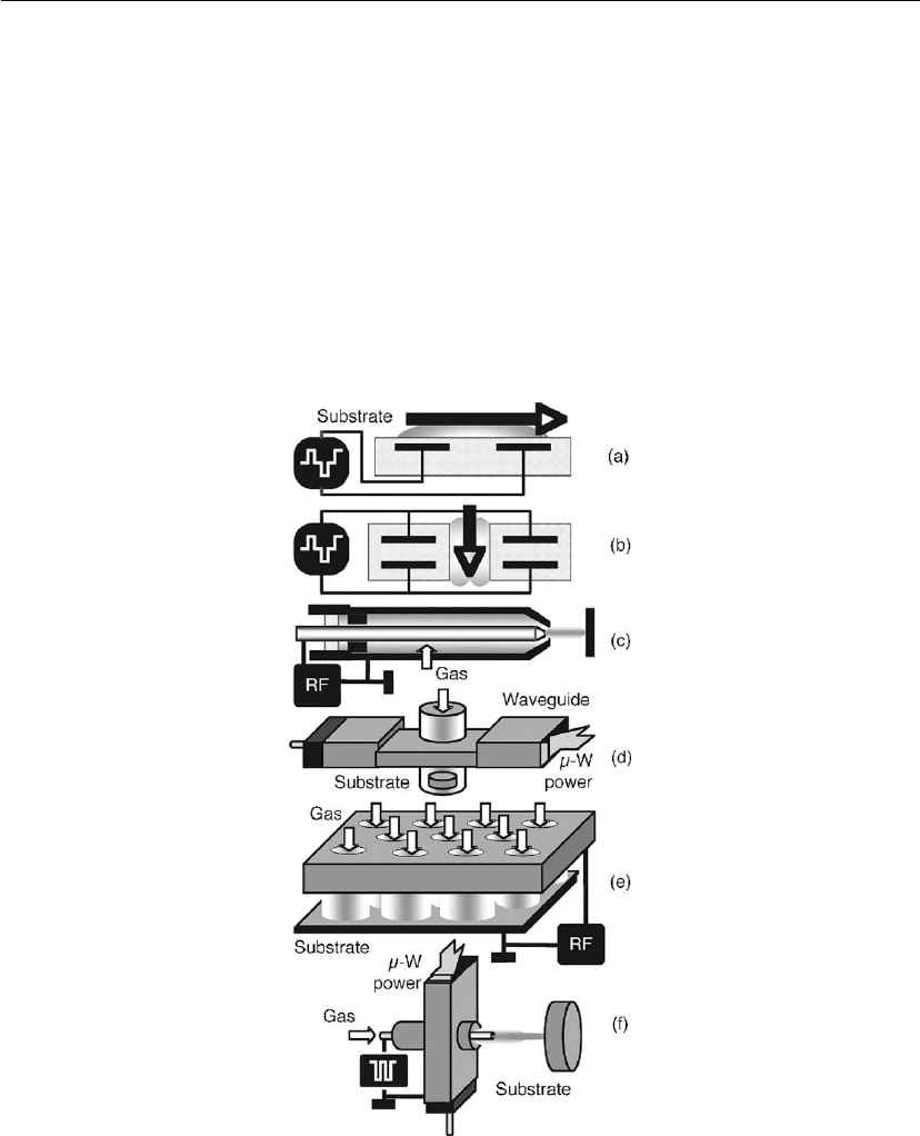
Measurements of the RF breakdown at atmospheric pressure in fused hollow cathode (FHC)
systems (see Section 17.3 and Figure 17.3e) with about a 1 mm gap between the cathode
structure and a grounded counter-electrode show peak-to-peak voltages of 1.2 kV in air, 0.4 kV
in Ar, and 0.18 kV in Ne [7]. The high voltages necessary to ignite atmospheric discharges
often lead to formation of hot filamentary arcs (streamers), as can be seen in later phases of the
Figure 17.3: Examples of cold atmospheric plasma sources: (a) Dielectric barrier discharge (DBD)
with electrodes inside a dielectric barrier; (b) DBD with parallel pairs of electrodes; (c)
radio-frequency (RF) plasma jet; (d) microwave waveguide plasma source; (e) fused hollow
cathode (FHC) plasma source; (f) hybrid hollow electrode activated discharge (H-HEAD) plasma
source.

870 Chapter 17
pulsed breakdown shown in Figure 17.1. This very common form of discharge appears more
frequently in molecular gases than in atomic gases [6]. In a realistic model of DC atmospheric
breakdown in CO
2
lasers by Palmer [8], the streamers appear mainly at large electrode
distances d when the volume ionization by a single avalanche forms the space-charge field
comparable to the total applied field. Then the secondary avalanches (caused e.g. by
photoionization) converge toward the primary avalanche and form highly conducting
filaments. This resembles a ‘micro-thunderstorm’ with a breakdown (lightning) between
clouds. A typical streamer has a diameter of about 0.2–0.4 mm in pure N
2
and about 1 mm in
air [9]. It can conduct up to several amperes of the DC current in air [13]. Both the streamer
diameter and the current depend on the applied voltage. It might be interesting to note that the
30 kA currents in a typical lightning flash in real thunderstorms with current channel diameters
of about 0.1 m (1–10 m is the light channel diameter) might be well compared with the
micro-thunderstorm in air with 3 A streamers having diameter of 1 mm. The current density in
both flashes is 1.2 kA/cm
2
. Also, shapes of the streamers often exhibit branching, similarly to
the lightning flashes, and they are quite noisy. Multiple plasma streamers are undesirable in
applications because they can cause uneven treatment and local damage to thermally sensitive
substrates, for example webs.
17.2.3 Suppression of Streamers in High-Pressure Plasmas
There are several options by which to suppress or even avoid arc streamers. For example, in a
small gap, d, the probability of space charges formation is lower, as could be understood from
Section 17.2.2, which limits the volume breakdown and formation of conductive channels with
streamers. In many atmospheric plasma systems helium is used because of its small atoms (He
has a diameter of ∼ 0.28 nm, while the air molecule has a diameter of ∼ 0.97 nm), high
diffusion coefficient D and a long mean free path compared to other gases. Helium ions are
very efficient in production of secondary electrons (high secondary electron emission
coefficient γ), which enables lower breakdown voltages.
Another option is the utilization of high-frequency power. For example, microwave power
(2.45 GHz) is often used because of its stabilizing effect in volume ionization without
the necessity for electrode-based secondary electrons. This enables the electric field to
be decreased. It has been shown experimentally that the breakdown voltage in He at
about 80 kPa (600 torr) decreases with the frequency in an interval between 5 and
30 MHz [10].
In the electrode systems powered by alternating current generators different dielectric barriers
covering the electrodes can be used to limit the current. In these dielectric barrier discharge
(DBD) systems only displacement current can flow and formation of the arc-based streamers is
naturally limited.
