Maier S.A. Plasmonics: Fundamentals and Applications. Майер С.А. Плазмоника: Теория и приложения
Подождите немного. Документ загружается.

54 Imaging Surface Plasmon Polariton Propagation
croscopy. The latter term highlights the conceptual similarity with the scan-
ning tunneling microscope (STM). In both cases, a sharp tip is brought into the
immediate vicinity of the surface under study (Fig. 4.1) using an appropriate
feedback-loop technique. Whereas a STM measures the current (induced by
an applied voltage) caused by electron tunneling between the surface and an
atomically sharp metal tip, a photon scanning tunneling microscope (PSTM)
collects photons by coupling the evanescent near field above the surface to
propagating modes inside a tapered optical fiber. The near-field optical tip
(also called the probe) is usually fabricated by pulling or etching an optical
fiber taper, and is often metalized at the end in order to suppress the coupling
of diffracted light fields. The resolution of this technique is limited by the size
of the tip’s aperture, which can reach dimensions of only 50 nm or even less
using etching (or more recently also microfabrication) techniques. In addition
to metal-coated probes, uncoated probes are also frequently used, which have
a higher collection efficiency and have been shown to image different com-
ponents of the electromagnetic field around nanostructures than probes coated
with a conductive layer [Dereux et al., 2001].
In order to study the confinement and propagation of SPPs using this scheme,
the tip has to be brought within a sufficiently close distance to the flat metal
surface so that it is immersed in the evanescent tail of the SPP field, i.e. within a
distance ˆz (calculated using (2.12)). For studies of gold or silver films at visible
frequencies, this requires a gap between the probe and the film on the order of
100 nm or less, which can be easily achieved using feedback techniques such
as non-contact mode atomic force microscopy, shear or tuning force feedback,
or by using the intensity of the collected light field itself as the feedback signal
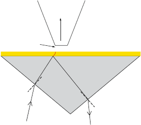
Apertured fiber probe
propagating
mode
evanescent
near-field
Figure 4.1. A typical setup for near-field optical imaging of SPP fields at a metal/air interface.
The evanescent tail of the fields penetrating into the air is coupled to propagating modes in a
tapered optical fiber tip. The SPPs can for example be excited via prism coupling (shown), a
tightly focused beam, or particle impact.
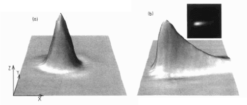
Near-Field Microscopy 55
Figure 4.2. Near-field image of a HeNe laser beam (λ = 633 nm) internally incident on an
uncoated (a) prism face and a prism face coated with a 53 nm thick silver film (b) at an angle
greater than the critical angle (scan range 40x40 μm). The exponentially decaying tail in (b)
is due to SPP propagation away from the excitation region. Reprinted with permission from
[Dawson et al., 1994]. Copyright 1994 by the American Physical Society.
(akin to the STM, where the tunneling current proportional to the amount of
collected electrons is used for this purpose).
In order not to interfere with the detection scheme, optical excitation of SPPs
takes usually place via either prism coupling (Fig. 4.1) or tightly focused beams
using an oil-immersion objective of high numerical aperture on the bottom side
of the substrate. We note that the prism coupling scheme is not suitable for
exciting SPPs of all possible propagation constants β, but only those within
the window of leaky modes as discussed in chapter 3.
The very first studies of the physical properties of SPPs using near-field op-
tical microscopy investigated the mode confinement at the interface of a thin
silver film with air. SPPs were excited in prism coupling geometry, and the
evanescent field on the air side probed via an apertured optical fiber tip. With-
out actually imaging the propagating fields using raster scanning, approaching
and retracting the fiber probe confirmed the localization and corresponding
enhancement of the electromagnetic field in the near-field region close to the
surface [Marti et al., 1993]. Monitoring of the collected signal intensity at dif-
ferent heights above the surface allowed the determination of the penetration
of the SPP fields into the air above the surface, confirming the spatial extent of
the exponentially decaying field [Adam et al., 1993].
In addition to the investigation of out-of-plane confinement, the combina-
tion of near-field collection with raster scanning techniques enables the direct
visualization of propagating SPPs. Dawson and co-workers used a PSTM to
spatially image the propagation of SPPs excited using prism coupling on a thin
silver film [Dawson et al., 1994]. Fig. 4.2b shows a three-dimensional render-
ing of the intensity collected in the near-field above the film surface. An excita-
tion wavelength λ
0
= 633 nm in the visible regime ensured good confinement
to the interface (ˆz ≈ 420 nm calculated using (2.12)). As a control experiment,
56 Imaging Surface Plasmon Polariton Propagation
Fig. 4.2a shows the evanescent field above a bare prism surface under the same
excitation conditions. Clearly, for the silver-coated prism propagation of elec-
tromagnetic energy away from the excitation spot is visible. Experiments such
as this enable the direct determination of the SPP propagation length L by fit-
ting the exponential tail starting at the SPP launching point. In this case, the
propagation length of the silver/air SPP was determined to be 13.2 μm, in good
agreement with theoretical modeling. Also, the in-plane spread of the SPP as
it propagates away from the excitation region can be monitored.
Collection-mode near-field optical microscopy has ever since these initial
investigations been extensively employed for studies of SPP propagation, most
prominently in a context of waveguiding along metal stripes, where the trans-
verse extent of the SPP is limited by the stripe width (chapter 7). This has
enabled the determination of the trade-off between propagation length and
out-of-plane as well as lateral confinement, and additionally investigations of
functional waveguide devices such as reflectors or Bragg mirrors. For exam-
ple, near-field imaging allowed the direct visualization of interference patterns
between co- and counterpropagating SPP waves. Some of these studies will be
presented in chapter 7 on plasmon waveguides.
Near-field probing has also proved very useful for the assessment of scat-
tering losses on structured metal surfaces [Bouhelier et al., 2001] as well as
for the determination of the dispersion properties of SPPs at curved surfaces
[Passian et al., 2004]. It has to be noted that the presence of the probing tip can
influence the dispersion, but for dielectric tips this effect can often be neglected
[Passian et al., 2005].
As might be expected, near-field optical microscopy is also often the method
of choice for studies of localized surface plasmons in metal nanoparticles or
ensembles of metal nanostructures (chapter 5). In these experiments, the light
path is usually reversed: By not collecting but illuminating the metal structure
under study via light emanating through the sub-wavelength aperture of a fiber
tip, near-field optical spectroscopy of the localized modes is possible, in addi-
tion to imaging of the spatial field distribution. Examples will be presented in
chapter 10 on spectroscopy and sensing.
In this illumination mode, the fiber probe effectively acts as a local dipolar
source for the excitation of surface plasmons (or propagating SPPs as described
in the previous chapter). Information about the electromagnetic structure of
the surface can be extracted from the transmitted or reflected light collected
using an objective in the far field. Apart from photon collection in the far field,
the metal film structure under investigation can also be directly mounted on
the photodiode itself, as shown by Dragnea and co-workers , which used this
geometry for the study of SPP propagation in sub-wavelength slits on a flat
metal film [Dragnea et al., 2003].
Fluorescence Imaging 57
4.2 Fluorescence Imaging
Instead of locally collecting the optical near field of SPPs using the aper-
tured fiber tip of a near-field optical microscope, emitters such as quantum
dots or fluorescent molecules can be directly placed into the evanescent tail of
the SPP field. If the frequency of the propagating SPPs lies within the broad
spectral absorption band of the emitters, their excitation via SPPs is possible,
and the intensity of the emitted fluorescence radiation is proportional to the
intensity of the local field at the position of the emitters. Therefore, SPP prop-
agation on a metal/air interface can be mapped by coating the surface with a
dielectric film doped with emitters. If the layer is sufficiently thin and of low
refractive index (e.g. quantum dots embedded in a polymer, or monolayers of
fluorescent molecules), the alteration of the SPP dispersion due to the covering
layer is small.
As will be discussed in more detail in chapter 9, fluorescent molecules
placed into the near field of propagating SPPs (and also that of localized plas-
mons) show an enhancement of their fluorescence yield if care is taken to coun-
teract non-radiative quenching. This can be achieved by inserting a thin spacer
layer on the order of a few nanometers between the metal film sustaining the
SPPs and the fluorescent molecules to inhibit non-radiative energy transfer.
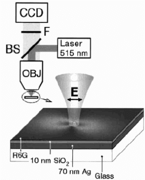
Ditlbacher and co-workers used this concept for the imaging of SPPs excited
on a 70 nm thin silver film by focusing a laser beam (λ
0
= 514 nm, P =
Figure 4.3. Fluorescence imaging of SPP fields. A SPP on a 70 nm silver film is excited via
illumination of a nanoparticle (phase-matching via a defect) using a 100× objective, and the
field distribution of the mode imaged by detecting the fluorescent emission of a coating layer
doped with Rhodamin 6G. Reprinted with permission from [Ditlbacher et al., 2002a]. Copyright
2002, American Institute of Physics.

58 Imaging Surface Plasmon Polariton Propagation
5 mW) on wire or nanoparticle surface defects created using electron beam
lithography (Fig. 4.3) [Ditlbacher et al., 2002a]. The metal film was coated
with a sub-monolayer of Rhodamine 6G molecules to enable the determination
of the spatial structure of the SPP fields. In order to reduce quenching due to
intermolecular interactions and non-radiative transitions to the metal film, the
molecular density was chosen to be sufficiently small and a 10 nm thin SiO
2
spacer layer inserted between the molecular film and the silver substrate. CCD
images of the fluorescence signal collected via a dichroic mirror are shown in
Fig. 4.4. The intensity distribution correlates well with the pathways expected
for SPPs excited via surface defects (compare with [Hecht et al., 1996] and
Fig. 3.13 of chapter 3).
Using this scheme, information about the lateral spatial confinement, the
propagation distance and interference effects can be extracted in analogy to the
direct probing of the near field using an apertured probe, albeit with a resolu-
tion of at best the diffraction limit. However, the effect of bleaching in regions
of high field intensity has to be carefully taken into account for quantitative
analysis.
Figure 4.4. Fluorescence images of the intensity distribution of SPPs excited by illumination
of (a) a silver nanoparticle (diameter 200 nm, height 60 nm), and (b) a silver nanowire (width
200 nm, height 60 nm, length 20 μm). The particles are situated on a continuous silver film
supporting SPPs. Reprinted with permission from [Ditlbacher et al., 2002a]. Copyright 2002,
American Institute of Physics.
Leakage Radiation 59
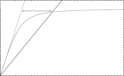
4.3 Leakage Radiation
The dispersion curve of SPPs excited at the air interface of a metal film
lies outside the light cone defined by k = n
air
ω/c, and the mode does not
suffer radiation loss into the air region (for a perfectly flat interface neglecting
roughness). However, energy can be lost into a supporting substrate of higher
index n
s
. This radiation loss occurs at all points of the dispersion curve that
lie to the left of the light line of the substrate k
s
= n
s
ω/c, as indicated in
Figure 4.5. Therefore, for SPP excited in the region of propagation constants
β defined by
k
0
<β<k
0
n
s
, (4.1)
leakage radiation into the substrate provides a second loss channel in addition
to the inherent absorptive losses.
We have seen in the preceding chapter that leaky SPPs are inherently excited
using prism coupling, and that the leakage radiation into the prism interferes
with the directly reflected beam. As pointed out, a zero reflection ensues only
under the condition of critical coupling (see (3.1)), when the absorptive losses
exactly equal the radiative losses, and all power is absorbed in the metal film.
This is only achieved for a critical thickness of the metal film.
Apart from monitoring the efficiency of prism coupling, leakage radiation
collection can be used for investigating SPPs excited by other means, such as
tightly focused beams or gratings, as long as the excited wave vectors β lie
within the substrate light cone, fulfilling (4.1).
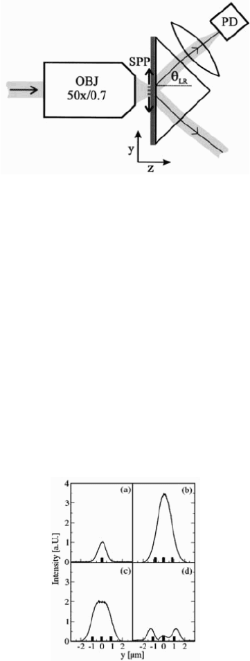
A typical setup for the collection of leakage radiation is shown in Fig. 4.6
[Ditlbacher et al., 2003]. In this study, the intensity of the leakage radiation
Wave vector
Frequency
substrate
air
Region of
Leakage radiation
metal/air SPP
Figure 4.5. Generic dispersion relation of a SPP at a metal/air interface. In the region enclosed
by the light lines of air and of the higher index substrate, the propagating SPPs lose energy via
leakage radiation into the substrate light cone, which can be collected for imaging purposes.
60 Imaging Surface Plasmon Polariton Propagation
Figure 4.6. Experimental setup for leakage radiation imaging of SPP fields. Here, SPPs are
excited using grating coupling, and the ensuing leakage radiation into the underlying prism col-
lected using a photodiode. Reprinted with permission from [Ditlbacher et al., 2003]. Copyright
2003, American Institute of Physics.
was used to quantify the coupling efficiency of light to SPPs via a grating-like
excitation scheme with a variable number of ridges spaced by a lattice constant
. We note that in this collection geometry, only half of the ensuing leakage
radiation is collected via the underlying prism. With this technique, spatial
intensity profiles can be obtained by varying the position of the sample with
respect to the exciting laser beam. The amount of leakage radiation collected
for films with one (a) and three coupling ridges of different lattice constants (b-
d) is shown in Fig. 4.7. A maximum light-SPP coupling efficiency of 15% was
achieved for a three-ridge sample of appropriate lattice constant. Naturally,
Figure 4.7. Quantifying coupling efficiency of a finite metal grating via collection of leakage
radiation. The graphs show the experimentally observed distribution of leakage radiation vs.
sample position (Fig. 4.6) for a single-ridge sample (a) and samples with three ridges of different
lattice constants (b-d). The maximum intensity profile in (a) was normalized to 1. Reprinted
with permission from [Ditlbacher et al., 2003]. Copyright 2003, American Institute of Physics.
Leakage Radiation 61
the same setup can also be used to quantify the coupling efficiency of other
methods, such as highly focused beams or coupling via inherent or designed
surface roughness (chapter 3).
Leakage radiation also has to be considered in the design of plasmon
waveguides. For example, all studies of laterally confined SPP propagation
in metal stripes or nanowires where prism-coupling excitation has been em-
ployed inherently only investigate modes in the leaky region (4.1) described
above. These leaky waveguides will be discussed in detail in chapter 7.
Apart from the observation of SPP propagation, leakage radiation imag-
ing can also be used for the direct visualization of the SPP dispersion rela-
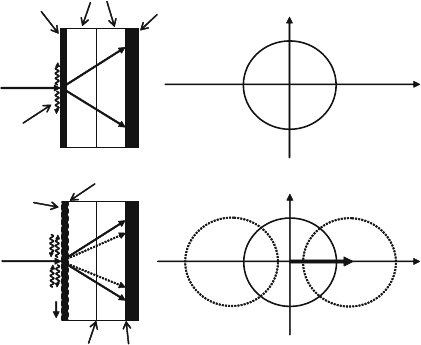
tion, which was demonstrated by Giannattasio and Barnes [Giannattasio and
Barnes, 2005]. In this work, SPPs at the air interface of a 50 nm thick silver
film were excited via a focused light beam using scattering from random sur-
face roughness for phase-matching (Fig. 4.8). Leakage radiation into the silica
substrate was directly imaged using a CCD camera glued to the underside of
the substrate. For a flat film (Fig. 4.8a), the radiation is emitted within a cone
of surface plasmon angle θ
SPP
defined by n
s
k
0
sin θ
SPP
= β, which intersects
the plane of the CCD in a circular pattern. Light of different frequencies can
be used for excitation, and the resulting wave vector β within the region (4.1)
determined by the computation of the angle θ
SPP
of leakage radiation from the
k
k+G
k
silver film
silica
substrates
matching fluid
CCD
k
x
k
x
k
y
k
y
k+G
k
k-G
G
G
laser
SPPs
k
-
G
silver film
grating
a)
b)
laser
matching glue
Figure 4.8. Experimental geometry of leakage radiation imaging for the determination of the
SPP dispersion relation. (a) Planar silver surface: a single cone of light is emitted into the silica
substrate. (b) Corrugated silver surface: the central cone is now intersected by other light cones
due to SPPs scattered by the grating with Bragg vector G. Reproduced with permission from
[Giannattasio and Barnes, 2005]. Copyright 2005, Optical Society of America.
62 Imaging Surface Plasmon Polariton Propagation
a)
b)
Scattered Ag/air SPP
Scattered Ag/glass SPP
Stop band
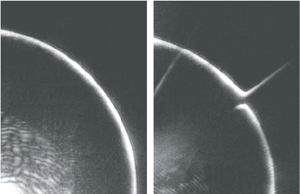
Figure 4.9. Direct image of the conical radiation in k-space sketched in Fig. 4.8 recorded by
a CCD array. (a) Planar sample. (b) Imaging of the stop band emerging at the intersection
between the two cones in k-space. Reproduced with permission from [Giannattasio and Barnes,
2005]. Copyright 2005, Optical Society of America.
radius of the imaged circle. Fig. 4.9a shows an image of parts of the circumfer-
ence of the cone obtained by collecting the leakage radiation, confirming the
usefulness of this method for the determination of the SPP dispersion relation.
This scheme allows a convenient way for the determination of the more
complex dispersion relation of a structured metal surface. For a surface with
regular, one-dimensional corrugations with grating constant a (corresponding
to a reciprocal grating vector G = 2π/a), perpendicular incidence of the excit-
ing laser light leads to leakage radiation into a central light cone (correspond-
ing to zero-order scattering) intersected by other cones ensuing from scatter-
ing SPPs with wave vectors k ± G (Fig. 4.8b). This leads to the formation of
band gaps for SPP propagation at the intersection of adjacent cones, which are
clearly visible in Fig. 4.9b as disruptions of the central circle. Additionally,
scattering pathways both into the air and substrate layers are visible in these
images, in the form of straight, jet-like lines.
4.4 Scattered Light Imaging
The propagation of SPPs at the air interface of metal films can often be sim-
ply imaged by collecting the light lost to radiation due to scattering at random
(or indeed designed) surface protrusions. Scattering at these localized bumps
allows SPPs with wave vector β>k
0
to acquire a momentum component
k
x
, which can lower β into the region within the air light cone (see equation
(3.3)), leading to coupling to the radiation continuum and thus the emission of
photons. For increasingly flat films with good surface quality, the amount of
scattering is reduced, making a detailed determination of the properties of the
SPPs such as their propagation length difficult.
Scattered Light Imaging 63
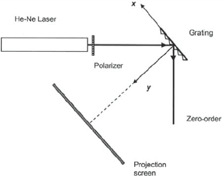
Figure 4.10. Experimental setup for the observation of the diffuse scattering background.
Reprinted with permission from [Depine and Ledesma, 2004]. Copyright 2004, Optical So-
ciety of America.
The observation of light scattering from random roughness can also be used
to map out the dispersion relation of SPPs on modulated surfaces. Depine and
Ledesma used this method to determine the band gaps of SPPs for a metal
surface corrugated with a blazed grating, by observing the so-called diffuse
light bands [Depine and Ledesma, 2004]. These arise due to scattering from
the random roughness of the grating. The experimental setup is very simple
and shown in Fig. 4.10. A SPP is excited by focusing a laser beam under
an angle θ to the surface normal onto the grating, and the scattered light is
projected onto a screen parallel to the substrate.
It has been shown that a blazed grating leads to polarization conversion of
the incoming and reflected light beam, mediated via SPPs, even when β is
completely parallel to the grooves of the grating [Watts and Sambles, 1997]. A
map of the reciprocal space (i.e., a two-dimensional plot of the in-plane com-
ponents of β ) is obtained by recording the intensity of the specular reflection
versus incidence angle θ and the angle φ between β and the Bragg vector of
the grating.
Depine and Ledesma have shown that the observation of the diffuse back-
ground does not necessitate such angular scanning in φ, which is now provided
by scattering at the inherent surface roughness.
The obtained intensity maps of the in-plane components of β are presented
in Fig. 4.11 both for light incident under TM (a) and TE (b) polarization. The
observed structure corresponds well to a calculation of the reciprocal space of
the electromagnetic modes sustained by this system, and to an experimental
determination using angular scanning [Watts and Sambles, 1997]. In these