Liu A.L., Tien H.T. Advances in Planar Lipid Bilayer and Liposomes. V.6
Подождите немного. Документ загружается.

emphasis on the potential of DSC to analyze the energetics of protein–lipid
association/insertion reactions.
Phospholipids can exist in solvent in a ‘gel-like’ (ordered) as well as ‘fluid-like’
(disordered) phase. The change from a gel to fluid-like phase of a solvated membrane
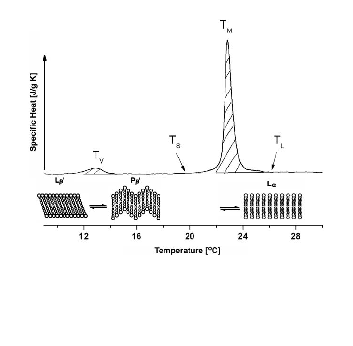
is called its phase transition or (melting point). As shown in Fig. 7, this change in
specific heat profile has a calorimetric maximum. Before reaching the main phase
transition temperature (T
M
), some lipids show a pre-transition phase (T
V
) at which
the ‘gel-like’ membrane changes from a lamellar (L
b
0
) to a ripple (P
b
0
) phase and
then proceeds into the fluid phase (L
a
).
The saturated covalent bonds in the alkyl chains of lipids can assume many
torsion angles. The flexibility of these covalent bonds provides many degrees of
freedom. High-energy conformations reduce significantly the all-trans configura-
tions and allow any angle of rotation. The phase change from ordered to disordered
behavior (melting) is regarded as first-order phase transition that follows Gibb’s law:
DG ¼ DH T
M
DS ¼ 0; or T
M
¼ DH=DS. (5)
where, DS ¼ entropy, DH ¼ enthalpy and DG ¼ free (Gibb’s) energy. Using an
experimentally determined heat capacity (C
p
), it allows the determination of the
phase change enthalpy,
DH ¼
Z
fluid
gel
C
p
DT; DS ¼ DH=T
M
. (6)
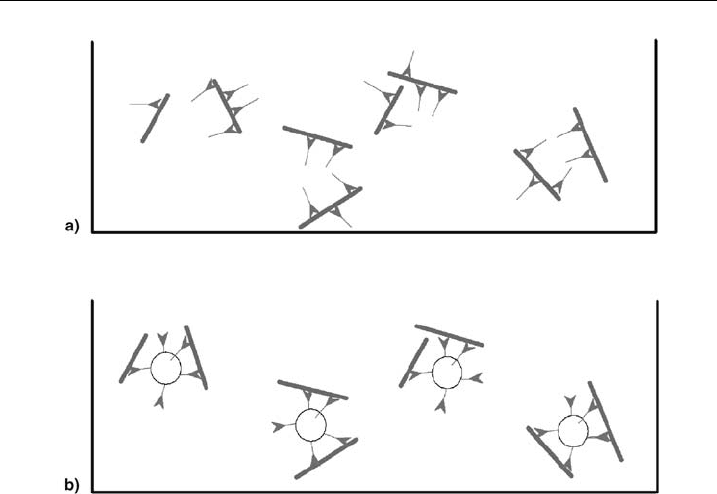
Figure 6 Schematic representation of protein^lipid interaction. Schematic view of (a) myosin II
(green) and putative binding to actin (red) and (b) myosin II (bound to lipid vesicles) and actin
(please see plate no. 5 in the color section).
W.H. GOLDMANN et al.234
From a kinetic view, the melting point is the state where the gel and fluid phase
are in equilibrium,
K 1 ¼
P
fluid
ðT
M
Þ
P
gel
ðT
M
Þ
(7)
The equilibrium constant, K is determined by the relationship,
K ¼ e
DG=RT
¼ e
ðDHTDSÞ=RT
(8)
where K ¼ 1atDG ¼ 0. Since the heat capacity is at a maximum at the phase
transition (melting) point, the enthalpy fluctuation is therefore also at a maximum.
Enthalpy also fluctuates with the surface area, i.e., ‘fluid-like’ lipids 4 ‘gel-like’
lipids and the size, i.e., the volume of lipid molecules is assumed to be constant. The
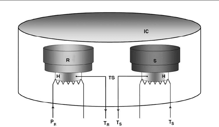
principle of the DSC apparatus is shown in Fig. 8.
The heating of the sample and reference solution is performed at a preset
heating rate, b ¼ DT/Dt, where the temperature of the system is determined by
T ¼ T
0
+bxt;(T
0
is the temperature at t ¼ 0). The principle of DSC requires the
temperature of the sample (probe, T
P
) and reference (T
R
) solution to remain
constant, i.e., T ¼ T
P
¼ T
R
. At an endothermic phase transition of the sample
solution, this has to be heated to a higher degree compared to the sample solution
to keep both temperatures at the same level. The heat output for the sample (P
P
)
will be larger than for the reference (P
R
), therefore the difference of the heat
Figure 7 Di¡erential scanning calorimetry. Heat capacity curves and t hermotropic phase
transitions of DMPC/DMPG vesicles.
Cytoskeletal Proteins at the Lipid Membrane 235
output, DP equals P
P
–P
R
. This is reflected in the heat capacities DC(T) between
sample and reference which is proportional to
DCðT Þ¼C
p
C
R
¼ DPðT Þ=b. (9)
In DSC thermograms, the difference of heat output DP is plotted against the
temperature T. At the known heat rate, b the heat capacity difference DC(T)
between the sample and reference solution as well as the partial dissipation of energy
in molar heat capacity can be determined. Figure 7 shows an example of a
thermogram for a DMPC and DMPG vesicle solution: pre-transition (T
V
)at131C
and main transition (T
M
)at231C. Integrating between the start (T
S
) and endpoint
(T
L
) of the DSC temperature signals determine the change in enthalpy
Z
T
S
T
L
C
U
DT ¼ D
U
H (10)
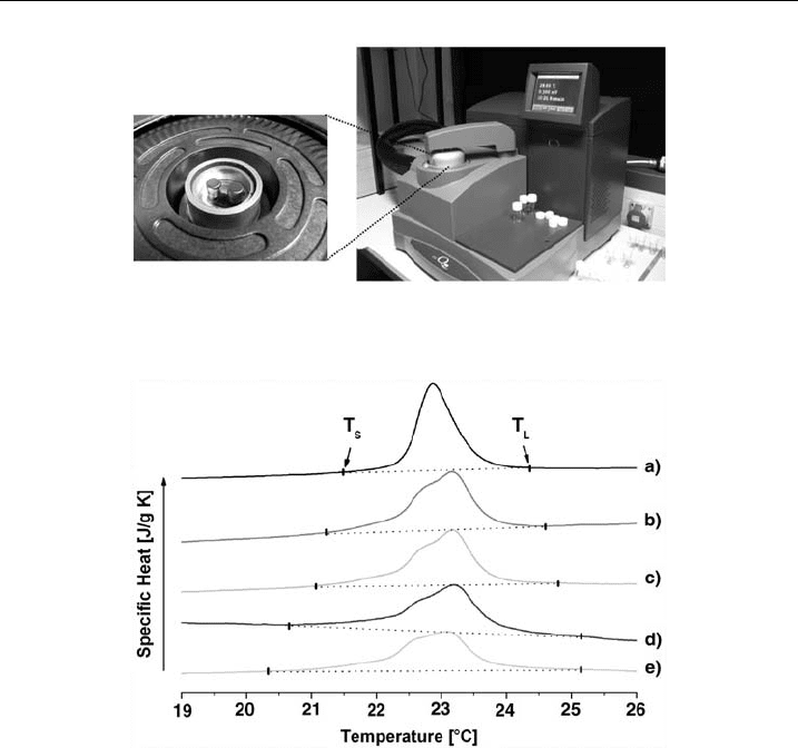
A differential scanning calorimeter Q100 from TA Instruments (Fig. 9) was used
and the reservoirs for the sample and reference solution are made of stainless steel
and to hold a volume of 100 ml each. Lipid-buffer solutions were placed in the
reference cell and the lipid-myosin-II-buffer solutions in the sample cell. Under
sealed conditions, both solutions were heated/cooled at a rate, b at 0.51C/min
between +71C and +351C in six cycles until the equilibrium of the phase transition
enthalpy was reached, using a mixture of DMPC and DMPG at a molar ratio 50:50.
A phase transition was observed at 231C. Data analysis was performed using the
software from Universal Analysis 2000 (TA Instruments) and Origin 7G.
Figure 8 Schematic representation of the DSC apparatus. S, sample cell; R, reference cell; H,
heating coil; IC, insulating casing;TS, temperature sensor;T
S
and T
R
are the currently measured
temperatures in sa mple- and reference cell and (P
R
;left)and(T
S
¼ P
S
; right) are the heat output
for the reference and sample cell.
W.H. GOLDMANN et al.236
3.1. Results
Myosin II insertion into phospholipid membranes was demonstrated using calor-
imetric measurements. The measurements were performed with multilamellar ves-
icles (MLVs) at 10 mg/ml consisting of DMPC/DMPG at a molar ratio of 50:50.
Using increasing myosin concentrations, the changes in main phase transition were
recorded (P
b
0
2 L
a
) as shown in Fig. 7. Adding increasing myosin II concentration
(traces b-e; 0.62-6.24 mM) to the lipid solution (a; no myosin II), a widening and
flattening of the peak curvature was observed (Fig. 10). The start (T
S
) and endpoint
(T
L
) of the phase transition are indicated by the arrows. The relative widening
calculated from the relation, ðDT
1=2
DT
0
1=2
Þ=DT
0
1=2
is shown in Fig. 11. For a
better comparison of the changes induced by the various myosin II concentrations,
the enthalpy changes, DH, were normalized to pure lipids, against DH
0
(Table 1).
Plotting the enthalpy changes DH/DH
0
against the molar ratios of myosin II and
Figure 9 The calorimeter. Image of a calorimeter fromTA Instruments (right) and sample and
reference cell (arrows, left).
Figure 10 T hermograms. DSC thermogram of DMPC/DMPG wit hout (a) myosin II and with
myosin II (b-e). Conditions: Lipid and myosin II co ncent ration: 14.62 mM, and (b) 0.62 mM,
(c) 1.45 mM, (d) 3 .12 mM and (e) 6.24 mM, respectively.
Cytoskeletal Proteins at the Lipid Membrane 237
lipids, an initial linear relationship followed by a saturation behavior of the lipid
vesicles for myosin II was observed. The control protein BSA showed no changes
(Fig. 12). Thermodynamic measurements (DSC) proved sufficient to determine the
insertion behavior of myosin II into lipid membranes composed of DMPG/DMPC
in vitro and the light scatter (stopped-flow) method confirmed these findings. The
binding affinity of myosin II associated with and without lipids and actin was of a
similar order of magnitude, confirming the previous observations of other mem-
brane-interacting proteins [16].
4. Solid-State NMR Spectroscopy
Among the biophysical techniques that allow the investigation of peptides and
proteins in bilayer environments solid-state NMR spectroscopy has proven to be a
valuable tool. Recently, magic angle sample spinning solid-state NMR has resulted
in the first NMR structures in the solid state of proteins in a microcrystalline
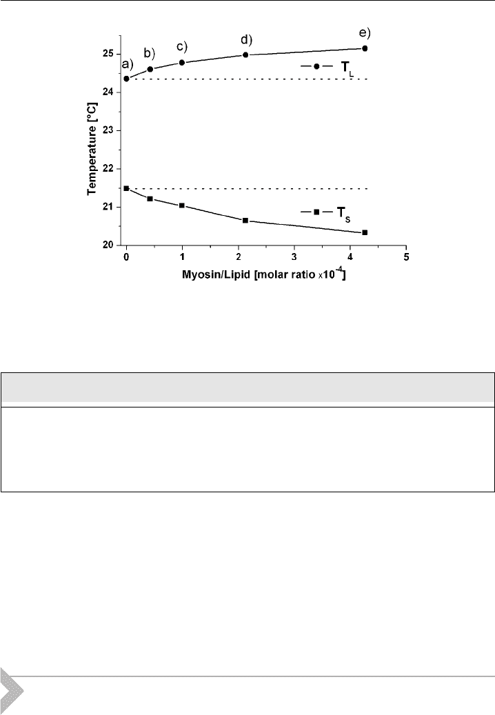
Figure 11 Phase transitions. A plot of T
S
(solidus poin ts) and T
L
(liquidus points) taken from
Fig. 10 as a function of myosin II^lipid molar ratio.
Table 1 Normalizing myosin II concentration against constant lipid concentration and
enthalpy changes DH against DH
0
(lipids only).
Lipid Myosin (mM) Myosin/Lipid DH/DH
0
Trace
10 mg/mlffi14.62 mM 0 0 1 a
0.62 1/23580 0.91251 b
1.45 1/10080 0.86383 c
3.12 1/4690 0.74985 d
6.24 1/2345 0.70055 e
W.H. GOLDMANN et al.238

environment [22] or when exhibiting a highly ordered microenvironment [23].
Furthermore, the technique makes accessible the structure, dynamics and topology
of membrane-associated polypeptides (reviewed, e.g., in Refs. [24–27]). Using
static-oriented samples, the tilt angles of helices with respect to the bilayer normal
have been determined [28], and by measuring a large number of conformational
constraints this approach has also been shown to be suitable for the complete
structure determination of membrane-bound peptides [23,29]. In this chapter, we
demonstrate how the orientation-dependence of NMR interactions is used to
extract angular constraints from such static-oriented samples.
Proton-decoupled
15
N solid-state NMR spectroscopy of peptides labeled at the
backbone amides with
15
N has been proven particularly convenient as this method
provides the approximate tilt angle of membrane-associated helices in a direct
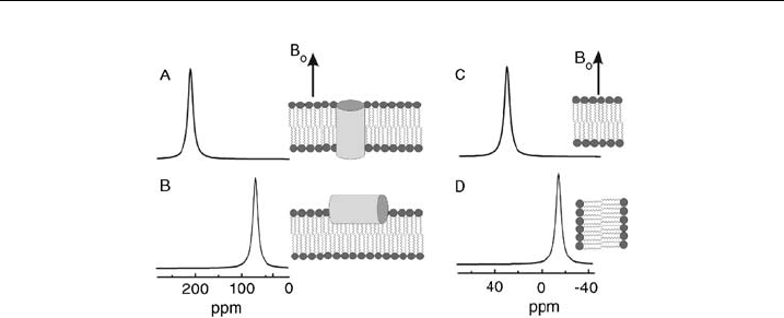
manner [27,28]. Whereas transmembrane helical peptides exhibit
15
N chemical
shifts around 200 ppm, sequences oriented parallel to the surface resonate at fre-
quencies o100 ppm (Figs. 13A and B).
In a similar manner the deuterium quadrupole splitting of the alanine –C
2
H
3
groups is dependent on the alignment of the polypeptide relative to the membrane
normal [30]. The technique has been used to study the membrane-channel
domains of the viral proteins Vpu [31] and M2 [32,33] also in the presence of the
channel blocker amantadine [34]. Furthermore antibiotic peptides have been stud-
ied in some detail using oriented solid-state NMR spectroscopy, including prote-
grin 1 [35], pardaxin [36], peptaibols [37–39], melittin [40] or magainins [41].
A family of designed histidine-containing antimicrobial peptides, which is also
efficient during the transfection of nucleic acids into cells [42], exhibits trans-
membrane alignments at neutral pH but reorients to the membrane surface at acidic
conditions [43]. By using solid-state NMR spectroscopy it was possible to show
that the peptide exhibits its most pronounced antimicrobial properties when ori-
ented parallel to the membrane surface [44] suggesting that the detergent-like
properties of amphipathic peptides are essential for membrane permeation [45].
Figure 12 DSC plots. A plot of the changes in enthalpy DH/ DH
0
against myosin II^lipid molar
ratio. Bovine serum albumine (BSA) was used as a control protein.
Cytoskeletal Proteins at the Lipid Membrane 239
4.1. Theory: The Anisotropy of Interactions Measured in Solid-State
NMR Spectroscopy
The nuclear interactions with the magnetic field are inherently anisotropic and,
therefore, dependent on the orientation and conformation of the molecule
with respect to the magnetic field direction [46–49]. Whereas in solution fast
molecular tumbling ensures isotropic averaging of the nuclear interactions, the re-
orientational correlation times of molecules that are associated with extended
phospholipid bilayers are slow. Therefore, the anisotropic properties of these in-
teractions are reflected in the NMR spectra of membrane-bound peptides or lipids.
The anisotropic chemical shift interaction is mathematically expressed by second
rank tensors, which in the principal axis system is described by three orthogonal
components s
11
, s
22
and s
33
(for a more detailed explanation see Ref. [28]). This
tensor can be transformed into other coordinate systems by successive rotations.
The component of the chemical shift tensor in direction of the magnetic field
direction (z-direction) corresponds to the measured NMR chemical shift value.
When expressed in terms of the Euler angles (Y and F) and the principal elements
of the chemical shift tensor s
11
, s
22
and s
33
, the measureable s
zz
amounts to
s
ZZ
¼ s
11
sin
2
Ycos
2
F þ s
22
sin
2
Ysin
2
F þ s
33
cos
2
Y (11)
Whereas the static
15
N chemical shift tensor of the amide bond exhibits s
22
and s
11
values in the 85 ppm and 65 ppm range, respectively, its s
33
component is char-
acterized by a much different value of approximately 230 ppm [50–54].Ina-helical
peptides the NH vector and the s
33
component cover an angle of about 181 and
both are oriented within a few degrees of the helix long axis. Due to the unique
size of s
33
and its orientation almost parallel to the helix axis it is possible to
measure an approximate alignment of the helix within oriented phospholipid
bilayers merely by recording the
15
N chemical shift interaction. Therefore, in
Figure 13 Simulated solid- state NMR spectra. (A) and (B) show simulated
15
N solid-state
NMR spectra of an a-helical polypeptide oriented with the helix long axis perpendicular (A)
or parallel (B) relative to the bilayer surface. The memb ranes are aligned with their normal
parallel to the magnetic ¢eld of the NMR spectrometer (B
0
). (C) and (D) show
31
P solid-state
NMR spectra of liquid crystalline phosphatidylcholine membranes oriented with the lipid long
axes parallel (C) or perpend icular (D) to B
0
.
W.H. GOLDMANN et al.240

samples uniaxially oriented with the membrane normal parallel to the magnetic
field transmembrane a-helical peptides exhibit
15
N resonances 4200 ppm
(Fig. 13A). In contrast, they resonate in the s
11
–s
22
range (i.e.,o100 ppm) when
aligned parallel to the membrane surface (Fig. 13B). To arrive at a detailed structural
analysis of solid-state NMR spectra from oriented samples, motional averaging
and its effects on the chemical shift anisotropy have to be taken into consider-
ation, however, the above analysis suffices for a semi-quantitative first analysis of
polypeptide–membrane interactions.
Furthermore the deuterium spectra of methyl group labeled alanines in oriented
membrane polypeptide sample have been analyzed. The methyl group of alanine
exhibits fast rotational motions around the C
a
–C
b
bond. As a result the
2
H tensor
is axially symmetric with respect to the C
a
–C
b
bond vector, and the measured
splitting Dn
Q
is directly related to the orientation of the C
a
C
b
bond:
Dn
Q
¼
3
2
e
2
qQ
h
ð3cos
2
Y 1Þ
2
(12)
where Y is the angle between C
a
–C
b
bond and the magnetic field direction and
e
2
qQ=h the static quadrupolar coupling constant [55].AsC
a
is an integral part of
the polypeptide backbone, the orientation of the C
a
–C
b
bond also reflects the
overall alignment of the peptide.
Due to fast axial rotation of the phospholipids around their long axis the
31
P
chemical shift is characterized by an averaged symmetric tensor. The singular axis
(s
||
) coincides with the rotational axis, i.e., the bilayer normal. In the
31
P solid-
state NMR spectra of pure liquid crystalline phosphatidylcholine bilayers the signal
at 30 ppm is thus indicative of phosphatidylcholine molecules with their long axis
oriented parallel to the magnetic field direction (Fig. 13C), whereas a –15 ppm
31
P
chemical shift is obtained for perpendicular alignments (Fig. 13D). In perfectly
aligned samples the phospholipid bilayer spectra consists of a single line. Intensities
to the right of this peak can arise from phospholipids with molecular orientations
deviating from parallel to the magnetic field direction. In addition, signals in this
region (o30 ppm) can be due to local conformational changes of the phospholipid
head group, for example due to electrostatic interactions of the (–HPO
4
–CH
2
–
CH
2
–N
+
(CH
3
)
3
) dipoles of the phosphocholine head group, hydrogen bonding
and/or electric dipole–dipole interactions [56,57]. We routinely record
31
P NMR
spectra of phospholipid bilayers also of the peptide carrying samples to test for the
quality of order and alignment of phospholipid bilayers.
4.2. Experimental Considerations
The peptides investigated by solid-state NMR investigations can be made available
either by biochemical overexpression or by chemical solid-phase peptide synthesis.
Whereas the former technique is well suited for uniform or selective labeling
schemes, the chemical approach allows for specific labeling of one or a few amino
acid residues. For example the talin peptide H17 with the sequence GEQI
AQ-
LIAGYIDIILKKKKSK-amide was prepared using automatic solid-phase peptide
synthesis. At the underlined positions the
15
N-labeled analogue of alanine was
Cytoskeletal Proteins at the Lipid Membrane 241
incorporated. The peptide synthetic products are commonly analyzed and purified
using reversed phase high-performance liquid chromatography and their identity
confirmed by mass spectrometry.
Typically, 10–15 mg of the polypeptide is reconstituted into about 100–200 mg
of phospholipid by co-dissolving the compounds in organic solvents or organic
solvent–water mixtures. For sample preparations encompassing the talin peptide
hexafluoroisopropanol has proven a good choice. On the other hand, the dena-
turation of larger proteins should be avoided by the usage of aqueous buffers during
the reconstitution process. Typically the mixtures are dried onto 30 ultra-thin cover
glasses (9 22 mm), where applicable, the organic solvents completely removed and
the samples equilibrated at 93% relative humidity. The glass plates are then stacked
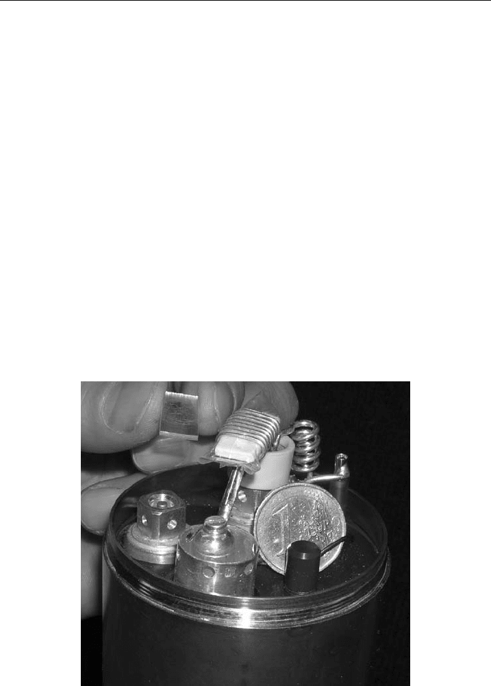
on top of each other, which results in small brick-shaped samples of 3–4 mm
thickness (Fig. 14). These are stabilized and sealed with teflon tape and plastic
wrappings. To ensure an optimal filling factor special NMR coils have been de-
veloped and tested for these samples [58]. These are flattened in such a manner to
reduce the empty space within the coil (Fig. 14). Considerable improvements in
signal-to-noise ratio can be achieved by this modification when compared to
standard commercial solid-state NMR coils [58]. The membrane normal is aligned
parallel to the magnetic field direction but alternative sample alignments have also
been investigated, e.g., when the dynamic properties of the membrane-associated
peptide are of interest [33,40,59]. Cross-polarization or Hahn echo NMR pulse
Figure 14 Solid-state NMR probe. The coils geometry has been adapted to the sample
geometry. A stack of glass plates wit h several thousand lipid bilayers in between each pair is
shown to the top left. The samples are protected and sealed before insertion into the £attened
coil of the NMR probe. Before acquisition the NMR probe is introduced into the NMR
magnet with the normal of the glass plates being oriented parallel to the magnetic ¢eld
direction (Reproduced with permission).
W.H. GOLDMANN et al.242

sequences are typically used to acquire
15
N,
2
H and
31
P NMR spectra with the
details given in previous publications, for example in Ref. [59,60].
4.3. Results and Discussion
Previous studies indicate the H17 exhibits membrane association predominantly
driven by hydrophobic interactions and with partitioning constants in the 10
4
M
1
range thereby being comparable to that of posttranslationally attached membrane
anchors [61]. During the transfer from the aqueous to the membrane-associated
state H17 undergoes a conformational transition from random coil to 86% a -helix
[61]. The proton-decoupled
15
N solid-state NMR spectrum of the talin peptide
H17 labeled with
15
N at the alanine-5 position exhibits a chemical shift of 80 ppm
(Fig. 15). This value is indicative of an alignment of the peptide helix at the labeled
site approximately parallel to the membrane surface [28]. The range of
15
N chem-
ical shift values that is obtained from a-helices oriented at tilt angles of 601,701,801
or 901 (perfect in-plane alignment) are shown below the spectrum (Fig. 15A). This
comparison indicates that the peak maximum of the
15
N maximum is in agreement
with tilt angles of 701–901. This slightly oblique alignment is in excellent agreement
Figure 15 Solid-state NMR spectra of the talin peptide in oriented phospholipid membranes.
(A) Proton-decoupled
15
N solid-state NMR spectra of 11mg of the talin peptide reconstituted
into 200 mg of 1-palmitoyl-2- oleoyl-sn-glycero-3-phosphocholine (POPC) membranes. The
mixture has been applie d onto gl ass surfaces, which are oriented with their norma l parallel to
the magnetic ¢eld direction. The bars indicate the calculated
15
N chemical shift range for
peptides oriented at the indicated tilt angles relative to the membrane normal [27]. For the
simulations the static main tensor elements were 223, 75 and 61 ppm w ith an alignment of the
tensor elements as described in Ref. [54]. (B) Proton-decoupled
31
P solid-state NMR spectrum
of the sample shown in panel A. The
31
P NMR line shape represents the distribution of
alignments of the phospholipid head group in the sample.
Cytoskeletal Proteins at the Lipid Membrane 243
