Liu A.L., Tien H.T. Advances in Planar Lipid Bilayer and Liposomes. V.6
Подождите немного. Документ загружается.


in the statistical mechanical model of Jacobs and co-workers, only W
f
(S)isan
expression with a genuine physical basis, while the rest are polynomial regressions
to the experimental data. In addition, the model contains several parameters with
completely unknown actual values, and by adapting these one gets an arbitrarily
good agreement with the experimental data. The specific formulae of the phase
transition model are thus of little interest here, so we henceforth focus on the
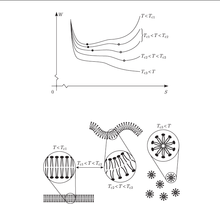
qualitative description it provides. Figure 15 illustrates this description by plotting
the free energy per molecule as a function of the average area occupied per mol-
ecule, with the temperature serving as a parameter, and with arbitrary units on both
axes.
As shown in Fig. 15, at temperatures below T
c1
, the dependence of free energy
on molecular area has a single minimum, which corresponds to the solid (gel)
phase, which is the only possible stable state (Fig. 16, left). Between T
c1
and T
c2
,
a second minimum occurs on the curve, corresponding to the liquid phase. In this
temperature range, both phases can exist, but the one corresponding to the global
minimum is more frequent. Above T
c2
, the first minimum disappears, and the
membrane can only persist in the liquid phase (Fig. 16, center). Finally, at
Figure 15 Free energy of a lipid molecule in the bilayer as a function of the molecul ar area, at
¢ve di¡erent temperatures.The units on both axes are arbitrary.
Figure 16 Membrane deformation and breakdown according to the phase transition model.
M. Pavlin et al.184

temperatures above T
c3
, the remaining minimum also vanishes, and the membrane
dissolves in the surrounding water, forming small micelles (Fig. 16, right). As an
example, for a DPPC bilayer, T
c1
E251C, T
c2
E401C and T
c3
E1651C [1].
In the presence of a transmembrane voltage, the model described above has to
be expanded by an additional component of the free energy, the electrical energy
W
e
, and the total free energy now becomes
W ðT; S; UÞ¼W
f
ðSÞþW
c
ðT; SÞþW
ic
ðT; SÞþW
ih
ðT; SÞþW
e
ðT; S; UÞ (7)
where we assume that the electrical energy depends not only on the transmembrane
voltage, but also on the temperature and the molecular surface area. In his model
[72], Suga
´
r derived the following approximation for W
e
ðT; S; UÞ:
W
e
ðT; S; UÞ¼kT log
YS
lpkT
2
m
S
128l
3
Y
U
4
(8)
where k is the Boltzmann constant, T the absolute temperature, Y the elasticity
module of the molecules in the direction of their hydrocarbon chains, S the average
area of the molecules, l is the length of the hydrocarbon chains, e
m
is the dielectric
permittivity of the molecules and U the transmembrane voltage. From this formula
it is evident that at a sufficiently high transmembrane voltage, W
e
becomes negative,
shifting the entire free energy curve down, and this shift is more pronounced at
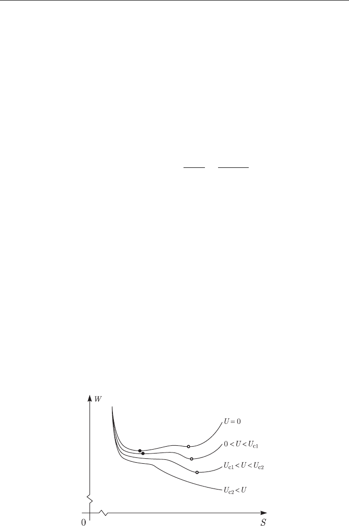
higher molecular areas. Figure 17 shows this effect at a physiological temperature
T
c1
oToT
c2
(for reasons described above, we again use arbitrary units for both
axes). At a voltage U
c1
, the first minimum of the free energy disappears, forcing the
membrane into the liquid phase state. At a somewhat higher voltage U
c2
, the
remaining minimum also ceases to exist, leading to the breakdown of the mem-
brane. The phases of the lipid membrane at various voltages are thus analogous to
those at various temperatures presented in Fig. 16. Using numerical values for all
the parameters of his model, Suga
´
r calculated that for a DPPC bilayer,
U
c1
E260 mV and U
c2
E280 mV.
Comparison between Figs. 15 and 17 shows that according to the phase tran-
sition model, the presence of transmembrane voltage has an effect similar to
Figure 17 Free energy of a lipid molecule in the bilayer as a function of the molecular area, at
four di¡erent transmembrane voltages.The units on both axes are arbitrary.
Electroporation of Planar Lipid Bilayers and Membranes 185

heating, causing a transition to the liquid phase, and eventually to decomposition
similar to a high-temperature disintegration. Such a description is very unrealistic,
since the permeabilized membrane is far from complete disintegration, and often
returns to the nonpermeabilized state.
For an impartial evaluation of the predicted value of the critical transmembrane
voltage, the parameters of the model which are at present arbitrary will have to be
determined experimentally. In addition, this model is only provisional until the
polynomial expressions obtained by regression are replaced by physical laws.
Still, the phase transition model meets several requirements at which all
the previous models fail. The permeabilized state is the minimum of free energy,
and the return to the nonpermeable state requires a sufficient input of energy,
which offers a possible explanation of the observed durability of the permeabilized
state. Similarly, the transition to this state requires a sufficient input of energy,
which could explain the dependence on pulse duration. Above the second critical
voltage, the downward slope of the free energy is never reversed, leading to a
breakdown and explaining the limited reversibility of electropermeabilization.
Except for the explanation of the dependence on the number of pulses, this model
thus meets all the qualitative requirements from the list at the beginning of this
chapter. This suggests that the approach based on the free energy could be a
promising one.
3.6. The Domain-Interface Breakdown Model
The domain-interface breakdown model takes into account the fact that cell
membranes can consist of distinct domains which differ in their lipid structure,
particularly in their content of cholesterol. According to this model, elect-
ropermeabilization is localized to the boundaries between the domains [88,89],as
Fig. 18 schematically shows.
Figure 18 Membrane breakdown according to the domain-interface breakdown model.
M. Pavlin et al.186

Similarly to the viscohydroelastic model, in the domain-interface breakdown
model the increased permeability is a result of fractures, with the difference that in
the former model they occur along the ripples, while in the latter they form along
the domain interfaces. As with the viscohydroelastic model, this description remains
questionable, as such fractures have never been observed. In addition, while the
model describes permeabilization as localized to the domain interfaces, the phe-
nomenon is also observed experimentally in bilayers and vesicles with homoge-
neous lipid structure. Thus the domain-interface breakdown can only serve as an
additional mechanism, perhaps enhancing permeabilization in cell membranes with
respect to that in artificial bilayers and vesicles.
3.7. The Aqueous Pore Formation Model
The first four models treated here – the hydrodynamic, the elastic, the viscoelastic
and the viscohydroelastic model – view electroporation as a large scale phenom-
enon, in which the molecular structure of the membrane plays no direct role.
1
The
next two – the phase transition model and the domain-interface breakdown
model – represent the other extreme, trying to explain the phenomenon by the
properties of individual lipid molecules and interactions between them.
A compromise between these two approaches is offered by the model of pore
formation, according to which electropermeabilization is caused by formation of
transient aqueous pores (electroporation) in the lipid bilayer. In this model, each
pore is formed (surrounded) by a large number of lipid molecules, but the shape,
size and stability of the pore are strongly influenced by the structure of these
molecules and their local interactions.
The model of pore formation is the last one to be described here, and in its
present form, it is considered by many as the most convincing explanation of
electropermeabilization. Therefore, in the following paragraphs an attempt will be
made to follow its development rather comprehensively, from the first designs up to
its current appearance.
The possibility of spontaneous pore formation in lipid bilayers was first analyzed
in 1975, independently by two groups [90,91]. According to this analysis (which
did not yet account for the effects of transmembrane voltage) formation of a
cylindrical pore of radius r changes the free energy of the membrane by
DW ðrÞ¼2gpr Gpr
2
(9)
where g is the edge tension and G the surface tension of the membrane. The first
term, often termed the edge energy, is positive, since a pore creates an edge in the
membrane, with a length corresponding to the circumference of the pore. The
second term, the surface energy, is negative, as a pore reduces the surface area of the
membrane. According to the above expression, the change of free energy is positive
for small pores, and negative for sufficiently large pores. This implies that
1
This point should not be obscured by the figures accompanying the models, in which separate lipid molecules are depicted.
However, these figures combine the macroscopical description, which is actually provided by these models, with the existing
knowledge of molecular structure of the lipid membrane.
Electroporation of Planar Lipid Bilayers and Membranes 187

spontaneous pore formation is inhibited by an energy barrier, explaining the sta-
bility of the membrane in the physiological conditions. The critical radius at which
the energy barrier reaches a peak and the height of this peak are given by
r
c
¼
g
G
; DW
c
¼ DW ðr
c
Þ¼
pg
2
G
. (10)
Typical parameter values (Ta ble 4 )giver
c
E10 nm. If a larger pore is artificially
created (e.g. by piercing the membrane), this energy barrier is overcome, and since
no stable state exists at larger pore radii, the membrane breaks down.
In the presence of a transmembrane voltage, formation of a pore also affects the
capacitive energy of the system. By accounting for this Abidor and co-workers
obtained a more general expression for the change of the free energy [47],
DW ðr; UÞ¼2gpr Gpr
2
ð
e
m
Þpr
2
2d
U
2
. (11)
where U is the transmembrane voltage, while e
e
and e
m
are the dielectric permit-
tivities of the aqueous medium (in approximation, that of water) in the pore and the
membrane. The transmembrane voltage reduces both the critical radius of the pore
and the energy barrier, which are now given by
r
c
ðUÞ¼
g
G þ
e
m
2d
U
2
; DW
c
ðUÞ¼DW ðr
c
; UÞ¼
pg
2
G þ
e
m
2d
U
2
. (12)
Applying parameters values from Tabl e 4 , Fig. 19 shows the free energy curves in
absence and in presence of transmembrane voltage (solid) The voltage reduces the
critical radius from r
c
E10 nm (outside the graph range) to r
c
E1.93 nm, i.e. to less
than 20% of the value in the absence of transmembrane voltage, and the energy
barrier is decreased in the same proportion.
Figure 19 Free energy change due to the occu rrence of a hydrophilic pore in the lipid bilayer,
plotted as a function of the pore radius.The solid curves show the case of a constant edge tension,
and the dashed curve the case where the edge tension increases as the pore radius decreases.
M. Pavlin et al.188

Like many of the models presented before, this version of the model of pore
formation fails to describe a permeabilized membrane, since according to it, an
above-critical voltage causes a complete breakdown of the membrane. Another
shortcoming of this model becomes evident from the structure of an aqueous pore
(Fig. 20, bottom), where the lipids adjacent to the aqueous inside of the pore are
reoriented in a manner that their hydrophilic heads are facing the pore, while their
hydrophobic tails are hidden inside the membrane. Weaver and Mintzer argued that
such a reorientation requires an input of energy which is larger for smaller pores
[92], correspondingly increasing the free energy of the membrane at small pore radii
Figure 20 Formation of an aqueous pore according to the model of electroporation. From top to
bottom: the intact bilayer; the fo rmation of a hydrophobic pore; t he transition to a hydrophilic
pore and a limited expansion of the pore radius corresponding to a reversible breakdown;
unlimited expansion of the pore radius corresponding to an i rreversible breakdown.
Electroporation of Planar Lipid Bilayers and Membranes 189

(Fig. 19, dashed). They suggested that this effect can be accounted for by treating
the edge energy as a function g(r) the value of which becomes larger as the pore
radius decreases,
DW ðr; UÞ¼2gðrÞpr Gpr
2
ð
e
m
Þpr
2
2d
U
2
, (13)
but they did not provide an expression for g(r).
The next step in the revision of the model of pore formation was a crucial one,
as it described the stable state of a pore [46]. The argument that led to this revision
is again illustrated in Fig. 20, where the hydrophilic structure of the pore is reached
through a transition from an initial, hydrophobic state, in which the lipids still have
their original orientation. The expressions for DW given so far do not deal with this
transition, and at all radii they treat the pore as fully formed, i.e. hydrophilic. Glaser
and co-workers argued that up to the pore radius r
p
at which the hydrophilic state
forms, the free energy change due to a hydrophobic pore must be analyzed instead,
for which they derived the expression (see Appendix A.5)
DW ðr; UÞ¼2pdrG
h
I
1
ðr=lÞ
I
0
ðr=lÞ
, (14)
where G
h
is the surface tension at the interface of the hydrophobic internal surface
of the pore and water, l is the characteristic length of hydrophobic interactions, and
I
k
denotes the modified Bessel function of k-th order. As in the hydrophilic case,
the total change of free energy also reflects the decrease of the membrane surface
and the electric energy. The change of free energy of the membrane accompanying
electroporation can thus be described by the system of two equations
DW ðr; UÞ¼
2pdrG
h
I
1
ðr=lÞ
I
0
ðr=lÞ
Gpr
2
ð
e
m
Þpr
2
2d
U
2
; ror
p
2prgðrÞGpr
2
ð
e
m
Þpr
2
2d
U
2
; r4r
p
8
>
>
>
<
>
>
>
:
(15)
The pore radius r
p
at which the transition from the hydrophobic to the hydrophilic
state occurs corresponds to the intersection of the hydrophilic and the hydrophobic
branch of DW,butasg(r) is not defined, it is also impossible to give an explicit
formulation of r
p
.
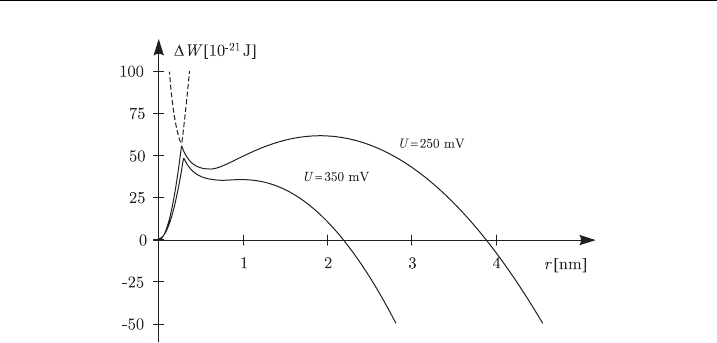
Using l ¼ 1nm [93], G
h
¼ 0.05 N/m [46] and other values as in Ta ble 4,we
can plot both branches of DW on the same graph, as shown in Fig. 21.For
transmembrane voltages of 250 and 350 mV, the solid lines give the curve into
which the hydrophobic and the hydrophilic branch combine, and the dashed lines
are the extrapolations of these two branches beyond their actual domains. Regret-
tably, a local minimum of free energy only occurs if the hydrophilic branch contains
a suitable form of the (unknown) function g(r).
The model of pore formation as illustrated by Figs. 20 and 21 represents what is
today referred to as the aqueous pore formation model (or simply as ‘‘the standard
model of electroporation’’), where the phenomenon is defined as formation of
aqueous pores in the presence of transmembrane voltage. In this model,
M. Pavlin et al.190

transmembrane voltage decreases the energy input necessary to induce a transition
from the hydrophobic
2
to the hydrophilic state. The hydrophilic pores correspond
to a local minimum of free energy, and are thus stable, which could possibly explain
the experimentally observed durability of the permeabilized state. Reversibility of
this state is limited as at voltages above the critical value there is an irreversible
breakdown of the membrane. Qualitatively, also the dependence of elect-
ropermeabilization on pulse duration is explained, since pore formation requires
a sufficient input of energy for the transition to the hydrophilic state. In the models
of pore formation, including the aqueous pore formation model of electroporation,
the transmembrane voltage does not cause, but only facilitates the formation of
hydrophilic pores, which can account for the stochasticity of electropermeabilizat-
ion. As Figs. 19 and 20 testify, the values at which this facilitating effect becomes
pronounced are in hundreds of millivolts, and thus in relatively good agreement
with the experiments.
Still, the aqueous pore formation model of electroporation has two significant
shortcomings. The first is clearly observable in Fig. 21; namely, with realistic pa-
rameter values applied, the transmembrane voltage reduces the energy barrier of the
hydrophobic–hydrophilic transition, but it reduces the barrier of an irreversible
breakdown to a much larger extent. For example, a transmembrane voltage above
361 mV reduces the energy barrier of aqueous pore formation by only several
percent, but once the pore is formed, this voltage imminently leads to the break-
down. This effect is independent of the choice of the arbitrary function g(r), since it
is governed exclusively by the contribution of the electric energy. Nonetheless, the
Figure 21 Free ene rgy change due to the formation of an aqueous pore, plotted as a function of
the pore radius.The initial increase corresponds to the formation and expansion of a hydrophilic
pore, the local maximum to the transition to a hydrophilic pore, and the subsequent local
minimum corresponds to the radius of a (semi-) stable hydrophilic pore. At su⁄ciently high
transmembrane voltages, this minimum transforms into a monotonic decrease, which
corresponds to an irreversible breakdown.
2
Because of the lateral thermic fluctuations of the lipid molecules, hydrophobic pores, with lifetimes in the picosecond range, are
in certain extent always present in the membrane.
Electroporation of Planar Lipid Bilayers and Membranes 191

undefined functional form of g(r) is the second shortcoming of the aqueous pore
formation model of electroporation. In absence of its definition, the expressions for
the energy barrier that impedes the hydrophobic–hydrophilic transition, as well as
the minimum radius of a hydrophilic pore r
p
also remain undefined. This short-
coming will probably be addressed in the future, either theoretically, by a derivation
of a physical law which would define g (r), or experimentally, by a measurement of
its values. On the long term, the former alternative is definitely preferred.
Subsequent paragraphs contain a short overview of various extensions of the
aqueous pore formation model that have been proposed by different authors.
3.8. Extensions of the Aqueous Pore Formation Model
Several approaches have been proposed for improving the aqueous pore formation
model presented in the preceding section. Two of these [94,95] addressed its du-
bious prediction that transmembrane voltage strongly facilitates an irreversible
breakdown. Barnett and Weaver accounted for the fact that a pore alters not only
the capacitive, but also the conductive energy of the membrane, and reformulated
the electric energy as
3
DW
e
¼
ð
e
m
Þp
d
U
2
Z
r
0
r dr
ð1 þ lðrÞÞ
2
(16)
with
lðrÞ¼
prs
p
2ds
e
(17)
where s
e
and s
p
are the electric conductivities of the aqueous medium outside and
inside the pore. The exact value of s
p
depends on the properties of the lipid
headgroups forming the surface of the pore, as well as on the properties of the
aqueous medium inside the pore, but in the first approximation it is reasonable to
assume that s
p
Es
e
, so that
lðrÞ¼
pr
2d
(18)
and the electric energy is given by
DW
e
¼ð
e
m
Þ U
2
4d log 1 þ
pr
2d
p
2r
1 þ
pr
2d
. (19)
Figure 22 compares, for a transmembrane voltage of 350 mV, the free energy
change as a function of pore radius with (solid) and without (dashed) the described
modification of the electric energy. The revised curve of free energy change shows
a significantly broadened range of stable pore radii. The voltage which leads to an
irreversible breakdown is shifted up to E458 mV, which is an improvement with
respect to the previous value of E361 mV, but is still rather low. Also in the revised
3
Note that using l(r) ¼ 0, we get DW
e
of the standard model (i.e. the result derived by Abidor and co-workers).
M. Pavlin et al.192

model, the facilitating effect of transmembrane voltage on formation of aqueous
pores remains very weak.
Freeman and co-workers [95] made an attempt to enhance this model further by
accounting for the energy needed by charged particles to traverse the pore
(the Born energy), which led them to a more complicated expression for l(r)
expressed as a polynomial regression of experimental data.
Another study has addressed the effect of the difference between the osmo-
larities of the extracellular medium and the cytoplasm [32]. To address this effect,
the change of free energy must incorporate the change of osmotic energy caused by
the pore formation,
DW
osm
¼
p
e
p
i
R
2
pr
2
(20)
where p
e
and p
i
are the osmotic pressures in the extracellular medium and inside the
vesicle or cell, respectively, and R is the radius of the vesicle or cell. The osmotic
energy thus acts similarly to the surface energy and the electric energy – it reduces
the free energy of the membrane, and is proportional to r
2
. This implies that a
difference between the osmolarities makes the membrane more susceptible to the
effects of the transmembrane voltage, which seems to be in agreement with ex-
periments [96,97].
Finally, it was also analyzed how electroporation is affected by the curvature of
the membrane [98]. Unlike in planar bilayers, in vesicles and cells the membrane is
inherently curved, with the curvature increasing with a decreasing cell radius.
According to the calculations by Neumann and co-workers, the change of cur-
vature energy caused by the pore formation is
DW
crv
¼
64Y
Rd
p
2
r
2
(21)
Figure 22 Free energy ch ange as a function of the pore radius, without (dashe d) and with
(solid) the modi¢cation of the conductive electric energy as formulated by Barnett a nd Weaver
[94] .
Electroporation of Planar Lipid Bilayers and Membranes 193
