Liu A.L., Tien H.T. Advances in Planar Lipid Bilayer and Liposomes. V.6
Подождите немного. Документ загружается.


variation of the local composition at certain temperature can be checked. The latter
is physiologically relevant since the same happens when spin probe or any other
membrane-soluble substance diffuses laterally through the regions with different
local compositions.
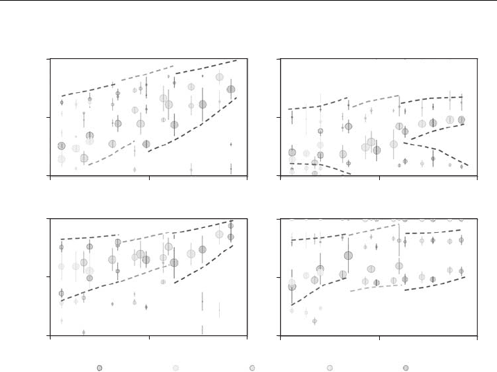
The most striking effect seen in Figs. 12 and 13 is the absolute character of
motional patterns that are available at certain temperature independently on the
local lipid composition. The most unrestricted and the most restricted patterns
follow the quasi-linear temperature dependence indicated with dashed colored lines
following the same color legend as in the presentation of the solutions in the same
graph. This fact is very surprising since the possible motional patterns are the same
at certain temperature for the lipids with long saturated acyl chains forming the S
phase or for the lipids with short or unsaturated chains that are in the Ld phase.
However, it is then exactly the composition which is responsible of the range of
patterns realized by a molecule embedded in the lipid bilayer. It should be stressed
that the lines of the most restricted motional patterns for all labels end up abruptly
just above the physiological temperatures without merging with the lines of the
most unrestricted motional patterns. It seems that this reflects the real molecular
picture since it does not depend on the type of spin probe.
HFASLMeFASL
13-doxyl 5-doxyl
0 % Chol
T [K]
270 305 340
270 305 340
T [K]T [K]
T [K]
Ω [ ]
270 305
DOPC
POPC
DMPC DPPC DSPC
340
0
0,5
1
Ω [ ]
0
0,5
1
Ω [ ]
0
0,5
1
Ω [ ]
0
0,5
1
270 305 340
Figure 12 Condensed results of the signi¢cant motional patterns of the 5-doxyl and 13-doxyl
MeFASL and HFASL spin probes in DSPC, DPPC, DMPC, POPC and DOPC membranes
without cholesterol. The temperature range was from 20 K below to 20 K above the main phase
transition temperature of the corresponding lipid. The free rotational space O is shown on the
y-axis, as the main characteristics of the rotational restriction and a measure of an isotropy of
rotational motion of a spin probe (please see plate no. 3 in the color section).
J. S
ˇ
trancar and Z. Arsov154

In addition, one can also see the well-known phenomena that the order of the
acyl chain, i.e., the restrictions of the rotational motion, decreases with the position
as detected in the cases of all the spin probes. This difference is even more striking
for the lipid bilayers with cholesterol as indicated by the line of the allowed most
restricted motional patterns (Fig. 13).
4.2. Effect of Spin Probe Partitioning on Membrane Heterogeneity
Characterization
The proportion of a particular spectral component, determined by EPR spectra
decomposition [13], is not necessarily the same as the fraction of a corresponding
lipid phase in a membrane, due to the partitioning properties of spin probe between
different lipid phases [56]. The DMPC/Chol system was studied. Spin probe
MeFASL(10,3) (methyl ester of 5-doxyl palmitate) was used.
From known phase diagrams fractions of particular lipid phases can be deter-
mined at certain temperature and composition by the so called lever rule [56].For
temperatures above the temperature of the main phase transition T
m
(T4T
m
), at
which the system is in the phase coexistence region Ld+Lo, it holds for the fraction
HFASLMeFASL
T [K]
T [K]T [K]
T [K]
Ω [ ]
0
0,5
1
Ω [ ]
0
0,5
1
Ω [ ]
0
0,5
1
Ω [ ]
0
0,5
1
270 305 340
270 305
DOPC
POPC
DMPC
DPPC
DSPC
340
270 305 340
270 305 340
13-doxyl 5-doxyl
40 % Chol
Figure 13 Condensed results of the signi¢cant motional patterns of the 5-doxyl and 13-doxyl
MeFASL and HFASL spin probes in DSPC, DPPC, DMPC, POPC and DOPC membranes with 40
molar% of cholesterol.The temperature range was from 20 K below to 20 K above the main phase
transition temperature of the corresponding lipid (without cholesterol).The free rotational space
O is shown on the y-ax is, as the mai n ch aracteristics of the rotational restriction and a measure of
anisotropy of rotational motion of a spin probe (please see plate no. 4 in the color section).
Application of Spin-Labeling EPR and ATR-FTIR Spectroscopies 155

of lipids in the Ld phase
f
Ld
ðTÞ¼
x
Lo
Chol
ðTÞx
0
Chol
x
Lo
Chol
ðTÞx
Ld
Chol
ðTÞ
(2)
where x
Lo
Chol
ðTÞ and x
Ld
Chol
ðTÞ represent cholesterol mole fractions at the corre-
sponding phase boundaries, and x
0
Chol
the mole fraction of cholesterol in the sample.
A spectrum I(B) is decomposed into a different number of spectral components
I
i
(B) related to the lipid phases (domain types)
IðBÞ¼
X
i
w
i
I
i
ðBÞ (3)
where w
i
is proportion of spectral component i and B the magnetic field. Since
phase diagrams for DMPC/Chol shows coexistence of two lipid phases at most
(Fig. 2), EPR spectra were decomposed into two spectral components (i ¼ 1, 2).
It can be seen from Fig. 14A that we can qualitatively describe a spectrum from
a two phase coexistence region, e.g., a spectrum of DMPC/Chol 0.2 sample,
which is at T4T
m
in the Ld+Lo coexistence region, as a superposition of spectra
from DMPC/Chol 0.1 and DMPC/Chol 0.4 samples that correspond to single-
phase regions. In order to make this procedure more precise, the spectra of samples
with composition corresponding to the mole fraction of cholesterol at the phase
boundary would have to be taken into account. An example of EPR spectra
decomposition of the same experimental spectrum to two spectral components
corresponding to the population of spin probes in disordered and ordered lipid
environment is shown in Fig. 14B.
Figure 14 Comparison of experimental and calculated EPR spectra. (A) Comparison of the
experimental spe ctrum of spi n-labeled DMPC/Chol 0.2 liposomes at temperature 33 1C
(T4T
m
), co rresponding to the coexistence region Ld+Lo, w ith a superposition of the spectra
corresponding to the single-lipid phase regions DMPC/Chol 0.1 (the Ld phase) and DMPC/
Chol 0.4 (the L o phase). (B) Comparison of the experimental EPR spect rum of DMPC/Chol
0.2 (T ¼ 33 1CK,T4T
m
) compared with simulated (¢tted) spectrum. The ¢tted spectr um is
decomposed into two spectral components corresponding to the coexisting population of
disordered (the Ld phase) and ordered lipids (the Lo phase).
J. S
ˇ
trancar and Z. Arsov156

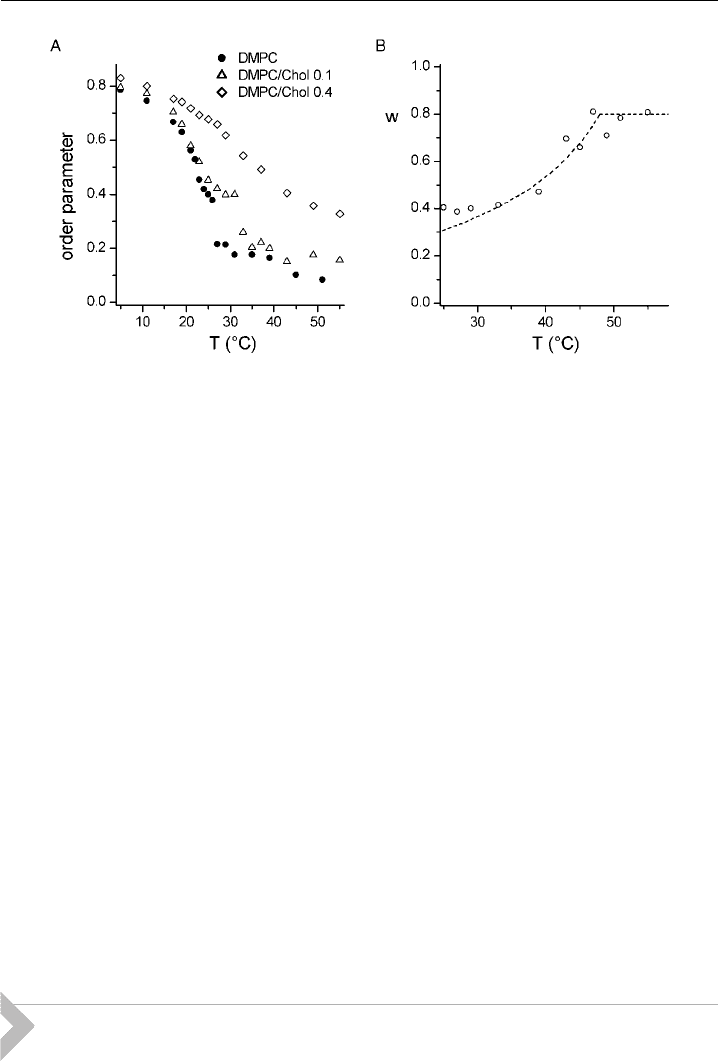
The temperature dependence of order parameter, reporting about the time-
averaged angular fluctuation of the acyl-chain segment where spin label (in our case
nitroxide radical) is attached to the spin probe molecule, was extracted from the
recorded EPR spectra by spectral simulations (Fig. 15A). Above T
m
¼ 24 1C for
pure DMPC [57] order parameter drops discontinuously as expected due to the
phase transition S-Ld. Similarly for DMPC/Chol 0.1 samples order parameter
shows a discontinuous drop close to the temperature corresponding to the com-
pletion of the phase separation Ld+Lo-Ld (Fig. 2). On the contrary, only a steady
decrease can be observed in the order parameter for DMPC/Chol 0.4, in accord-
ance with the presence of only one lipid phase for all temperatures (Fig. 2).
In Fig. 15B for DMPC/Chol 0.2 sample (at T4T
m
) the comparison between
the temperature dependence of experimentally determined proportion of spectral
component corresponding to the population of disordered lipids according to
equation (3) and calculated values of fraction for the Ld phase according to equation
(2) is shown. The temperature at which proportion increases to the maximum value
agrees well with the temperature corresponding to the completion of phase
separation Ld+Lo-Ld (Fig. 2). Since proportions agree relatively well with the
calculated fractions, it seems that our spin probe partitions approximately uniformly
between the Ld and the Lo phase [56].
5. Membrane Heterogeneity Characterization by
ATR–FTIR Spectroscopy
The IR spectral features most sensitive to phospholipid molecular structure
and interactions arise from the vibrations of the acyl chains. There are several modes
Figure 15 Temperature dependence of parameters obtained by EPR spectra decomposition for
di¡erent DMPC/Chol samples. (A) Order parameter of the spectral component corresponding to
the population of ordered lipids. Estimate of uncertainty in order parameter is 70.02. (B)
Measured proportions of the spectral component corresponding to the population of disordered
lipids (points) and c alcul ated fraction of lipids in the Ld phase (line) for the mixture DMPC/
Chol 0.2. Estimate of uncertainty in measured values of proportion is 70.05.
Application of Spin-Labeling EPR and ATR-FTIR Spectroscopies 157

sensitive to chain conformation and interaction. The methylene stretching
frequencies qualitatively monitor both lipid conformational order and acyl-chain
packing [58]. As these modes are among the most intense in the IR spectra of lipids
and membranes, they have been widely used for lipid conformational studies.
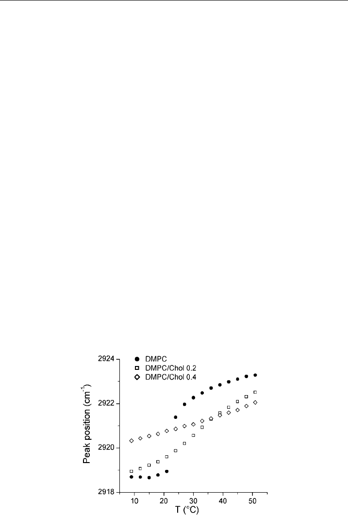
As an example, the temperature dependence of the methylene antisymmetric
stretching band peak position is shown in Fig. 16 for DMPC/Chol system. For
pure DMPC results show a strong jump in the peak frequencies above T
m
(Fig. 16).
For samples with Chol 0.2 the increase of the peak position with temperature
becomes steeper above T
m
and then less steep again close to the temperature
corresponding to the completion of phase separation Ld+Lo-Ld (Fig. 2). Samples
with Chol 0.4 show a linear increase in frequency (Fig. 16), due to the presence of
only one lipid phase, i.e., the Lo phase, for all temperatures (Fig. 2).
A potentially interesting region of the IR spectrum for studying properties of
different lipid phases is the carbonyl absorption band arising from the stretching
vibrations of ester carbonyl groups of glycerolipids. The band is strong and occurs
in a region essentially free of significant absorptions by groups. Carbonyl bands are
conformationally sensitive, reflect the level of hydration at the membrane interface
and are influenced by hydrogen bonding [59]. It was noted before that the band
shape changes, if the lipid undergoes the main phase transition [60].
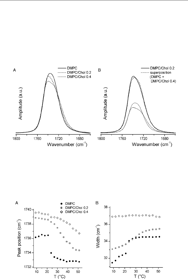
To highlight differences in the shape of the carbonyl band for samples with
different amounts of cholesterol, overlaid spectra normalized to the same area are
presented in Fig. 17A. The most obvious difference is the increased asymmetry in
the shape of the carbonyl band for samples with cholesterol and a much larger
carbonyl bandwidth for samples with Chol 0.4 compared to samples without
cholesterol. The increased asymmetry in the presence of cholesterol [61] and
broadening [62] was already noted before for the DPPC/Chol system.
Similarly to EPR spectra of spin-labeled liposomes, also ATR–FTIR spectra of
lipid multibilayers at appropriate temperature and composition are expected to
Figure 16 Temperature dependence of methylene antisymmetric stretching band peak position
for DMPC/Chol samples with di¡e rent mole fractions of cholesterol. Samples were prepared in
excess D
2
O. Estim ate of the uncertainty in the peak position is 70.1 to 70.2 cm
^1
.
J. S
ˇ
trancar and Z. Arsov158

reflect the coexistence of different lipid phases. As shown in Fig. 17B a spectrum
from a sample exhibiting phase coexistence can be qualitatively described as a
superposition of spectra from samples that correspond to single phases. If the ATR–
FTIR spectra of samples with composition corresponding to the mole fraction of
cholesterol at the phase boundary were taken into account, the agreement between
experimental and calculated spectrum should be better.
The temperature dependence of the carbonyl band peak position for samples
with different mole fractions of cholesterol is shown in Fig. 18A. For pure DMPC
Figure 17 Dependence of carbonyl band shape in DMPC/Chol sa mples on mole fraction of
cholesterol. Samples were prepared in excess D
2
O, T ¼ 9 1C(ToT
m
). (A) Comparison of t he
carbonyl band shape fo r samples with di¡erent amou nt of cholesterol. Absorption bands were
normalized to the same area. (B) Comparison of the carbonyl band shape of DMPC/Chol 0.2,
corresponding to the coexistence region S+Lo, with a superposition of the carbonyl bands
corresponding to the single-lipid phase regions DMPC (the S phase) and DMPC/Chol 0.4 (the
Lo phase ).
Figure 18 Temperature dependence of carbonyl band (A) peak position and (B) half-
bandwidth for DMPC/Chol samples with di¡erent mole fractions of cholesterol. Samples were
prepared in excess D
2
O. Estimate of the uncertainty for both parameters is around 70.3 cm
^1
.
Application of Spin-Labeling EPR and ATR-FTIR Spectroscopies 159

a jump of the peak position to lower frequencies occurs near the temperature of
the main phase transition T
m
(Fig. 18A). For DMPC/Chol 0.2 samples the slope
changes its steepness above T
m
again (Fig. 18A). Samples with Chol 0.4 show more
or less steady decrease of the peak position with temperature (Fig. 18A). Similar
lipid-phase-specific behavior can also be observed for the temperature dependence of
the half-bandwidth of carbonyl band (Fig. 18B). It can be concluded from com-
parison of Figs. 16 and 18 that the shapes of the curves of the temperature depend-
ence of the peak position and of the half-bandwidth of the carbonyl band report on
the lipid phase of the sample and reflect conformational changes in the bilayer.
6. Conclusions
It was shown that SL-EPR in combination with sophisticated simulations
and data analysis techniques developed in the recent decade became a powerful
methodology for membrane exploration. In addition, ATR–FTIR method was
demonstrated to be suitable for examining hydrated lipid bilayers.
The results obtained by SL-EPR on supported membranes or dehydrated
membranes indicate that the generalization to the biological membranes has to be
done carefully. Full interaction scheme at the local molecular level has to match the
one in fully hydrated lipid bilayer system prepared in excess free water to enable
direct predictions in the more complex environments like biological membranes.
Furthermore, the ATR–FTIR spectroscopy can be used to examine water structure
adjacent to lipid bilayers, while it is possible to appreciate the presence of bound
water.
It can be concluded that the molecular picture of lipid bilayer is more complex
than a simple classification to different lipid phases. The molecular probing by
SL-EPR reveals much more, from the local interactions to partitioning and com-
position, making this technique a powerful tool in exploration of membrane
heterogeneity. Moreover, also ATR–FTIR spectroscopy is appropriate and com-
plementary for this purpose. It was shown that in mixtures of phosphatidylcholines
and cholesterol beside the acyl-chain modes also the carbonyl band is useful to
reveal the presence of different lipid phases.
ACKNOWLEDGEMENTS
The authors acknowledge the financial support from the state budget by the Slovenian Research
Agency (programme no. P1-0060, project no. J1-6581 and Z1-9502). The part of the work dealing
with the ATR–FTIR spectroscopy was carried out with the financial support of the Sincrotrone
Trieste. We thank Prof. Peter Laggner and Dr. Heinz Amenitsch for valuable suggestion regarding the
work on SL-EPR on oriented membranes. We thank Iztok Urbanc
ˇ
ic
ˇ
and Nace Zidar for carrying out
the experimental work concerning detection of biased spin probe reporting. We thank Dr. Luca
Quaroni for the help on the research conducted with infrared spectroscopy. Some of the work was
done in collaboration with different research groups enabled by the financial support from EC COST
D22 and COST P15 Actions.
J. S
ˇ
trancar and Z. Arsov160

REFERENCES
[1] M. Bloom, J.L. Thewalt, Time and distance scales of membrane domain organization, Mol.
Membr. Biol. 12 (1995) 9–13.
[2] K. Jorgensen, J.H. Ipsen, O.G. Mouritsen, M.J. Zuckermann, The effect of anaesthetics on the
dynamic heterogeneity of lipid membranes, Chem. Phys. Lipids 65 (1993) 205–216.
[3] K. Jørgensen, O.G. Mouritsen, Phase separation dynamics and lateral organization of two-
component lipid membranes, Biophys. J. 69 (1995) 942–954.
[4] C. Leidy, W.F. Wolkers, K. Jørgensen, O.G. Mouritsen, J.H. Crowe, Lateral organization
and domain formation in a two-component lipid membrane system, Biophys. J. 80 (2001)
1819–1828.
[5] E.J. Shimshick, H.M. McConnell, Lateral phase separation in phospholipid membranes,
Biochemistry 12 (1973) 2351–2360.
[6] P.F.F. Almeida, W.L.C. Vaz, T.E. Thompson, Lateral diffusion in the liquid phases of dim-
yristoylphosphatidylcholine/cholesterol lipid bilayers: a free volume analysis, Biochemistry 31
(1992) 6739–6747.
[7] B.R. Copeland, H.M. McConnell, The rippled structure in bilayer membranes of
phosphatidylcholine and binary mixtures of phosphatidylcholine and cholesterol, Biochim.
Biophys. Acta 599 (1980) 95–109.
[8] T.P.W. McMullen, R.N. McElhaney, New aspects of the interaction of cholesterol with
dipalmitoylphosphatidylcholine bilayers as revealed by high-sensitivity differential scanning
calorimetry, Biochim. Biophys. Acta 1234 (1995) 90–98.
[9] S. Karmakar, V.A. Raghunathan, Cholesterol-induced modulated phase in phospholipid mem-
branes, Phys. Rev. Lett. 91 (2003) 098102.
[10] R. Welti, M. Glaser, Lipid domains in model and biological membranes, Chem. Phys. Lipids 73
(1994) 121–137.
[11] M. Edidin, Lipid microdomains in cell surface membranes, Curr. Opin. Struct. Biol. 7 (1997)
528–532.
[12] F.R. Maxfield, Plasma membrane microdomains, Curr. Opin. Cell Biol. 14 (2002) 483–487.
[13] Z. Arsov, M. Schara, J. S
ˇ
trancar, Quantifying the lateral lipid domain properties in erythrocyte
ghost membranes using EPR-spectra decomposition, J. Magn. Reson. 157 (2002) 52–60.
[14] M. Ge, K.A. Field, R. Aneja, D. Holowka, B. Baird, J.H. Freed, Electron spin resonance
characterization of liquid ordered phase of detergent-resistant membranes from RBL-2H3 cells,
Biophys. J. 77 (1999) 925–933.
[15] D.A. Brown, E. London, Structure and function of sphingolipid- and cholesterol-rich mem-
brane rafts, J. Biol. Chem. 275 (2000) 17221–17224.
[16] G.J. Schu
¨
tz, G. Kada, V.P. Pastushenko, H. Schindler, Properties of lipid microdomains in a
muscle cell membrane visualized by single molecule microscopy, EMBO J. 19 (2000) 892–901.
[17] J. S
ˇ
trancar, M. Schara, S. Pec
ˇ
ar, New EPR method for cellular surface characterization,
J. Membr. Biol. 193 (2003) 15–22.
[18] F.J. Sharom, C.W.M. Grant, A model for ganglioside behaviour in cell membrane, Biochim.
Biophys. Acta 507 (1978) 280–293.
[19] M.W. Peters, K.R. Barber, C.W.M. Grant, Lateral distribution of gangliosides in bilayer mem-
branes: lipid and ionic effects, J. Neurosci. Res. 12 (1984) 343–353.
[20] L.A. Bagatolli, E. Gratton, G.D. Fidelio, Water dynamics in glycosphingolipid aggregates studied
by LAURDAN fluorescence, Biophys. J. 75 (1998) 331–341.
[21] E. Bertoli, M. Masserini, S. Sonnino, R. Ghidoni, B. Cestaro, G. Tettamanti, Electron par-
amagnetic resonance studies on the fluidity and surface dynamics of egg phospatidylcholine
vesicles containing gangliosides, Biochim. Biophys. Acta 467 (1981) 196–202.
[22] H. Beitinger, V. Vogel, D. Mo
¨
bius, H. Rahmann, Surface potentials and electric dipole moments
of ganglioside and phospholipid monolayer: contribution of the polar headgroup at the water/
lipid interface, Biochim. Biophys. Acta 984 (1989) 293–300.
[23] M. Shinitzky, Membrane fluidity and cellular functions, in: M. Shinitzky, (Ed.), Physiology of
Membrane Fluidity, Vol. 1, CRC Press, Boca Raton, 1984, pp. 1–51.
Application of Spin-Labeling EPR and ATR-FTIR Spectroscopies 161

[24] D. Marsh, Lipid-protein interactions and heterogeneous lipid distribution in membranes, Mol.
Membr. Biol. 12 (1995) 59–64.
[25] Z. Arsov, M. Schara, M. Zorko, J. S
ˇ
trancar, The membrane lateral domain approach in the
studies of lipid-protein interaction of GPI-anchored bovine erythrocyte acetylcholinesterase,
Eur. Biophys. J. 33 (2004) 715–725.
[26] H. Schindler, J. Seelig, EPR spectra of spin labels in lipid bilayers, J. Chem. Phys. 59 (1973)
1841–1850.
[27] S.P. Van, G.B. Birelli, O.H. Griffith, Rapid anisotropic motion of spin labels: models for motion
averaging of the ESR parameters, J. Magn. Reson. 15 (1974) 444–459.
[28] J.R. Lakowicz, Fluorescence spectroscopic investigations of the dynamic properties of proteins,
membranes and nucleic acids, J. Biochem. Biophys. Methods 2 (1980) 91–119.
[29] C. Ho, B.W. Williams, C.D. Stubbs, Analysis of cell membrane micro-heterogeneity using the
fluorescence lifetime of DPH-type fluorophores, Biochim. Biophys. Acta 1104 (1992) 273–282.
[30] H.J. Steinhoff, A. Savitsky, C. Wegener, M. Pfeiffer, M. Plato, K. Mo
¨
bius, High-field EPR
studies of the structure and conformational changes of site-directed spin labeled bacteriorhodop-
sin, Biochim. Biophys. Acta 1457 (2000) 253–262.
[31] J. S
ˇ
trancar, M. S
ˇ
entjurc, M. Schara, Fast and accurate characterization of biological membranes
by EPR spectral simulations of nitroxides, J. Magn. Reson. 142 (2000) 254–265.
[32] B. Filipic
ˇ
,J.S
ˇ
trancar, Tuning EPR spectral parameters with a genetic algorithm, Appl. Soft
Comput. 1 (2001) 83–90.
[33] J. S
ˇ
trancar, T. Koklic
ˇ
, Z. Arsov, Soft picture of lateral heterogeneity in biomembranes, J. Membr.
Biol. 196 (2003) 135–146.
[34] J. S
ˇ
trancar, T. Koklic
ˇ
, Z. Arsov, B. Filipic
ˇ
, D. Stopar, M.A. Hemminga, Spin label EPR-based
characterization of biosystem complexity, J. Chem. Inf. Model. 45 (2005) 394–406.
[35] D.J. Schneider, J.H. Freed, Calculating slow motional magnetic resonance spectra: a user’s guide,
in: L.J. Berliner, J. Reuben (Eds.), Biological Magnetic Resonance, Vol. 8: Spin Labeling,
Theory and Applications, Plenum Press, New York, 1989, pp. 1–76.
[36] A.A. Kavalenka, B. Filipic
ˇ
, M.A. Hemminga, J. S
ˇ
trancar, Speeding up a genetic algorithm for
EPR-based spin label characterization of biosystem complexity, J. Chem. Inf. Model. 45 (2005)
1628–1635.
[37] J. S
ˇ
trancar, EPRSIM-C Version 6.2., 1996–2007, http://www.ijs.si/ijs/dept/epr/EPRSIMC_
overview.htm.
[38] J. Mravljak, J. Konc, M. Hodos
ˇ
c
ˇ
ek, T. S
ˇ
olmajer, S. Pec
ˇ
ar, Spin-labeled alkylphospholipids in a
dipalmitoylphosphatidylcholine bilayer: molecular dynamics simulations, J. Phys. Chem. B Con-
dens. 110 (2006) 25559–25561.
[39] N.J. Harrick, Internal Reflection Spectroscopy, John Wiley & Sons, New York, 1967.
[40] U.P. Fringeli, The structure of lipids and proteins studied by attenuated total reflection (ATR)
infrared spectroscopy, II. Oriented layers of a homologous series: phosphatidylethanolamine to
phosphatidylcholine, Z. Naturforsch. 32c (1977) 20–45.
[41] J. Grdadolnik, J. Kidric, D. Hadzi, Hydration of phosphatidylcholine reversed micelles and
multilayers – an infrared spectroscopic study, Chem. Phys. Lipids 59 (1991) 57–68.
[42] H. Binder, The molecular architecture of lipid membranes – new insights from hydration-tuning
infrared linear dichroism spectroscopy, Appl. Spectrosc. Rev. 38 (2003) 15–69.
[43] E. Sackmann, Supported membranes: scientific and practical applications, Science 271 (1996)
43–48.
[44] S.A. Tatulian, Structural effects of covalent inhibition of phospholipase A2 suggest allosteric
coupling between membrane binding and catalytic sites, Biophys. J. 84 (2003) 1773–1783.
[45] S.A. Tatulian, Attenuated total reflection Fourier transform infrared spectroscopy: a method of
choice for studying membrane proteins and lipids, Biochemistry 42 (2003) 11898–11907.
[46] J. Milhaud, New insights into water-phospholipid interactions, Biochim. Biophys. Acta 1663
(2004) 19–51.
[47] S. Leikin, V.A. Parsegian, D.C. Rau, R.P. Rand, Hydration forces, Annu. Rev. Phys. Chem. 44
(1993) 369–395.
J. S
ˇ
trancar and Z. Arsov162

[48] K. Gawrisch, D. Ruston, J. Zimmerberg, V.A. Parsegian, R.P. Rand, N. Fuller, Membrane
dipole potential, hydration forces, and the ordering of water at membrane surfaces, Biophys. J.
61 (1992) 1213–1223.
[49] J.N. Israelachvili, H. Wennerstro
¨
m, Role of hydration and water structure in biological and
colloidal interactions, Nature 376 (1996) 219–225.
[50] M. Rappolt, H. Amenitsch, J. S
ˇ
trancar, C.V. Teixeira, M. Kriechbaum, G. Pabst, M. Maj-
erowicz, P. Laggner, Phospholipid mesophases at solid interfaces: in-situ X-ray diffraction and
spin-label studies, Adv. Colloid Interface Sci. 111 (2004) 63–77.
[51] J.L.R. Arrondo, F.M. Goni, J.M. Macarulla, Infrared spectroscopy of phosphatidylcholines in
aqueous suspensions. A study of the phosphate group vibrations, Biochim. Biophys. Acta 794
(1984) 165–168.
[52] L. Ter-Minassian-Saraga, E. Okamura, J. Umemura, T. Takenaka, Fourier transform infrared-
attenuated total reflection spectroscopy of hydration of dimyristoylphosphatidylcholine
multibilayers, Biochim. Biophys. Acta 946 (1988) 417–423.
[53] J. Grdadolnik, J. Kidric, D. Hadzi, An FT-IR study of water hydrating dip-
almitoylphosphatidylcholine multibilayers and reversed micelles, J. Mol. Struct. 322 (1994)
93–103.
[54] H. Binder, Water near lipid membranes as seen by infrared spectroscopy, Eur. Biophys. J.,
DOI:10.1007/s00249-006-0110-6.
[55] D. Cringus, J. Lindner, M.T.W. Milder, M.S. Pshenichnikov, P. Vo
¨
hringer, D.A. Wiersma,
Femtosecond water dynamics in reverse micellar nanodroplets, Chem. Phys. Lett. 408 (2005)
162–168.
[56] Z. Arsov, J. S
ˇ
trancar, Determination of partition coefficient of spin probe between different lipid
membrane phases, J. Chem. Inf. Model. 45 (2005) 1662–1671.
[57] J.M. Holopainen, J. Lemmich, F. Richter, O.G. Mouritsen, G. Rapp, P.K.J. Kinnunen,
Dimyristoylphosphatidylcholine/C16:0-ceramide binary liposomes studied by differential scan-
ning calorimetry and wide- and small-angle X-ray scattering, Biophys. J. 78 (2000) 2459–2469.
[58] R. Mendelsohn, D.J. Moore, Vibrational spectroscopic studies of lipid domains in biomembranes
and model systems, Chem. Phys. Lipids 96 (1998) 141–157.
[59] R.N.A.H. Lewis, R.N. McElhaney, W. Pohle, H.H. Mantsch, Components of the carbonyl
stretching band in the infrared spectra of hydrated 1,2-diacylglycerolipid bilayers: a revaluation,
Biophys. J. 67 (1994) 2367–2375.
[60] H.L. Casal, H.H. Mantsch, Polymorphic phase behaviour of phospholipid membranes studied by
infrared spectroscopy, Biochim. Biophys. Acta 779 (1984) 381–401.
[61] J. Umemura, D.G. Cameron, H.H. Mantsch, A Fourier transform infrared spectroscopic study of
the molecular interaction of cholesterol with 1,2-dipalmitoyl-sn-glycero-3-phosphocholine,
Biochim. Biophys. Acta 602 (1980) 32–44.
[62] O. Reis, R. Winter, T.W. Zerda, The effect of high external pressure on DPPC-cholesterol
multilamellar vesicles: a pressure-tuning Fourier transform infrared spectroscopy study, Biochim.
Biophys. Acta 1279 (1996) 5–16.
Application of Spin-Labeling EPR and ATR-FTIR Spectroscopies 163
