Lima J.J.Pedroso, de (ed.). Nuclear Medicine Physics
Подождите немного. Документ загружается.


418 Nuclear Medicine Physics
or
F =
q
∞
0
C
0
(t)dt
(7.205)
Thus, one can calculate the blood flow from the total quantity injected and
from the area under the tracer time concentration curve, no matter over what
period of time the tracer was injected. However, we cannot calculate the vol-
ume of the system without knowing more about the input function. Unless
the injection is truly instantaneous, the mean transit time of the concentration
function at the output is not the same as the frequency function of transit
times through the system.
The true mean transit time through the system is the difference between the
mean transit time of the concentration function at output and the mean time
of the tracer input
¯
t =
¯
t
o
−
¯
t
i
(7.206)
7.2.7 Regeneration of the Frequency Function of Transit Times by
Deconvolution
We are now going to consider some applications of convolution and decon-
volution methods.
These are important operations concerning the applicability of the input–
output problems of systems to any biological system (not only to dilution sys-
tems), provided that certain conditions are satisfied by the system (linearity
as well as time invariance, that is, constant behavior over time).
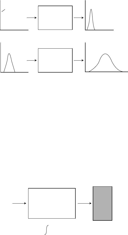
Figure 7.45 is a schematic representation of the functions involved in a
process in which a system responds to stimuli.
Function h(t) is the response of the system to a unit impulse, that is, a delta
function of unit amplitude at zero time, or in the case of NM, for example, a
very fast injection of unit activity at the beginning of the experiment.
The convolution of functions h(t) and I(t) is represented by the integral
R(t) =
t
0
I(t −τ)h(τ)dτ = h(t)∗I(t)
where h(t) is an operator that converts an object or input function I(t) into
an output function R(t). In the above equation, t is the sole variable, and τ is
used as an auxiliary variable.
There can be any variables and they may be applied in convolution in 2D
or multidimensional spaces.
Through convolution, a response function can be calculated if the input
function and the unit response function are known (Figure 7.46).
Systems in Nuclear Medicine 419
δ(t)
I(t) R(t)
h(t)
tt
tt
FIGURE 7.45
h(t) is the response of the system to a unit impulse, transit time distribution (or retention function
or residence times function). I(t) is the input function and R(t) is the response function (or a
residue function).
In most cases, however,the important problemis the inverse of convolution,
thatis, I(t) iswanted when R(t) andh(t) areknown, or h(t) is wanted whenR(t)
and I(t) are known. In such cases, the unknown integrand function is said to
be deconvoluted from the known functions, and the necessary mathematical
procedures are called deconvolution. Deconvolution is, then, an operation
defined as the inverse of convolution.
The shape of the activity dilution curve of an organ depends on the organ,
the blood flow, and also the shape of the tracer bolus applied. In principle, the
deconvolution of these curves will remove the effects of the bolus shape, but
in many practical situations the results have been shown to be too unstable
for reliable use. Continuous deconvolution after fitting with known curves
often leads to integral equations with general solutions only in constrained
I
(t) h(t)
R(t)
R(t) = h(t–τ) I(τ) dτ
t
0
R(t) = h(t)
*
I(t) = I(t)
*
h(t)
FIGURE 7.46
Input I(t) and response R(t) functions of a system portrayed by a unit response function h(t).

420 Nuclear Medicine Physics
conditions or somewhat special cases. In metabolic processes, the organ is
often regarded as the system, the input as the time-activity function, and the
response(or retentionfunction) asthe time-activityfunctionin theorganitself.
Using deconvolution, the residue function of the organ can be recovered.
Biomedical uses of 1D and 2D deconvolution in dynamic and metabolic
studies have been reported since the 1950s, particularly in cardiovascu-
lar, renographic, brain, kidney, and gastroenterology applications; in image
processing; and in reconstruction and restoration algorithms.
Discrete deconvolution is carried out through polynomial division, matrix
division, fast Fourier transform algorithms, and numerical function mini-
mization methods. When noise affectsthe data, the deconvolution procedures
are strongly perturbed. Deconvolution is an ill-conditioned procedure; this
means that small errors in data obtained for the two measured functions can
give rise to large deviations in the solution. The perturbation due to noise in
these techniques can be studied by adding known noise content to the func-
tions under deconvolution and using these data to develop efficient methods
of noise filtering.
Assuming an output concentration C
0
(t) in response to an arterial concen-
tration at input C
a
(t) and that these functions are known, then
C
0
(t) =
t
0
C
a
(t −τ)h(τ)dτ (7.207)
Now we want to calculate h(t).
The Laplace transform of Equation 7.207 is
C
0
(s) = C
a
(s)H(s) (7.208)
and the inverse transform of H(s) is
L
−1
{H(s)}=L
−1
0
C
0
(s)
C
a
(s)
1
= h(t). (7.209)
In general, C
0
(s) and C
a
(s) are only empirically determined and have no
recognizable, simple, analytical form for formal inverse transformation.
There are two ways of overcoming this difficulty. In the first one, the convo-
lution integral is considered a simple summation and h(t) is calculated either
numerically or on a digital computer. In the second, the curves C
a
(t) and C
0
(t)
are empirically fitted to arbitrary formal expressions that can be more easily
deconvoluted.
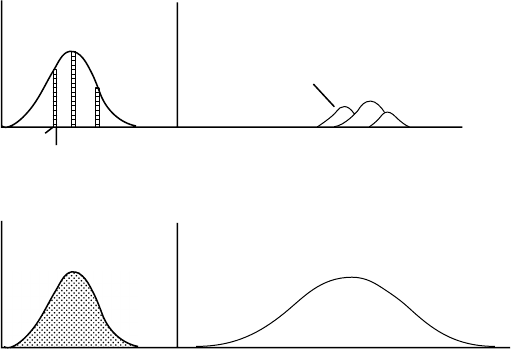
The convolution integral in summation form becomes (Figure 7.47)
C
0
(t) =
t
k=1
h(t +1 −k)C
a
(k) (7.210)
Systems in Nuclear Medicine 421
C
a
(k)
(a)
(b)
C
a
(t)
k–1
k
k
h[t–(k–1)]
1
1
2
2
3
3
C
0
(k)
C
0
(t)
C
0
(t) =
t
t
∑
k = 1
h(t+1–k)C
a
(k)
Δk = 1
FIGURE 7.47
(a) Input function is decomposed in a sequence of time shifted delta functions represented by
small rectangles whose area is equal to the mean value of the ordinate. To each of these functions
corresponds an output numbered as 1, 2, and 3. (b) The summation of all input delta functions
is C
a
(t) and of all the output curves is C
0
(t).
where k takes only integer values for integer values of t.
Theconvolution processcan bevisualized if theinput functionI(t) isdecom-
posed in an infinite number of time shifted delta functions with amplitude
equal to the value of the function in each time and considering as final result
the summation of the responses to all these delta functions.
Most of the situations in practical NM need a discrete approximation of con-
volution. When the functions involved in convolution are sampled functions,
the convolution integral becomes
R(k) =
k
n=0
h(k −n)I(n) (7.211)
where R(k), h(k − n), and I(n) are the sampled values of the functions.
Equation 7.211 can be written in matrix form as
R = H × I (7.212)
where I is a triangular matrix with elements (k + 1) (k +1) with n +1 line
consisting of terms I(n), I(n −1), I(n −2),…, I(0) followed by (k − n) zeros.
H and R are column matrixes with k + 1 elements.
In Figure 7.47a, the input function is decomposed into a sequence of delta
functions represented by small rectangles whose area is equal to the mean
422 Nuclear Medicine Physics
value of the ordinate. To each of these functions corresponds an output func-
tion numbered as 1, 2, and 3. The summation of all input delta functions is
C
a
(t) and the summation of all the output curves is C
0
(t).
For example,
C
0
(t) = h(1)C
a
(1) (7.213)
C
0
(2) = h(2)C
a
(1) +h(1)C
a
(2) (7.214)
.
.
.
C
0
(t) = h(t)C
a
(1) +h(t −1)C
a
(2) +...+ h(1)C
a
(t) (7.215)
This gives a system of t simultaneous linear equations, the solution of which
yields t values for h.
The fitting procedures have included several functions (sum of exponen-
tials, Fourier series, etc.) to fit the frequency functions to formal expressions.
7.2.8 Data Analysis and Models in PET Studies
Immediately after reconstruction, SPECT and PET images are static maps of
values proportional to activity in units of counts per second per pixel.
These values are generally converted to units of counts per second per
milliliter of tissue by calibration with a known volume of tracer activity.
NM images can be used as static, dynamic, or parametric entities [30–32].
When the biological processes under analysis evolve slowly over time, static
images may be good enough to supply the wanted information (e.g., tumor
detection). A color look-up table is often used to assign a color to each pixel
value in static images.
When functional information is wanted, dynamic imaging acquisition
is required. For example, it may be necessary to estimate the parameters
of a compartmental model that supposedly fits data from PET or SPECT.
These kinetic parameters are important, because they quantify physiological
processes.
In dynamic imaging, series of scans are acquired by continuous scanning,
often for long times; and after the reconstruction of sequences of PET images,
some kind of spatiotemporal quantification is associated with the activity
data.
One common approach, for a sequence of PET or SPECT images, is to get
the variation over time of the mean value of pixels in ROIs that have been
previously rescaled and aligned. To help with the definition of anatomical
ROIs in the brain, the higher-resolution MRI or CT is commonly used. The
TACs are generated with data points that correspond to the mean pixel value
in a specific ROI at a given time.
Systems in Nuclear Medicine 423
These TACs can be further normalized by plotting, from point-to-point, the
ratio of the values of one ROI TAC to the values of another ROI TAC that is
taken as a reference.
For quantification some reference is needed, and this can be the tracer con-
centration in arterial plasma (blood sampling required) or some other tissue
(a different area of the same image).
In PET studies, the tracers are introduced into the body by intravenous
injection or inhalation. The tracers are mixed with blood in the heart chambers
to give a nearly constant concentration in arterial blood (model input). The
concentration of the tracer delivered to the tissue capillaries can be obtained
from any peripheral artery, and the amount of tracer delivered to the tissue
is proportional to the blood flow (perfusion).
Some of the possibilities for finding the input function to an organ are by
external measurement in the left ventricle, aorta, or large artery as a function
of time (i.e., the respective TAC). This method, which requires fast response
equipment to cope with the temporal sampling requirements of the particular
input function, is also limited to those cases in which the left ventricle or a
great vessel is located within the limits of the image [33,34].
Statistical methods that will be discussed later can also be used to effectively
obtain these input functions.
Depending on their biochemical properties, the radiotracers used in PET
studies are grouped in two broad categories.
The first category comprises the nonspecific radiotracers that trace a bio-
chemical path and lead to the measurement of the extraction parameters
of a tissue which describe its uptake and metabolism. Examples of these
radiotracers are
15
OH
2
, which is an inert, freely diffusible tracer used to mea-
sure cerebral blood flow;
18
FDG, which traces the initial phases of glucose
metabolism but does not follow Kreb’s cycle after phosphorylation and is,
therefore, effectively retained in cells, permitting the evaluation of glucose
metabolism in tissue; and
18
F-fluoromisonidazole, which is a bioreductor
drug that follows an intracellular path of reduction and can be used to localize
viablehypoxic tissue.The kineticsof theradiopharmaceuticals ofthis kindcan
be evaluated through simple systems consisting of one or two compartments
apart from plasma.
The second category includes specific radiotracers involved in interactions
with a receptor, a carrier, or a specific interaction site. Examples of these radio-
tracers are
11
C-flumazenil, which is an antagonist with high affinity and selec-
tivity for central benzodiazepine receptors, and
11
C-SCH23390, an antagonist
withhigh affinityand selectivityfor dopamine D1receptors. Both radiotracers
are used to study alterations in the density and affinity of central recep-
tors. The behavior of the radiopharmaceuticals in this category is generally
assessed by means of three-compartment models: free, nonmetabolized lig-
and in plasma; free ligand in tissues; and ligand specifically bound to tissues.
In tracer studies, the data acquisition methodology and the recommended
data analysis method depend on the aim and type of study being performed.
424 Nuclear Medicine Physics
Different approaches have been used for the kinetic modeling of images
from SPECT and PET tracer studies, with the purpose of estimating quanti-
tative biological parameters.
Despite their many differences, these methods—mostly only used for brain
PET studies—can be roughly placed into general categories; for example, in
those based on data (data-driven), where no models or compartments are
assumed, and those that use models (model-driven) which tend to assume a
compartmental system.
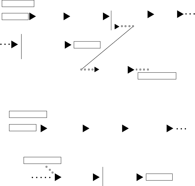
The data-driven methods require no previous decision about a model
associated with the system (Figure 7.48).
After reconstruction, the images are used to draw ROIs and obtain TACs
that will be subjected to one of several graphical procedure sequences. These
correspond to the path 3-4-5-6 in Figure 7.48.
Tracer ROI curves can also be used for modeling. This corresponds to the
path 3-4a in Figure 7.48 and 4a-5a-6a in Figure 7.49.
Data driven
Raw data
MTGA
compart. models
spect. Analysis
SUV
Model
calculations
(blood data)
Reconst.
12 3
3
3
4
4a
5
5
6
PET image
Drawing ROIs
Regional
TACs
To model driven
Results
FIGURE 7.48
Data-driven methods for data analysis in NM.
Model
calculations
(blood data)
1a 2a 3a 4a
4a 5a
Paramatric
sinogram
Paramatric
images
Reconstruction
Drawing ROIs
SPM
Model driven
From data driven
Sinogram
Results
FIGURE 7.49
Model-driven methods of analyzing NM data.
Systems in Nuclear Medicine 425
The ROI TACs are fitted to mathematical models to estimate parameters
that are biochemically or physiologically meaningful (path 4-5). These, when
the input function is known, can be rate constants, receptor densities, or blood
flows. The input function is usually the arterial plasma activity that is sampled
during the PET study and counted in the well counter. This approach allows
comparison of the model parameters in PET studies.
Multiple time graphical analysis methods and spectral analysis are gener-
ally used to quantify the dynamics of the processes.
The MTGA is a series of data-driven graphical procedures used to quan-
tify PET data that involve the study of the distribution and accumulation
of radioactive tracers in organs and tissues over time. The technique is an
alternative to compartmental models, as it does not assume an imposed
chain of predetermined events and it is suitable for both homogeneous and
nonhomogeneous tissues.
The data are usually acquired as a series of dynamic frames, beginning with
the tracer injection and continuing until either the statistics of the images is
compromised by prolonged decay or there is a limitation in memory storage
capacity.
The MTGA methods (Patlak and Logan plots) employ a transformation of
the data such that a linear regression of the transformed data yields macrosys-
tem parameters which are computed with better reproducibility from fewer
data than the separate rate constants [35,36]. By plotting these macroparam-
eters, the functional parameters are directly estimated from the slope of the
linear phase of the curves obtained.
Some negative points about these methods are the uncertainty about the
choice of the point in the plot when linearity begins, the possibility of bias
introduced by statistical noise [29], and the failure to return any information
about the underlying compartmental structure.
Spectral analysis [35] characterizes the system’s impulse response function
(IRF) as a positive sum of exponentials and uses nonnegative least squares to
fit a set of these basic functions to the data. The macro system parameters of
interest are then calculated as functions of the IRF [34,35]. Spectral analysis
also returns information on the number of tissue compartments evident in
the data and is defined as a transparent technique.
Schmidt [30] showed that for the majority of plasma input models the obser-
vation of all compartments led to only positive coefficients; and, as such, the
spectral analysis [35] solution using nonnegative least squares is valid.
The ROI-based methods may be further classified into linear and nonlinear
methods. The former transform the data so that the parameters of interest
can be estimated by linear regression methods, whereas nonlinear techniques
generally estimate the kinetic parameters by iterative minimization.
The two different types of analysis (Patlak and Logan) are used according
to the type of situation.
The Patlak-plot applies to irreversibly binding radiotracers and involves
creating an ordinate and an abscissa using combinations of the input function
426 Nuclear Medicine Physics
and image data at each of the observed time points for every PET frame. A
TAC is obtained for a particular ROI.
For each time point, the ROI values represent the overall concentration of
radioactivity in the tissue, which can differ from the tracer concentration.
A TAC is created for the input function Cp(t), the concentration of tracer
in the plasma, by interpolating the measured plasma curve to the middle of
each frame.
For every image frame, the ratio of the ROI value in that frame divided by
the plasma concentration is plotted as a function of the integral of the plasma
curve (from t = 0 to the current time) divided by the plasma concentration at
that time point, that is,
ROI(t)/C
p
(t) versus
t
0
C
p
(t)dt/C
p
(t) (7.216)
where ROI(t) is the concentration of a tracer in a tissue ROI, t is the time
elapsed from injection to a particular frame, and Cp(t) is the concentration of
tracer in plasma. In these instances, the net influx rate K
i
(min
−1
) equals the
slope of the Patlak plot, that is, k
1
×k
3
/(k
2
+k
3
).
This method can be thought of as a way of scaling the tracer concentration
in a region of tissue by the capacity of the tissue to absorb the tracer over time.
However, it was noticed that this plot becomes linear at those time instants
when the transport of tracer trapped into the compartment is essentially
unidirectional.
Patlak showed that the slope of a straight line fitted to this linear region
is equivalent to the influx constant, K
i
, for the system. K
i
can be defined as
the ratio of the total amount of tracer accumulated in a tissue region after an
infinite amount of time, divided by the integral of the plasma TAC from t = 0
to infinity.
Graphical methods are easy to perform and are generally considered more
robust than kinetic analysis with (full) compartmental models, especially for
noisy data sets.
The traditional linear regression model takes into account only the errors
in the Y variable.
The bias in the (reference input) Gjedde–Patlak slope may be diminished
by using a linear regression model that accounts for errors in both variables
[32]. This method can also take into account separate weights for both
variables.
In another work (1985), Patlak has shown that the technique still applies
when the tracer is simply “nearly” irreversibly bound and that a tissue ref-
erence region, with no specific tracer uptake, can be used as input function
instead of the measured blood TAC.
Systems in Nuclear Medicine 427
This analysis is used in neurotransmitters ([
18
F]-F-DOPA, [
18
F]-FMT, [
11
C]-
raclopride, etc.), metabolism markers ([
18
F]-FDG), and even for cardiac
applications.
The Logan plot is a graphical linearization method that applies to reversibly
bound tracers which have a constant net efflux that is nonnegligible when
compared with the constant influx. The approach involves creating a plot
with the following coordinate axis:
0
ROI(t)dt/ROI(t) versus
T
0
C
p
(t)dt/ROI(t) (7.217)
where ROI(t) is the concentration of a tracer in a tissue ROI, t is the time
elapsed from injection to a particular frame, and Cp(t) is the concentration of
tracer in plasma [33].
Similar to the Patlak plot, the slope and intercept of a Logan plot have
distinct interpretations depending on the actual model one chooses to ascribe
to the underlying system.
The straight line in a Logan plot should be fitted through those frames that
are linear; these frames usually occur somewhere in the middle of the plot.
This is another way of saying that data should be collected over a long enough
period to see a net efflux of tracer from the ROI.
A formal analysis of the Logan plot shows that it is valid for an arbitrary
number of compartments for both plasma and reference input models when
the data are free from noise.
7.2.9 Parametric Analysis
Parametric analysis is required when the studies involve complex analy-
sis and advanced statistical methods to localize the regional distribution of
functions, particularly in situations in which input function is affected by
noise. Parametric images incorporate the highest degree of quantification and
require highly developed statistical methods that are mostly used in brain
studies.
In parametric analysis, information is obtained up to the image resolution
level,that is, the parameters arecalculated for each image voxel and the results
are represented as maps of the parameter.
The values bound to the image voxels are the physiological parameters
studied (perfusion, glucose consumption, receptor density, etc.).
Existing methods for computing kinetic parametric images work by first
reconstructing a sequence of PET images and then estimating the kinetic
parameters for each voxel in the imaged volume. Special computer programs
convert the dynamic information to parametric functional information.
Parameter estimates are obtained from a priori specified compartmental
structures using oneof a variety of least-squaresfittingprocedures:linearleast