Lima J.J.Pedroso, de (ed.). Nuclear Medicine Physics
Подождите немного. Документ загружается.


158 Nuclear Medicine Physics
5.2 Properties of Image Detection Systems
The quality of medical images is mainly related to the capability of detecting
relevant information. The image quality depends on its information content
and how accessible the information is.
The properties of imaging systems are usually expressed in physical terms.
Several concepts are used to describe imaging systems in terms of quality of
the response: spatial resolution, sensitivity, contrast, and noise.
There are complementary physical quantities, which are used together with
these basic concepts to enhance their objectivity. The most important com-
plementary quantities are the point spread function (PSF), the modulation
transferfunction (MTF), the Wiener spectrum, andsignal-to-noise ratio(SNR).
In addition, we have the Rose model with the contrast–detail curves and
ROC analysis (receiver–operatorcharacteristics) asintermediaries, which also
unify concepts that include basic properties.
The detector is obviously one of the most important parts of imaging
systems, which, in the case of NM, are gamma radiation detectors.
Some desirable properties of γ-ray detectors are the high effective atomic
number, high density, high photopeak fraction, and short decay time.
∗
The
first two properties are related to the sensitivity of the detector, the third to
selectivity in photon detection, and the fourth to the ability to process high
activities [34].
Other properties, such as high luminosity efficiency, large detector area,
and the low refractive index, may be important in specific contexts.
Some of the generally accepted parameters for measuring the properties of
the image detection systems are briefly considered in the next section.
5.2.1 Distance of Resolution: PSF
The spatial resolution is related to the sharpness or detail of the images.
The performance of the devices for medical imaging, in terms of sensitivity
and spatial resolution,can be entirely defined by the PSF, which is the function
that describes the image when the object is a point.
If B(x, y) is the PSF for a 2D imaging system and is symmetric about a central
axis, then the response of the system can be fully described by the curve B(x)
in an axial plane that intersects B(x, y).
The volume under the surface B(x, y) is proportional to the system sen-
sitivity.
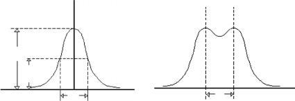
For a system that gives an isotropic response, the width at half maximum
of the curve B(x) is the distance of resolution, d (Figure 5.14a).
∗
In the case of scintillation detectors, the time decay is the time required for the scintillation
emitted to fall by a factor equal to e
−1
.
Radiation Detectors and Image Formation 159
B
(a) (b)
d d
X
X
h
h/2
FIGURE 5.14
(a) The distance of resolution, d, is the width at half maximum of the curve B(x), that is, the PSF.
(b) Two points to the distance d are separated with difficulty in the image.
Two points located at distance d are separated with difficulty in the image
(Figure 5.14b).
For digital systems, the resolution can be associated with the number of
pixels representing the image (typically 128 ×128, 512 ×512, etc.) when d is
smaller than twice the side of the pixel.
If the PSF is a Gaussian function with standarddeviation σ, then the distance
of resolution is d = 2.35σ.
A perfect 2D imaging system would present a point for each object point.
Degradation occurs in real devices, and images of points appear as an area
whose intensity decreases from the central region to the periphery. Consid-
ering that each activity distribution can be represented by a set of points, the
loss of sharpness for the images of large sources is caused by the overlapping
of the images of the numerous points of the object.
The function that represents the 2D response of an imaging system to a
point object of unit intensity is the function of the degradation of a point or
the normalized point spread (PSF), h(x, y).
Since the PSF can vary with direction, the line spread function (LSF) is often
used along an axis of an orthogonal coordinate system.
The LSF hL(x) can be calculated from h(x, y),ifh(x, y) is symmetric and
independent of the direction.
In general, for gamma cameras the PSF is approximately constant in planes
perpendicular to the collimator axis. Therefore, for a 2D image at a distance
z in front of the collimator, the image g(x, y) is obtained by 2D convolution of
the object function f (x, y) with h(x, y), that is,
g(x, y) =
+∞
−∞
+∞
−∞
f (x −x
, y −y
) h(x
, y
) dx
dy
. (5.21)
The output is therefore, an overlap of displaced versions of the PSF
weighted for each position to the input amplitude in that point.
Equation 5.21 can be applied if the image is linear and invariant to dis-
placements. For NM systems, linearity and invariance in space are only
160 Nuclear Medicine Physics
approximately verified owing to the effects of dead time and nonuniformity
of response.
Equation 5.21 can also be presented as
g(x, y) = f (x, y) ∗h(x, y), (5.22)
where * means convolution.
Equation 5.21 can be applied to sections of homogeneous objects provided
that the PSF, corrected for attenuation, is known in a number of planes
perpendicular to the center line of the collimator.
Equation 5.21 takes into account the degradation factors of the imaging
system, such as those that depend on the device characteristics, from the
acquisition to the interpolation in the screen display. However, it does not
consider noise. A more objective approach to modeling the process of images
would be
g(x, y) = f (x, y) ∗h(x, y) + η, (5.23)
where η represents the noise.
The increase of the detector thickness in scintillation cameras causes degra-
dation of the intrinsic spatial resolution. This can be improved, however, by
increasing the energy of the photons and increasing the number of PMTs.
5.2.2 Modulation Transfer Function
The information contained in the PSF, defined above, is rarely used directly;
another quantity is used instead, the modulation transfer function (MTF),
which is more convenient for some purposes. The MTF is based on the con-
cept that all radioactivity distribution in a plane normal to the axis of the
collimator can be expressed by a series of sinusoidal components, relative
to two rectangular coordinate axes, and having their own spatial frequen-
cies, amplitudes, and phases. The frequencies of the various components are
integer multiples of a frequency usually termed fundamental.
Activity distributions with high variations or activity that show fine detail
have a large contribution from high-frequency components in their spectral
content, whereas images that involve only low spatial variation essentially
have only low-frequency components [35].
Based on this interpretation and using the Fourier analysis, it is assumed
that the images obtained by a particular device are approximate descriptions
of the objects in terms of sinusoidal components with features that depend
on the device.
Hence,images ofobjects witha simplesinusoidal structurecan beemployed
to assess the response of imaging systems.
The MTF expresses the response of the system, the system components,
or the modulated sinusoidal distributions of the object parameter when the
frequency varies.
Radiation Detectors and Image Formation 161
Y
o
X
Y
Act.
Y
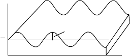
FIGURE 5.15
Object consisting of the sum of a constant value of activity, with activity sinusoidally modulated
in the xx direction.
An instrument to study the response of an NM imaging system could be a
set of activity distributions consisting of sums of sinusoidal modulations in
the direction of the xx axis, with different spatial frequencies summed to a
constant value (y;
−
) as shown in Figure 5.15.
For each spatial frequency the MTF is the ratio of the contrast in the image
to the contrast in the object and represents the fraction of the information of
the object that is retained in the image. An ideal system has a unit value for
MTF at all frequencies.
For a sinusoidal activity variation, the modulation or contrast is the ratio
between the AC component (half of the peak-to-peak value of activity) and
the average activity, or DC component.
The image of a sinusoidally modulated object is supposed to be a sinu-
soidally modulated image with the same frequency, although the amplitude
and phase can be different.
Ageneral expression for MTF can be obtained from Equation 5.24 by apply-
ing the 2D Fourier transform to both members of the equation and performing
some transformations.
Since the Fourier transform of the convolution between two functions is
equivalent to the product of the Fourier transform of the two functions, we get
G(ω
x
, ω
y
) = F(ω
x
, ω
y
)H(ω
x
, ω
y
), (5.24)
where the functions G and g, F and f , and H and h are pairs of Fourier trans-
forms. G represents the output spectrum, F the input spectrum, and H a
characteristic function of the system defined in the frequency domain.
The angular frequencies ω
x
and ω
y
are related to the spatial frequencies v
x
and v
y
in directions x and y by
ω
x
= 2πvx and ω
y
= 2πvy. (5.25)
The DC contribution to the frequency spectrum of f and g are the values F
and G when ω
x
= ω
y
= 0, that is, F (0,0) and G (0,0). Then
G(0, 0) = F(0, 0)H(0, 0). (5.26)

162 Nuclear Medicine Physics
The Fourier transform of the function of response to a point h (x, y) is by
definition
H(ω
x
, ω
y
) =
+∞
−∞
+∞
−∞
h(x, y)e
−j(ω
x
x+ω
y
y)
dx dy. (5.27)
The Fourier transform of h (x, y), H (ω
x
, ω
y
), is usually called the transfer
function of the imaging device, which is a quantitative measure of the ability
of the system to preserve the contrast and sharpness of the image.
The Fourier transform of the PSF when ω
x
= ω
y
= 0 is given by Equation
5.28.
H(0, 0) =
+∞
−∞
+∞
−∞
h(x, y) dx dy = ξ (5.28)
H(0,0) is consequently the total information transferred in 1 s to the plane
of the image when the object is a source point with a disintegration speed of
one disintegration per second, that is, the camera efficiency ξ.
The MTF value (Equation 5.29) is the ratio between the image and object
modulations, considering that they are sinusoidal functions (second term
of equation). The third member of Equation 5.29 is given by applying
Equation 5.24:
MTF =
G(ω
x
, ω
y
)
G(0, 0)
F(ω
x
, ω
y
)
F(0, 0)
=
H(ω
x
, ω
y
)
H(0, 0)
(5.29)
The final result for the MTF can be obtained as the quotient between the
H(ω
x
, ω
y
) and H(0, 0), that is, the transfer function divided by the system
efficiency.
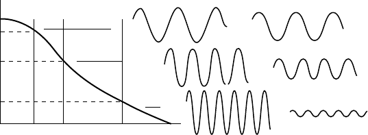
The MTF is defined for all frequencies as the ratio of the modulations or
contrasts in the image and in the object (Figure 5.16).
We can also write
MTF =
Image Modulation
Object Modulation
. (5.30)
The MTF is a decreasing function of frequency. For spatial frequencies
higher than c, called the cutoff frequency, the contrast of the object is not
transmitted to the image, and the MTF is zero.
Cutoff frequencies for some imaging diagnostic methods are
• Film/screen: 10–20 line pairs/mm
• CT scanner: 1–2 line pairs/mm
•
Gamma camera: 0.3 line pairs/mm
• Image intensifier tube with CSI screen: 4–5 line pairs/mm
Radiation Detectors and Image Formation 163
100
MTF %
Object
Lines/mm
Image
80
60
10
v
1
v
2
v
3
v
c
FIGURE 5.16
Modulation transfer function vs. spatial frequency.
5.2.3 Detection Efficiency or Sensitivity
The efficiency of detection or sensitivity applies to the specific energy of the
radiation used and measures the fraction of energy emitted by the source that
is actually employed by the detector to produce the image. In general, the
efficiency of a detector can be divided into three parts: geometric efficiency,
quantum (or intrinsic) efficiency, and conversion (or extrinsic) efficiency.
The first is related to the geometric scheme exploited in detecting the radi-
ation. The second is the fraction of incident energy that is absorbed in the
detector. The thirdmeasures the yield from the transformation of the absorbed
energy by the detector into useful signals.
5.2.3.1 Geometric Efficiency
The geometric efficiency, E
g
, is the relationship between the solid angle seen
by the detector and solid angle of emission. Essentially, it depends on the size
of the detector and on the source–detector distance. The presence of dead
regions in detectors must often be considered: at the edges of the detectors or
between detectors that have been combined. The last effect is very important
in detectors whose large area of detection is achieved by joining up small
detector units. In these cases, the fill factor is defined as the quotient between
the effective area of detection and total area.
5.2.3.2 Quantum Efficiency
The quantum efficiency, E
Q
, measures the probability of interaction, that is,
the fraction of energy of incident photons that is absorbed by the detector:
E
Q
= 1 −e
−μx
d
, (5.31)

164 Nuclear Medicine Physics
where x
d
is the thickness of the detector and μ is the linear attenuation
coefficient of the detector material.
Therefore, the quantum efficiency depends on the μ of the detector material,
the thickness of the detector, and the photons’ energy. Quantum efficiency is
modified by absorbent materials that are placed in the path of incident rays,
suchas theprotectionthat enclosesthe detectorssuch aswindows (aluminum,
glass, titanium, etc.), grids, and collimators.
Equation 5.31 presupposes that any energy transferred by incident photons
to the detector produces a usable signal, despite a fraction being discarded
when there is energy selection.
The human eye operating in the visible range of the electromagnetic spec-
trum (λ from 400 to 700 nm) has an E
Q
of nearly 1%. For the same energy
range, the E
Q
of the film is typically 5–20%; and for CCDs, it is 50–90%.
5.2.3.3 Conversion Efficiency
The efficiency of conversion, E
c
, is the fraction of the photon energy that is
absorbed by the detector and that is converted into a measurable signal, either
electrical or luminous.
5.2.3.4 Total Efficiency
The total efficiency(or sensitivity) of a detector is the productof the geometric,
quantum, and conversion efficiency, E
t
= E
g
×E
Q
×E
c
.
In addition, the total efficiency of the detector depends on the dead time
of the system. This is the period after detection during which the detector is
unable to carry out a new detection.
A system with high sensitivity includes more information in the images in
the same time than a system with lower sensitivity can.
5.2.4 Noise
The LSF and MTF of the imaging systems previously described were
considered under noise-free conditions.
Due to the statistical nature of the production of gamma photons, random
fluctuations in the radiation intensity emitted by radionuclides are expected.
These are recognized as Poisson noise and cause degradation of the contrast.
The probability distribution for p photons in a time of T seconds, when the
average intensity is i photons per second, is
P(p; i, T) =
(iT)
p
e
−iT
p!
. (5.32)
The interaction with the detector can be considered binomial with an E
Q
success probability.

Radiation Detectors and Image Formation 165
The distribution of the photons that interact is the Poisson distribution with
standard deviation equal to
σ = (iTE
Q
)
1/2
, (5.33)
where iT is the number of incident photons. The SNR is
SNR = 10 log
10
(N
0
E
Q
) dB. (5.34)
If the detection process is followed by a process with gain g, the average
signal amplitude will be
S = N
0
E
Q
g. (5.35)
The standard deviation of this quantity should include the noise and the
noise gain in amplification process, g. Considering the two sources, the total
noise is
σ
s
=
N
0
E
Q
g
2
+σg
2
(1 +N
0
E
Q
)
1/2
. (5.36)
In modern systems, the noise introduced by the amplifiers is usually neg-
ligible. In order to completely define the noise, it must be known how the
signal and noise are affected by spatial frequency.
The power spectrum or Wiener spectrum, which describes the noise as a
function of the spatial frequency, allows a complete description of the noise.
It tells us what MTF frequencies contain the most contribution to noise.
White noise corresponds to equal power at all frequencies.
Imageprocessingsuchas filtering orreconstructionmaychange the noise
level.
5.3 Methods of Image Production in Nuclear Medicine
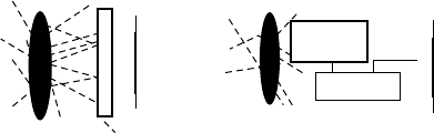
The two ways to obtain images in NM are considered in general in
Figure 5.17.
a. Includes all types of conventional NM imaging except for SPECT, that
is, static, whole body, and dynamic scintigraphy and scintigraphy
with synchronized acquisition
b. Includes the SPECT and PET techniques
The selective retention of compounds marked with γ emitters enables
images of the distribution of the tracer to be obtained that not only include
morphology aspects but also contain data on the metabolic capabilities of
166 Nuclear Medicine Physics
ObjectObject
Receptor
Image
Image
Collimator
(a) (b)
Computer
FIGURE 5.17
(a) Images obtained by direct emission. (b) Images obtained by indirect techniques.
the organs, with local and frequently quantitative information. The NM can
depict, through images, the spatiotemporal distribution variations of the
biomolecules that compose the human body.
These images are plane orthogonal projections of the local concentration
of a radiopharmaceutical that is present in a partial volume of the body, and
they are usually called planar images, or simply scintigraphies. The images
are similar to 2D maps where one dimension has been removed from the
original 3D distribution.
Dynamic studies can also be achieved by acquiring rapid sequences of pla-
nar images. This capability is important, because a static image of a biological
system obtained at a certain instant provides little information, as dynamic
processes are the essence of physiology.
The specific functional content of the NM techniques was a decisive step
forward in the study of the metabolic dynamics of numerous organs.
The radial acquisition around an object followed by reconstruction enabled
the acquisition of tomographic maps of radionuclide concentration. This
methodology, when used with the detection of a single photon, is known
as SPECT and offers a better contrast than planar scintigraphy. Moreover, the
acquisition of several transverse sections of the activity distribution allows
a reasonable reconstruction of its space location. The availability of rotating
gamma cameras at a reasonable cost has enabled SPECT to become a standard
technique in conventional NM.
The most important factors to be consideredwhen choosing the scintillation
detectors to detect gamma radiation for the purpose of obtaining images are
the field of view, the stopping power of the detector material, the efficiency,
the response time, and the energy resolution.
Scintillation cameras have steadily improved and they are now almost at the
top of their performance relative to cost. This performance has been achieved
even though the NaI (Tl) scintillator, universally used in gamma cameras,
does not offer good energy resolution or good time response, besides having
other undesirable physical properties such as high hygroscopicity. The main
strengthsof the NaI (Tl) are its high attenuation coefficient for medium energy
photons and high luminosity.
Radiation Detectors and Image Formation 167
5.3.1 Gamma Camera and SPECT
The scintillation camera, the imaging device restricted to conventional NM, is
a position sensitive gamma radiation detector; and it is used in more than 90%
of routine studies using γ rays of 140 keV from technetium 99m (
99m
Tc). This
is the radionuclide used in over 90% of diagnostic studies in NM, because
it emits virtually no particles; it has an almost optimal gamma energy for
detection by the NaI (Tl), because it has excellent properties with regard to
the chemical labeling of molecules; it has a short period; and, finally, because
it can be produced through generators.
The gamma camera was developed by Hal Anger [23] and has not under-
gone major changes since its creation in 1958. For this reason, the gamma
camera is also known as the Anger camera. The detection portion of the
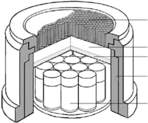
gamma camera, the head (Figure 5.18), consists mainly of a crystal of sodium
iodide activated with thallium, NaI (Tl), in the form of a disk or rectangle
30–50 cm wide and 0.9–1.2 cm thick. This is enclosed in a hermetic aluminum
cylinder, coated with a layer of a light diffuser and equipped with a trans-
parent base (Lucite), a set of (60–90) photomultiplier tubes optically coupled
to the transparent window of the crystal, a lead collimator, and some elec-
tronics. The thickness of the crystal is a compromise between intrinsic spatial
resolution and efficiency of detection.
The main function of the collimator is to limit the photons that interact with
the crystal to only those that have a particular path relative to the detector
surface, which allows 2D projections of the activity distribution of the tracer
molecule to be obtained. The selection of photons makes the detection less
efficient. Typically, 99% of the emitted photons are stopped at the collimator.
Nevertheless, it is essential to the formation of a projection that is spatially
correlated with the activity distribution of the radiolabeled molecule. The
collimatorsignificantly reducesthesensitivity ofthe camerawhile, atthe same
time, it affects the spatial resolution of the gamma camera and makes it depen-
dent on the distance of the object to the detector and on the characteristics of
the collimator.
Collimator
Crystal Nal(TI)
Optical interface
Shielding
Photomultiplier
tube
FIGURE 5.18
Diagram representing the components of the gamma camera.