Leroy C., Rancoita P.-G. Principles Of Radiation Interaction In Matter And Detection
Подождите немного. Документ загружается.

January 9, 2009 10:21 World Scientific Book - 9.75in x 6.5in ws-bo ok975x65˙n˙2nd˙Ed
770 Principles of Radiation Interaction in Matter and Detection
isotope
18
F, which has a half-life of 110 min (Table 11.2) and decays via positron
emission.
18
FDG is a sugar analogue, where one or several of the hydrogen atoms
are substituted by a
18
F atom. FDG accumulates in organs where glucose is used,
as the primary source of energy and therefore FDG is used for instance in studies of
the glucose metabolism of the brain and heart. The traditional method of producing
18
FDG consists of using a proton beam of about 10 MeV on a target of enriched
water H
18
2
O via the reaction
18
O(p,n)
18
F. Such proton beams are available at many
Van der Graaff tandem accelerators and cyclotrons lo cated close to where the pa-
tient is treated. This reaction cross-section has a threshold of around 2.57 MeV
and resonance around 5.13 MeV with a maximum cross-section of 697 mb. The rate
of production of fluorine-18 from 100%
18
O-enriched water targets can be calcu-
lated as a function of the proton energy. For instance, for a 10 MeV proton beam,
the proton range in water is 119 mg/cm
2
and the production rate of fluorine-18 is
39.1 mCi/Ah. Taken into account the energy dissipation in the front target foils
(estimated to 0.5 MeV), one has about 70 mCi/h fluorine-18 yield, for 2 µA beam,
corresponding to a
18
FDG yield of 20 mCi/h.
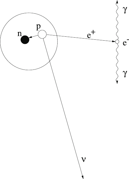
The positron emitted by the radioactive tracer or radiopharmaceutical, via the
decay p → n ν
e
e
+
([Z, A] → [Z − 1, A]ν
e
e
+
), annihilates very close to the emission
point (≤ 1 mm) with an electron of the body to pro duce a pair of 511 keV photons
emitted back-to-back (Fig. 11.5). The effective range of positrons and end point
energy for most used isotopes in PET are shown in Table 11.2. These photons
traverse the body and enter the active medium of the PET detector placed on a
ring.
The PET camera is detecting two photons emitted back-to-back in coinci-
dence. The observed pair of back-to-back photons defines an axis along which the
disintegration of the radioactive element has taken place. The line connecting two
detected photons is called a chord. The time correlation between detected photons
permits the selection of pairs in coincidence and their association to a chord. The
positron emitters can then be traced back as they participate in biological pro-
cesses. The simultaneous detection of several pairs of photons indicates the rate of
disintegration along different axes and enables one to determine the distribution of
the compound in the body and to draw conclusions as to the proper functioning of
tissues and organs. The time interval between the detection of these two photons
is a few ns F W HM, typically (2–5) ns. A good timing resolution minimizes the
accidental coincidence rate and permits the use of the arrival time difference to
determine the radioisotope position along the chord.
An excellent spatial resolution (< 5 mm F W HM [Moses, Derenzo and Budinger
(1994)]) is needed along the two directions corresponding to the axial and trans-
axial directions of the tomographic devices. The detector spatial resolution helps to
achieve the quality of spatial resolution in the reconstructed image.
The photon detector must combine an angular coverage, large enough to inter-
cept the photons of interest, and a high spatial resolution, in order to account for
January 9, 2009 10:21 World Scientific Book - 9.75in x 6.5in ws-bo ok975x65˙n˙2nd˙Ed
Medical Physics Applications 771
Fig. 11.5 The positron emitted by the radioactive tracer or radiopharmaceutical annihilates with
an electron of the body to produce a pair of 511 keV photons emitted back-to-back.
the details of the body area under investigation, as resolved as possible.
Optimized spatial, timing and energy resolutions are factors determining the
sensitivity of the detector and permit the reduction of the dose, injected into the
patient.
The use of readout electronic chain with low noise is of great importance in
order to optimize the signal-to-noise ratio. Good energy resolution typically means
< 100 keV [Moses, Derenzo and Budinger (1994)]. Good energy resolution helps the
rejection of secondary photons produced by Compton scattering of primary photons
from the source (the ones of interest) on biological matter ( tissues, bones, . . .).
Strict mechanical constraints apply to the building of imagers. The ensemble of
active elements has to be well mechanically adjusted, avoiding cracks. The absence
of collimator, an heavy metal layer, in PET imagers helps the mechanical stability
of the system.
The detectors used in P ET scanners are composed of scintillation blocks (or
other active materials) and systems placed on a ring. The diameter of the ring defines
January 9, 2009 10:21 World Scientific Book - 9.75in x 6.5in ws-bo ok975x65˙n˙2nd˙Ed
772 Principles of Radiation Interaction in Matter and Detection
the distance between detectors. This distance influences the spatial resolution of
the imaging system. Better resolution is obtained for smaller distance. However,
smaller diameter also favors higher number of random coincidences. The granularity
of the scintillating crystal assembly, defined by the size of the crystal elements, is
the primary factor determining the spatial resolution. Higher segmentation lead to
better spatial resolution. About fifteen rings can be put on top of each other to
form an array of detectors that permits three-dimensional imaging. A resolution of
about 6 mm in each direction is achieved.
The selection of a crystal is made according to its potential energy resolution,
which depends on the mean number of photoelectrons. The noise of the readout
electronic chain is a limiting factor [Leroy and Rancoita (2000)].
The use of high atomic number (high-Z) material as active medium of the de-
tector gives high photon detection sensitivity and an acceptable pulse-height resolu-
tion. Sensitive materials such as NaI(Tl), CsI(Tl) and BGO are used. The assembly
of these crystals along a ring structure allows the building of large active medium
volumes providing the necessary large angular coverage. The crystals must have
large light yield and high detection efficiency for photons of 511 keV of energy for
PET and of 140 keV or less for SPECT. The light yields of NaI(Tl) and CsI(Tl)
are large compared with BGO. It is standard to choose NaI(Tl) as reference. The
relative light yield of CsI(Tl) and BGO are 0.40 and 0.15, respectively. The crys-
tals must have large stopping power for photon energy ranges faced in SPECT
and PET. High-Z and high density materials have to be selected. BGO has higher
density (7.13 g/cm
3
) than NaI(Tl) (3.67 g/cm
3
) and CsI(Tl) (4.53 g/cm
3
). K-edge
location has to be taken into account and would favor the use of BGO. The high
counting rate and, in some cases, the relatively short lifetime of the radioisotope re-
quire minimal dead time, of the order of a few µs. The decay times of BGO (300 ns)
and NaI(Tl) (250 ns) are comparable and much lower than the CsI(Tl) decay time
(1000 ns).
The medical imagers represent a large volume of active material and therefore
the cost of the active material is an issue. Most of the time, the imagers in opera-
tion in the medical field are purchased from commercial company (not universities)
following market prices. The price of crystals ranges from a few dollars to (10–15)
dollars per cm
3
.
The radiation hardness of the detecting material is also an element of consi-
deration. Although the detector is exposed to doses much smaller than those en-
countered in other fields (space, accelerator and reactor environments) radiation
degradation can possibly be observed with time and lead to detecting material re-
placement. Good radiation hardness extends the lifetime of the detecting devices,
avoiding frequent replacements.
Therefore, from the point of view of best performance for a crystal to be used
as the active medium of a medical scanner, one is looking for a crystal having a
light yield comparable to that of NaI(Tl), a density comparable to that of BGO,

January 9, 2009 10:21 World Scientific Book - 9.75in x 6.5in ws-bo ok975x65˙n˙2nd˙Ed
Medical Physics Applications 773
but with a decay time much smaller than BGO, while remaining affordable. Several
new types of scintillating materials have been or are being developed for a new
generation of medical scanners. Among these, the yttrium aluminium perovskite
(YAP:Ce) [Baccaro et al. (1995)] has a light efficiency of about 40% relative to
NaI(Tl), a density of 5.37 g/cm
3
, lower than BGO but higher than NaI(Tl) and
CsI(Tl). YAP:Ce has a rather high-Z value (Z = 39) which guarantees good photon
absorption. YAP:CE has a decay time of 25 ns which is another advantage over BGO
(decay time of 300 ns). The detection of two 511 keV photons by coincidence by two
YAP:Ce crystal bundles (5×5 pillars of 0.2 × 0.2 × 3.0 cm
3
) coupled to position
sensitive photomultiplier tubes have given a spatial resolution of 1.2 mm F W HM,
a time resolution of 2.0 ns F W HM and a large efficiency of 70% with a threshold
of 150 keV [Del Guerra (1997)].
Following the development of YAP, the Crystal Clear Collaboration [Lecoq
(2000)] has developed Luthetium Aluminium Perovskite (LuAP) crystals. LuAP
has a high light yield and a very short decay time of 18 ns. The density of LuAP is
also high (8.34 g/cm
3
). The peak emission of 380 nm is well adapted to avalanche
photodio de readout, allowing in turn compact detecting system.
PET detector modules can also be built from photon converter, readout by Mul-
tiwire proportional chambers (MWPC). The converter consists of either a high-Z
metal such as lead or tungsten or a combination of crystal such as BaF
2
/TMAE
gas. The 511 keV photon are converted into photoelectrons, which are collected by
a MWPC, generating a timing pulse and identifying the interaction position. This
type of scanner offers the advantage to be of moderate costs, no photomultiplier
being used. However they also present several disadvantages like the photon conver-
ter lower efficiency to detect single 511 keV photons {(10–30)% as opposed to 90%
for BGO [Moses, Derenzo and Budinger (1994)]}, decreasing the coincident even
detection efficiency. The number of photoelectrons per 511 keV interaction is very
small, causing a poor energy resolution. Poor limited spatial resolution {(5–11) mm
F W HM [Moses, Derenzo and Budinger (1994)]} for BaF
2
and poor timing resolu-
tion (88 ns [Moses, Derenzo and Budinger (1994)]) for high-Z metal converters are
also disadvantages.
11.3 Magnetic Resonance Imaging (MRI)
This imaging technique has an advantage compared to SPECT and PET since it
does not require the use of any radioactive material. Instead, it uses an intrinsic
property found in some nuclei: the non-zero nuclear spin. MRI uses magnetic fields
varying from 0.2 to 2 T and radiofrequency (RF) waves, to observe the magneti-
zation change of the non-zero spin nuclei. The hydrogen isotope,
1
H, which has a
nuclear spin of
1
2
, is a major component of the human body and will be used as the
main source of information.

January 9, 2009 10:21 World Scientific Book - 9.75in x 6.5in ws-bo ok975x65˙n˙2nd˙Ed
774 Principles of Radiation Interaction in Matter and Detection
11.3.1 Physical Basis of MRI
Let us consider the behavior of the nucleus of
1
H under the influence of a magnetic
field [Desgrez, Bittoun and Idy-Peretti (1989)]. The proton has a spin of
1
2
and,
therefore, has two observable states S
z
= +
1
2
or S
z
= −
1
2
. The energy difference
between the two states is:
∆E = hγB, (11.32)
where γ is the gyro-magnetic ratio which is characteristic of each atom. In the case
of hydrogen, γ = 42.58 MHz/T. For a 2 T magnetic field, this gives an energy of
35.1 µeV. The related frequency is 85.16 MHz, in the RF range, since the resonance
or Larmor frequency is given by:
ν = γB. (11.33)
As for the individual magnetization held within the nucleus, it is expressed by:
~µ = 2πγ
~
S. (11.34)
We have
~
M =
P
~µ, where
~
M is the net magnetization of the system. When a
magnetic field is applied, the majority of the nuclei will align in the same direction,
giving
~
M ∝
~
B
0
, according to a Boltzmann distribution:
N
−
N
+
= exp
µ
−
∆E
kT
. (11.35)
To simplify, let us choose
~
B
0
= B
0
~z. As previously seen, it is possible to
change the magnetization of a single nucleus, if it is reached by a photon of energy
E = hγB. A RF wave that equalizes the populations N
−
= N
+
, giving a net
magnetization of M
z
= 0, is called a saturation pulse or 90
◦
impulsion. After that
impulsion, the system will return to its equilibrium according to [Hornak (2002)]:
M
z
= M
0
³
1 − e
−t/T
1
´
. (11.36)
T
1
is called the spin-lattice relaxation time. If it is a 180
◦
impulsion (complete
inversion of populations), then the equilibrium will be recovered like:
M
z
= M
0
³
1 − 2e
−t/T
1
´
. (11.37)
A 90
◦
impulsion brings the net magnetization in the XY plane. It then starts
to precess around the z-axis at the Larmor frequency. To reach equilibrium, it will
decrease as:
M
xy
= M
xy
τ
³
e
−t/T
2
´
, (11.38)
where T
2
is the spin-spin relaxation time and τ , the time marking the end of the
impulsion. It decreases as the spins of the individual nuclei dephase. There is a
dephasing because each nucleus has its own magnetic field affecting the surrounding
nuclei. Therefore, the precession around the z-axis is done at several slightly different
January 9, 2009 10:21 World Scientific Book - 9.75in x 6.5in ws-bo ok975x65˙n˙2nd˙Ed
Medical Physics Applications 775
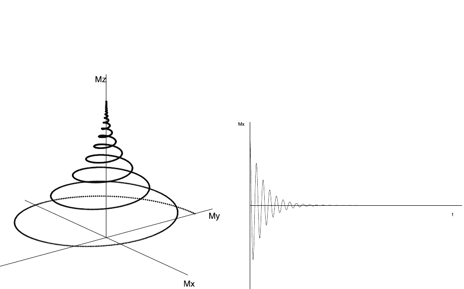
Fig. 11.6 Evolution of the net magnetization’s orientation and evolution of M
x
as a function of time after a saturation pulse.

January 9, 2009 10:21 World Scientific Book - 9.75in x 6.5in ws-bo ok975x65˙n˙2nd˙Ed
776 Principles of Radiation Interaction in Matter and Detection
resonance frequencies. There is an other source of dephasing: the non-uniformity
of the magnetic field. These two effects combine to change the observed spin-spin
relaxation time to T
∗
2
:
1
T
∗
2
=
1
T
2
+
1
T
2,inhomo
. (11.39)
When considering both z and XY plane magnetization, the orientation of the
net magnetization varies as illustrated in Fig. 11.6 (left side).
Figure 11.6 (right side) represents the signal collected around the x-axis as
a function of time. The exponential decrease of the signal is called a FID (Free
Induction Decay). To be able to reconstruct the image, the frequency ω , the spin-
lattice and spin-spin relaxation times have to be extracted. The exponential nature
of the signal and the response time required for the measurements make it hard to
gather all the information needed. That is why the echo is used instead.
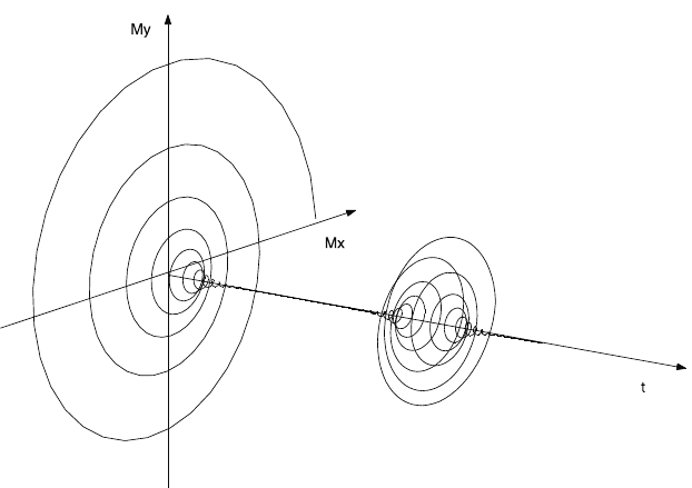
The echo of a signal is a re-phasing, which can be total or partial, of the trans-
verse magnetization. It can be done in two ways: applying an inversion pulse or a
field gradient.
Starting with a saturation pulse, we have a FID. Then, after a waiting period,
a 180
◦
impulsion, or the inverse of the field gradient, is applied as illustrated in
Fig. 11.7.
The echo lasts longer than the original signal, allowing the measuring equipment
to respond and gather enough information to reconstruct the image.
11.3.2 Forming an Image
To form an image, an echo has to be produced. Several methods can be used. Here
are four examples. They are divided in two categories: spin-echo and gradient-
echo [Sprawls (1993)].
11.3.2.1 Spin-Echo
The spin-echo methods use only RF waves to create the echo. A sequence used is a
90
◦
impulsion followed by a 180
◦
impulsion after t = T E/2, where T E is the echo
time. The measurements are taken after another wait of t = T E/2. The manoeuvres
are repeated at every repetition time T R. This method is called spin-echo. The
height of the echo signal will be:
S = kρ
³
1 − e
−T R/T
1
´
e
−T E/T
2
, (11.40)
where ρ is the non-zero spin nuclei density and k is a proportionality constant,
which depends on the measuring equipment.
The inversion-recovery method uses the same idea but inverses the sequence. Af-
ter waiting T R , an inversion pulse is applied. After t = T I, where T I is the inversion
January 9, 2009 10:21 World Scientific Book - 9.75in x 6.5in ws-bo ok975x65˙n˙2nd˙Ed
Medical Physics Applications 777
Fig. 11.7 Representation of the transverse magnetization orientation during a FID and an echo as a function of time.

January 9, 2009 10:21 World Scientific Book - 9.75in x 6.5in ws-bo ok975x65˙n˙2nd˙Ed
778 Principles of Radiation Interaction in Matter and Detection
time, a 90
◦
impulsion is given to the system and the measurement is done imme-
diately after. In this case, we have [Hornak (2002)]:
S = kρ
³
1 − 2e
−T I/T
1
+ e
−T R/T
1
´
. (11.41)
The advantage of the spin-echo methods is that it is independent of T
∗
2
and,
therefore, of the inhomogeneities of the magnetic field. Unfortunately, these methods
require longer acquisition time.
11.3.2.2 Gradient-Echo
The main goal of the gradient-echo methods is to reduce acquisition time. The
Small Angle Gradient Echo (SAGE) uses small angle RF pulses to accelerate the
longitudinal magnetization’s recovery. The signal will have the form [Hornak (2002)]:
S = kρ
¡
1 − e
−T R/T
1
¢
sin θ e
−T E/T
∗
2
¡
1 − cos θ e
−T R/T
1
¢
. (11.42)
Here, there is a dependence on T
∗
2
, which will require corrections in the data treat-
ment.
Another method that uses a gradient-echo is called magnetization prepara-
tion. The idea is to apply a saturation or inversion pulse to “prepare” the lon-
gitudinal magnetization to the gradient-echo acquisition.
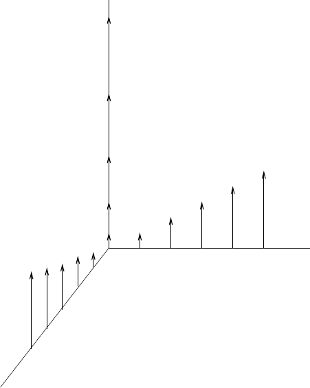
11.3.2.3 Space Positioning
First of all, the region that has to be scanned is divided into small volumes called
voxel s . Each volume enclosed in the width ∆z is a slice. Each slice will correspond
to an image that is divided in pixels (a voxel in the slice corresponds to a pixel in
the image). In order to be able to localize a voxel in space, the magnetic field is
different in each voxel (Fig. 11.8), giving a different Larmor frequency.
There are two methods of coding. In the first one, the three field gradients are
applied one after the other. First, a gradient defines the slice. Simultaneously, a 90
◦
impulsion is applied. The gradient is then turned off and a gradient defining x (or y)
is turned on. Every abscissa (or ordinate) has a different phase after this gradient is
turned off (phase encoding gradient). At last, a third gradient is applied. It gives to
every y (or x) a different Larmor frequency (frequency encoding gradient). During
the application of this gradient, the FID occurs. In the second method, there is also a
slice selection accompanied by a saturation pulse. It is followed by the simultaneous
application of the other 2 gradients accompanied by the FID.
The information gathered by the measuring instruments is a function of
time. Using a 2-dimensional Fourier transform, the information is translated in fre-
quencies, which are finally translated in spatial co ordinates. Several techniques can
be used in MRI. Research is still underway to discover the most efficient technique
time- and quality-wise.
January 9, 2009 10:21 World Scientific Book - 9.75in x 6.5in ws-bo ok975x65˙n˙2nd˙Ed
Medical Physics Applications 779
x
z
y
Fig. 11.8 The spatial positioning magnetic field gradients applied to encode each voxel with a
specific Larmor frequency. All the magnetic fields are in the z-direction. The specific magnetic field
applied to a point in space is the addition of the X-, Y- and Z-gradients.
11.3.2.4 Flows
Like all the other imaging techniques, MRI requires the immobility of the pa-
tient. However, the movement inside the patient cannot be controlled. With MRI,
this movement can be used to have an other type of imaging: flow imaging. This
technique is used for angiographies. Three types of sequences can be used: time-of-
flight, phase contrast and contrast enhanced angiographies [Hornak (2002)].
On a regular MRI image, blood vessels seem empty because the atoms that
receive the 90
◦
impulsion do not receive the 180
◦
and, therefore, no echo is
created. The idea behind the time-of-flight angiography technique is to follow the
atoms that received the saturation pulse, i.e., to do a second slice selection with the
inversion pulse in order to have an echo. The sequence used for this technique is a
spin-echo sequence with a 90
◦
and a 180
◦
impulsion with different frequencies.
The phase contrast angiography technique introduces a bipolar gradient in the
slice selecting gradients, i.e., between the saturation and inversion pulses in a spin-
echo sequence. This gradient is constituted of two inverse gradient lobes applied