Leroy C., Rancoita P.-G. Principles Of Radiation Interaction In Matter And Detection
Подождите немного. Документ загружается.

January 9, 2009 10:21 World Scientific Book - 9.75in x 6.5in ws-bo ok975x65˙n˙2nd˙Ed
780 Principles of Radiation Interaction in Matter and Detection
one after the other. Of course, for the immobile atoms, this gradient has no ef-
fect since each lobe will nullify the effect of the other one. But, a moving nucleus
will be affected. Therefore, to see the blood movement, one has to take the image
twice (once with and once without the bipolar gradient) and subtract one from
the other. The immobile matter will disappear, while the moving matter will have
different intensities depending on its velocity.
The last technique is the most used: the contrast enhanced angiography. It is
based on the principle that the relaxation time, T
1
, changes when a paramagnetic
contrast agent is injected into the blood. In brief, T
1
changes with the blood’s
surroundings. And as it changes, contrasts can be seen. A rapid data acquiring
sequence is used.
11.3.2.5 Functional MRI
Using a sequence of impulsions and gradients called the echo-planar imaging, it
is possible to gather the information needed for a whole image in the short time
perio d TR. This allows one to take several images pictures per second just like a
video. This technique opens new avenues to study the human body. For example, it
is possible to track the blood flows in the brain allowing an elaborate study of the
brain’s reaction to stimuli.
The idea behind echo-planar imaging is to sweep all sections of a k-space (the
Fourier transform of the image) [Hornak (2002)]. First, a saturation pulse and a slice
selective gradient are applied to the system. Then, simultaneously a phase and a
frequency encoding gradients are injected, bringing the initial data to a “corner” of
the k-space. A bit after, an inversion pulse is applied. Finally, the following sequence
is used to sweep the k-space: a phase gradient is applied, immediately followed by
a frequency gradient, during which data is gathered; then, another phase encoding
gradient is applied after which another echo is produced; and the previous steps are
repeated, until the entire k-space is swept.
11.4 X-Ray Medical Imaging with MediPix Devices
The MediPix device, discussed in Sects. 6.5.1-6.5.2.5, can be exploited for medi-
cal imaging. Medical imaging is typically in the 5–80 keV X-ray energy range. The
photoelectric effect is the dominant interaction between X-ray photons and detect-
ing material in that energy range. In this section, we look at the application of
MediPix-type devices in X-ray medical imaging. The MediPix-type devices, when
exposed to X-ray beams, produce real-time digital images that can be stored and
analyzed. These devices can improve the quality of image as the X-ray detection
is improved while the noise is reduced. This improvement of the image quality al-
lows the reduction of the radiation exposure to the patient. The image quality is
expressed in terms of several parameters. Some of the most important parameters

January 9, 2009 10:21 World Scientific Book - 9.75in x 6.5in ws-bo ok975x65˙n˙2nd˙Ed
Medical Physics Applications 781
are briefly reviewed below. However, this list is non-exhaustive and readers who
wants to complete their information are invited to consult [Mikulec (2000); Pfeiffer
(2004); Stoehr (2005); Norlin (2007)], for instance.
11.4.1 The Contrast
In the 5–80 keV energy range, tissue, bone and calcifications have very distinct
absorption coefficients and high-contrast images can be formed. The contrast, C,
is describing the difference in intensities I
1
and I
2
between two adjacent regions
(several pixels) which present different absorptions of X-rays. If one uses a photon
counting system, the contrast can be expressed in terms of counts between the
two adjacent regions. If N
1
and N
2
are the photons counted in the two regions
(normalized to the same area), the contrast is defined as [Mikulec (2000); Anton et
al. (2006)]:
C =
I
1
− I
2
I
1
+ I
2
=
N
1
− N
2
N
1
+ N
2
. (11.43)
Since the numbers of counted photons have a Poisson noise, one defines a signal-
difference-to-noise ratio (SDNR) which measures the visibility of a given con-
trast [Anton et al. (2006)]:
SDN R =
N
1
− N
2
σ
N
1
−N
2
= C
p
N
1
+ N
2
, (11.44)
where σ
N
1
−N
2
is the variance of the counts distribution. For a contrast C, a parti-
cular value of SDNR can be achieved by either long exposure to photon counting
(large incident photon fluence) or a large pixel-area [Anton et al. (2006)].
11.4.2 The Modulation Transfer Function
One defines the modulation transfer function (MTF) as the spatial frequency re-
sponse of an imaging system or a component. The spatial frequency is measured
in units of cycles per mm (c/mm) or also in units of line pairs per millimeter
(lp/mm). The MTF is the contrast at a given spatial frequency relative to low
frequencies and therefore measures the quality of transmission (in to out) of the
contrast of an object through an imaging chain for a given spatial frequency. High
spatial frequencies correspond to fine image detail. Signals which change often over
a given distance have a high spatial frequency. Signals with low spatial frequency,
i.e., which change slowly over the same distance are easier to detect than those of
high spatial frequency. The MTF at a spatial frequency ν is defined as the ratio
between the modulation of a sinusoidal pattern M
in
and the modulation of image
M
out
obtained after transmission [Pfeiffer (2004)]:
MT F (ν) =
M
out
(ν)
M
in
(ν)
. (11.45)

January 9, 2009 10:21 World Scientific Book - 9.75in x 6.5in ws-bo ok975x65˙n˙2nd˙Ed
782 Principles of Radiation Interaction in Matter and Detection
The M T F (ν) ranges between 0 and 1 for all frequencies. The MTF can be described
by analytical functions. In the case of square pixel (such is the case of MediPix1
and MediPix2) of size l
2
with uniform sensitivity over the whole pixel area, the
theoretical limit for MTF is [Pfeiffer (2004)]:
MT F (ν) =
sin(πνl)
π νl
. (11.46)
This parametrization comes from the possibility to decompose the information for a
given image into a set of sinusoidal functions of different amplitude. Therefore, one
can say that the MTF gives the spatial response of a detector to a sinusoidal input
stimulus. There are several factors affecting the MTF: a) focal spot blur: because
the X-ray source is not point-like and causes blur in the X-ray image. Standard
procedures to minimize this effect is to reduce the focal spot size, optimizing (in-
creasing) the distance between the source and the subject of imaging and decreasing
the distance between the subject of imaging and the X-ray detector (basically what
one would do with a camera); b) the pixel size: the pixel size has to be as small
as possible (55 µm × 55 µm for MediPix2) possibly smaller than the details of the
image; c) scattering of photons produced from the conversion of absorbed X-rays:
this effect can be minimized by the choice of an adequate conversion material.
11.4.3 The Detective Quantum Efficiency
The detective quantum efficiency (DQE) is the signal-to-noise ratio (SNR) transfer
function. It measures how the SNR at the input (SNR
in
, SNR of the incoming
X-ray flux) of an imaging system is transferred to the output (SN R
out
, SNR of
the image) and measures its possible degradation by the imaging system. Then, the
DQE is expressed as:
DQE(ν) =
SNR
2
out
(ν)
SNR
2
in
(ν)
. (11.47)
The noise is from various origins: the electronic, fixed pattern and quantum
noises. The electronic noise is produced by the read-out components of the imaging
system. The fixed pattern noise originates from gain and off-set value variations
among pixels. The so-called quantum noise is the result of the random nature of the
X-ray photons: the number n
in
of X-ray photons produced by the source incident
per unit area and unit time on the detector has fluctuations that follow a Poisson
distribution with a variance
√
n
in
. Then, with SNR
in
=
√
n
in
, Eq. (11.47) can be
written as
DQE(ν) =
SNR
2
out
(ν)
n
in
. (11.48)
SNR
out
is determined by using the noise power spectrum (NPS). The NPS describes
the noise transfer properties of the imaging system. The NPS is a quantity which
accounts for the distribution of noise variations with spatial frequency. Technically,

January 9, 2009 10:21 World Scientific Book - 9.75in x 6.5in ws-bo ok975x65˙n˙2nd˙Ed
Medical Physics Applications 783
the calculation of the NPS uses the Fourier transform of noise image to determine
the variance of noise power present at each spatial frequency. The shape of the NPS
shows where the noise power is concentrated in frequency space [Riederer, Pelc and
Chesler (1978); Boedeker, Cooper and McNitt-Gray (2007)]. In simpler terms, the
NPS gives the variance of a noise process, but distributes it as a function of the
spatial frequency. Hence, using the normalized NPS (NNPS) and the MTF, one
has [Pfeiffer (2004)]:
SNR
2
out
=
MT F
2
(ν)
NNP S(ν)
. (11.49)
Combining Eqs. (11.48), (11.49), one finds:
DQE(ν) =
MT F
2
(ν)
NNP S(ν) × n
in
. (11.50)
One can consult, for instance, [Mikulec (2000); Pfeiffer (2004); Stoehr (2005); Norlin
(2007)] for a review of results of the measurements of MTF, NPS and DQE done
with MediPix1 and MediPix2. A final remark for this section is the phenomenon of
charge sharing and its implication for X-ray imaging. As discussed in Sect. 6.5.2.5,
charge sharing results from several mechanisms with the consequence that deposited
charges are shared among adjacent pixels. Particle physics may use the charge shar-
ing effect for improved tracking. For medical physics charge tracking is viewed as an
adverse phenomenon as it decreases the photon detection efficiency. Depending on
the threshold set for the MediPix2-type device, incoming photons can be counted a
single time or several times. There are several ways envisaged to reduce the charge
sharing effect and even to suppress it. It has been proposed to develop a readout
electronics which can sum the charge of several pixels [Llopart et al. (2002)]. This
solution has however a drawback as summing charges collected in several pixels
yields additional noise. The problem of charge summing and adequate noise control
will be hopefully solved with MediPix3, the successor of MediPix2 [Medipix Collab.
(2008)]. Another solution advanced for suppression of the charge sharing effect is to
develop 3-D structure detectors in place of the standard planar pixel detectors. It
is advocated that the transverse electric field in a 3-D structure (perpendicular to
the incoming photon) will force the electron to drift to the correct pixel [Norlin
(2007)]. This development has to be accompanied with an increase of the silicon
thickness for maintaining a practical quantum efficiency.
January 9, 2009 10:21 World Scientific Book - 9.75in x 6.5in ws-book975x65˙n˙2nd˙Ed
This page intentionally left blankThis page intentionally left blank
January 9, 2009 10:21 World Scientific Book - 9.75in x 6.5in ws-bo ok975x65˙n˙2nd˙Ed
Appendix A
General Properties and Constants
785

January 9, 2009 10:21 World Scientific Book - 9.75in x 6.5in ws-bo ok975x65˙n˙2nd˙Ed
786 Principles of Radiation Interaction in Matter and Detection
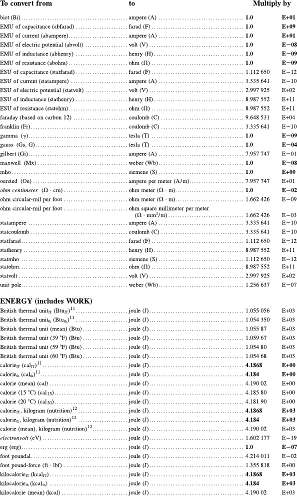
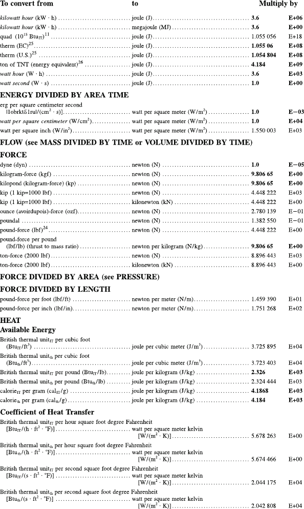
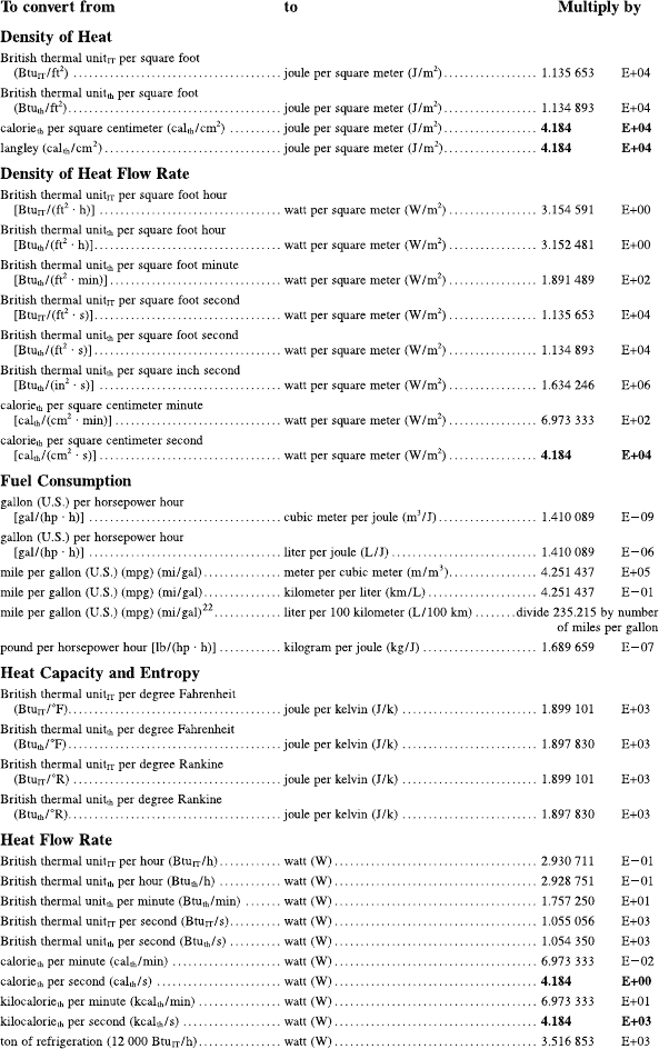
A.1 Conversion Factors
The conversion factors are from Appendix B.9 of [Taylor (1995)].
January 9, 2009 10:21 World Scientific Book - 9.75in x 6.5in ws-bo ok975x65˙n˙2nd˙Ed
Conversion Factors 787
January 9, 2009 10:21 World Scientific Book - 9.75in x 6.5in ws-bo ok975x65˙n˙2nd˙Ed
788 Principles of Radiation Interaction in Matter and Detection
January 9, 2009 10:21 World Scientific Book - 9.75in x 6.5in ws-bo ok975x65˙n˙2nd˙Ed
Conversion Factors 789