Lennarz W.J., Lane M.D. (eds.) Encyclopedia of Biological Chemistry. Four-Volume Set . V. 4
Подождите немного. Документ загружается.


are extended sufficiently to permit all of the data
required for structure determination to be collected
from one crystal.
DENOVOSTRUCTURE DETERMINATION
The result of a structure analysis is a 3-D electron
density map, which is computed from the X-ray
scattering by Fourier transformation. This calculation
requires not only the measured amplitudes (square roots
of the intensities) of the scattered X-rays but also the
assignment of phases to all of the reflected waves. These
phases are not determined by the X-ray experiments.
Two strategies, multiple isomorphous replacement
(MIR) and multiwavelength anomalous diffraction
(MAD), are commonly employed to assign phases for
structure analysis of a new or unknown protein fold. In
both strategies, contributions of added heavy atoms or
of incorporated selenomethionine are used to calculate
the phases of the protein by triangulation. The first step
in both methods is determination of the positions of the
heavy atoms. MIR requires measurements from native
crystals and from a series of derivative crystals modified
by addition of heavy atoms. These derivatives are
obtained by immersing preformed crystals in holding
solutions containing heavy atom complexes, or less
frequently, by co-crystallization. Typical reagents are
mercurials or platinum complexes. The method assumes
that X-ray scattering by the protein atoms is exactly the
same in the derivative as it is in the native crystals so that
all differences in the diffraction pattern can be attributed
to the added heavy atoms. Perturbation of the protein
induced by addition of heavy atoms (loss of isomorph-
ism) is the most common source of errors in MIR.
Multiwavelength anomalous diffraction exploits scat-
tering effects that occur at wavelengths near the X-ray
absorption edges of elements such as Se, Br, and
lanthanides. At the peak of the absorption edge of
these elements, the imaginary anomalous component
( f
00
) is large, producing differences between intensities of
h,k, l and 2h, 2 k, 2 l reflections. The real anomalous
component ( f
0
) is large (and negative) at the inflection
point of an absorption edge, but is small at wavelengths
remote from the edge. Effects of the real (dispersive)
component appear as wavelength-dependent differences
in intensities. Both f
0
and f
00
vary with wavelength, and
both components are used to determine phases. MAD
experiments thus require X-rays that can be tuned to
appropriate energies and are performed at synchrotron
X-ray sources. Although the changes in intensities that
arise from anomalous scattering effects are usually
smaller than for MIR, MAD has the advantage that
the requisite data are obtained from one crystal. Effects
of nonisomorphism are thus circumvented. With precise
measurements of the relevant intensity differences and
algorithms based on direct methods, it is currently
possible to locate , 100 Se atoms per asymmetric unit.
Structures as large as 300 kDa have been determined
from anomalous scattering by selenomethionine.
Experience demonstrates that measurements of anom-
alous diffraction at the peak wavelength of lanthanides
or even Se (single-wavelength anomalous diffraction, or
SAD) can be sufficient to determine a structure.
In the determination of the phases that are used
to calculate an initial experimental electron density map,
it can be advantageous to combine data from isomor-
phous replacement and anomalous scattering. Requiring
bulk solvent regions to have uniform electron densities
(solvent flattening) is a useful restraint that improves the
accuracy of phases. Computational suites such as CCP4,
CNS, SHARP, or SOLVE/RESOLVE are designed to
exploit all these and other sources of phase information.
FITTING ELECTRON DENSITY MAPS
Atomic models are built into electron density maps using
graphics displays that allow interactive and menu-driven
manipulations of the models. Modern software for
model fitting incorporates a number of features that
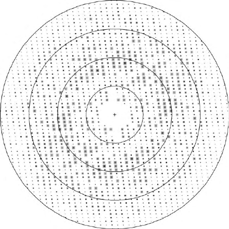
FIGURE 1 The definition of resolution in crystallography. This
figure shows the relative intensities of diffracted X-rays (reflections) in
a two-dimensional planar section through three-dimensional diffrac-
tion space. Each reflection occurs at a particular angle (2u) determined
by Bragg’s law,
l
=d ¼ 2sin
u
; where d is the distance between reflecting
planes; reflections farther from the center have larger scattering angles
and smaller d spacings. The superimposed circles are drawn with radii
corresponding to resolutions of 7.2, 3.6, 2.4, and 1.8A
˚
. All of the
unique reflections within a sphere swept out by the 1.8A
˚
circle will be
used to calculate the electron density at a resolution of 1.8A
˚
, whereas
only the smaller number of reflections within the second sphere will be
used for a 3.6A
˚
electron density map. As the limiting d spacing
becomes smaller, more data are incorporated in calculation of the
electron density, and the resolution is said to be higher.
X-RAY DETERMINATION OF 3-D STRUCTURE IN PROTEINS 423
simplify and speed this process, including local optim-
ization of models within the density and sampling of
likely side chain rotamers.
REFINEMENT
Initial atomic models that are constructed by interactive
fitting of experimental electron density maps are usually
incomplete and inaccurate. They serve as the starting
point for refinement, a process in which the agreement
between the observed and computed structure factors is
improved by adjustment of the atomic positional and
thermal parameters. Modified parameters are used in
turn to compute phases and new maps that permit
refitting of the model and addition of missing parts. This
cyclic procedure is repeated until the fitting appears to
have converged. It is a fortunate consequence of the
physical and mathematical relationship between a
crystal structure and its diffraction pattern that struc-
tures can be corrected or completed if most of the
scattering has been accounted for in an atomic model.
Missing and/or incorrectly placed atoms are detected in
difference maps that subtract the computed from the
observed structure. There are two fundamentally differ-
ent approaches to optimization of molecular models: the
first is straightforward minimization by algorithms such
as conjugate gradient and the second is sampling of
conformations, usually with molecular dynamics simu-
lations. Molecular dynamics calculations that begin
with atom velocities corresponding to elevated tempera-
tures (simulated annealing) are a very efficient way to
improve initial models. Standard minimizations are
performed after the annealing cycles.
Minimizations are large, iterative least-squares com-
putations whose convergence is hampered by small
ratios of data to adjustable parameters. There are at least
four parameters to be assigned to each atom: its
position, x, y, z, and a temperature factor (or displace-
ment parameter) that reports excursions of the atom
about its equilibrium position. Even at 2.0A
˚
resolution,
a typical data:parameter ratio for this simplest set of
parameters is about 2.5. To compensate for limitations
in the data, most refinements are conducted with
harmonic restraints that maintain standard polypeptide
geometries and in effect increase the number of
observations. Data:parameter ratios and robustness
can also be increased by replacing Cartesian with
torsional variables or by imposing strict constraints to
maintain the identity of chains that are repeated in the
asymmetric unit.
Various target functions can be chosen for minimiz-
ation in least-squares refinement. The conventional
target is the discrepancy between the observed ampli-
tudes of reflections and the amplitudes calculated
from the model. The corresponding reliability index,
or R-factor, is defined as SkF
obs
l 2 lF
calc
k
SlF
obs
l, where
the sums are taken over all reflections. It thus resembles
an average fractional error. Alternative targets substitute
intensities for amplitudes or utilize correlation func-
tions. R
free
, an agreement index calculated with a subset
of reflections that is never sampled in refinement, is an
important monitor of refinement.
STRUCTURE DETERMINATION
BY
MOLECULAR REPLACEMENT
When the protein of interest is related to a known
structure, coordinates for the known homologue may be
employed to solve the unknown. The initial compu-
tation is usually carried out in two stages, referred to as
rotation and translation searches. The model is oriented
and then positioned in the unit cell of the unknown
crystal by correlating observed intensities or structure
factor amplitudes with those calculated from the
oriented model. It is then subjected to refinement.
There is no hard-and-fast rule about the level of
sequence similarity that augurs success in molecular
replacement (MRP), or about the fraction of the
structure to be used in searches. A standard practice
has been to truncate side chains that are not identical in
the two proteins. For multidomain proteins it may be
desirable to use individual domains, rather than the
intact protein, as search models.
The principal hazard in MRP is bias from the model
structure. Although incorrect features should appear at
lower-than-average electron densities, they do not
disappear completely. One effective strategy to minimize
bias is the computation of “omit” maps, in which local
regions of the model that need adjustment or verification
are not included in the phase calculations.
Similarity of structures is exploited in another form of
molecular replacement in which the electron densities
corresponding to structural repeats are averaged to
generate modified maps and modified phases. This
approach assumes that the multiple copies of chains or
subunits found in an asymmetric unit (the smallest motif
from which the crystal can be generated by translation
and rotation) adopt identical conformations. These
copies are related by coordinate transformations that
are local and do not obey crystallographic symmetry;
hence they are referred to as non-crystallographic
symmetry operations. Implementation of this mode of
molecular replacement was the key to the pioneering
structure analyses of spherical viruses.
Studies of Molecular Complexes
and Conformational Changes
The classic comparisons of deoxy- with methemoglobin
and the difference Fourier analyses that revealed azide
424
X-RAY DETERMINATION OF 3-D STRUCTURE IN PROTEINS
bound to myoglobin presaged the central role of
crystallography in the descriptions of conformation
changes and molecular interactions. Subsequent struc-
tures have firmly established the plasticity of proteins
and have demonstrated that a surprising variety of
conformational states can be accessed by a particular
polypeptide.
LIGAND BINDING IN SITU
Myoglobin–azide and lyzozyme–tetrasaccharide com-
plexes were the prototypes for experiments in which
structures of molecular complexes have been determined
from preformed crystals after immersion in holding
solutions containing ligands. This approach is feasible
because the extensive solvent-filled channels within the
crystals allow small ligands or substrates to diffuse to
their binding sites. Density corresponding to the ligands
is found in maps computed with amplitudes measured
from the complex and phases from the ligand-free
protein. Subsequent refinement often reveals local
changes in the protein, elicited by interactions with
the ligands. If the binding sites are not saturated, the
electron densities will include images of both the ligand-
free and ligand-bound species, and it may be necessary
to refine the occupancies of the ligands and to include
alternate conformations for parts of the protein.
LARGE CONFORMATION CHANGES
In contrast, major conformation changes such as those
accompanying oxygenation of hemoglobin cannot be
accommodated by the original crystal lattice and either
are suppressed by competing crystal-packing forces or
disrupt packing interactions and disorder the crystals.
Descriptions of these larger structural changes therefore
require analysis and comparison of different (non-
isomorphous) crystals obtained under conditions that
favor one or another conformation. Determination of
the alternate conformation entails solution of a new
structure by de novo or molecular replacement methods.
Dramatic conformation changes are not uncommon and
can include displacements of loops or flaps, swinging or
pivoting of domains, and even remodeling of secondary
structures. Numerous examples have been gathered in
compendia of molecular motions.
STUDIES OF REACTION INTERMEDIATES:
T
RAPPING METHODS
Structures related to intermediates in enzyme-catalyzed
reactions can be obtained from complexes with unreac-
tive substrate analogues or transition-state analogues.
More elegant experiments are also feasible, as many
enzymes are active catalysts in the crystalline state.
With sufficient knowledge of the reaction mechanism, it
is possible to design experiments in which true
intermediates accumulate in the crystal, or to trap
structures of these intermediates at appropriate times by
flash-cooling. In studies of isocitrate dehydrogenase,
Stoddard and co-workers exploited mutations to favor
accumulation of intermediates either prior to or follow-
ing hydride transfer. The analysis of several intermedi-
ates in catalysis by P450-cam was accomplished by
controlling addition of reactants. Data were collected
from the ferrous enzyme–substrate complex after
chemical reduction, and crystals were then allowed to
react with oxygen to form the subsequent dioxygen
intermediate, which was in turn reduced by
X-irradiation to yield a putative activated-oxygen
intermediate.
TIME-RESOLVED CRYSTALLOGRAPHY
The most challenging experiments are those that aim to
determine structure as a function of time. They require a
narrow time window for initiating reactions and very
rapid data collection, usually by the Laue method, which
employs broadband radiation to capture most of the
reflections in a single image of the diffraction pattern.
The mixtures of structures that are present vary with
time and must be sorted out. Time-resolved structural
studies of the photodissociation of CO from crystals of
myoglobin have revealed non-heme binding sites for CO
and established the nature of relaxations in the heme and
globin that follow photolysis.
Accuracy of X-Ray Structures
and Metrics of Reliability
THE IMPORTANCE OF RESOLUTION
The exact definition of resolution in crystallography
is illustrated in Figure 1, which depicts a plane from the
3-D lattice in diffraction (reciprocal) space. Resolution is
probably the most important parameter in any assess-
ment of a structure determination. The dramatic effects
of resolution on the appearance of electron density
maps have been illustrated in several texts and reviews.
The number of reflections included in a structure
determination increases as (1/d
min
)
3
. Resolution thus
controls the data:parameter ratios that are critical in
refinement and is a primary determinant of positional
accuracy. The choice of a resolution limit for a structure
analysis is dictated by the completeness of the outermost
shell of data and the agreement between measurements
of symmetry-related reflections. These metrics are
usually displayed in tables describing the structure
determination.
X-RAY DETERMINATION OF 3-D STRUCTURE IN PROTEINS 425
UNCERTAINTIES IN COORDINATES
One would like to obtain estimates of the positional
uncertainties for each atom and the derived uncertainties
of bond lengths, bond angles, and torsions. The diagonal
elements of the inverse matrix that is calculated in least-
squares refinement provide these uncertainties in math-
ematically rigorous fashion. However, for proteins the
size of the least-squares matrices (at least 3N by 3N,
where N is the number of atoms in the asymmetric unit)
have generally precluded this computation. Approxi-
mations that derive global estimates of average coordi-
nate errors from R-factors versus resolution are partly
flawed because they assume the same temperature
factors for all atoms. One compromise has been to
invert large diagonal blocks taken from the full matrix.
Evaluation of the estimated standard deviations arising
from experimental errors is complicated by the inclusion
of restraints in most refinements. The average errors in
bond lengths are often dominated by the deviations
chosen for restraints.
Refinements of a few structures have included
inversion of the full matrix. Analysis of concanavalin
A at 0.94A
˚
resolution provided details of the geometry
at the manganese- and calcium-binding sites with
accurate estimates of the deviations of bond lengths
and angles in the metal cluster, information that is
critical for understanding the properties of the metal
center. Comparison of rigorous with approximate error
estimates for this and other ultra-high-resolution struc-
tures demonstrates, as might have been expected,
that positional uncertainties depend strongly on the
values of the temperature factor (displacement par-
ameter). This dependence is especially pronounced in
unrestrained refinements.
Solvent molecules (usually water) that occupy defined
sites are included in archived coordinate lists (.pdb files).
They are important for structure and function, and they
contribute significantly to X-ray scattering even though
their residence times may be short. Waters in buried or
active sites may be very well-defined, but in general
water oxygens are the least well-positioned atoms and
the most subject to error. Some publications report
refinement of both occupancy and isotropic B values for
solvents, but because these parameters are highly
correlated, others choose to allow only B to vary in
refinement. It is customary to verify that waters are
placed at canonical distances from neighbors and
make reasonable interactions with protein atoms or
other solvents.
RELIABILITY INDICES (R-FACTORS)
R-factors are widely cited criteria of the accuracy and
reliability of a structure and are used to judge the
progress and convergence of refinements. Readers of
structure reports need to be aware of their shortcomings.
The actual values will depend on the refinement
algorithm, the choices of restraints or constraints,
the omission or inclusion of weak reflections, and on
the resolution (minimum d-spacing). The con-
ventional R-factor, SkF
obs
l2 lF
calc
k
SlF
obs
l,canbe
decreased artificially by overfitting, e.g., by includ-
ing many solvents with small scattering contributions
or by using anisotropic temperature factors at too
low a resolution.
The introduction of R
free
in the early 1990s provided
a better index of reliability and accuracy. R
free
is
calculated from a subset of data, randomly chosen
with respect to intensity and resolution, that is never
used in refinement. Acceptable refinement strategies
such as inclusion of additional solvents should produce
decreases not only in the conventional R but also in the
R
free
value.
OTHER QUALITY INDICES:
S
TRUCTURE VALIDATION
Crystal structures are full of details, and models are built
interactively and subjectively into the electron densities.
It is useful to have objective methods to flag possible
errors and regions that deserve further adjustment. Very
occasionally, incorrect folds have been fit to electron
densities, and wrong connections between pieces of the
polypeptide have been introduced in maps at relatively
low resolution. More common errors are mistaken
registration of sequences and misoriented peptide
planes. It is important to examine Ramachandran plots
of F, C angles for outliers with unusual (high-energy)
backbone conformations; side-chain conformations
should resemble one of the common rotamers. Com-
parisons of model density with experimental or
omit maps identify residues that may be incorrectly
modeled because they are mobile or adopt multiple
conformations. The Protein Data Bank routinely sub-
jects submitted coordinates to examination by PRO-
CHECK, which analyzes the stereochemistry and
other properties, flags possible errors, and assigns a
quality index.
STRUCTURES AT VERY
HIGH RESOLUTION
The library of structures at resolutions beyond 0.9A
˚
is
small but is growing steadily. Data:parameter ratios for
these analyses allow unrestrained refinement and
anisotropic modeling of the temperature factors,
which requires specification of nine parameters per
atom rather than four. In these structures it is
possible to see what had to be surmised or inferred
426
X-RAY DETERMINATION OF 3-D STRUCTURE IN PROTEINS

FIGURE 2 Stereoviews of electron density from the crystal structure determination of TEM-1
b
-lactamase at 0.85A
˚
resolution. At this resolution, densities corresponding to individual atoms are
apparent (cyan), and difference densities (red) identify the positions of hydrogen atoms. The data:parameter ratio was 6:1 in refinement with anisotropic temperature factors; alternate conformations
were included for 169 residues; R
free
is 0.112. Reproduced from Minasov, G., Wang, X., and Shoichet, B. K. (2002). An ultrahigh resolution structure of TEM-1 beta-lactamase suggests a role for Glu
1 66 as the general base in acylation. J. Am. Chem. Soc. 124, 5333–5340.
from lower resolution structures—many of the hydro-
gen atoms, alternate conformations, and distinc-
tions between oxygen and nitrogen atoms. Direct
observation of hydrogen bonds is especially valuable
for enzymologists, as is resolving ambiguities about
the orientations of Asn, Gln, and His. As more high-
resolution structures are completed, it should be
possible to document true deviations of geometries
from the canonical values embedded in restraint
libraries. The example shown in Figure 2 illustrates
the clear definition of densities corresponding to
individual atoms and the assignments of hydrogens
from difference maps.
Displaying and Comparing
Structures
The computing power of current desktop machines
allows the non-crystallographer to display and analyze
structures that have been deposited in the Protein Data
Bank. Particularly useful features of available programs
are algorithms that align structures for comparisons of
conformations, facile analysis of noncovalent inter-
actions, routines for mutation and model building, and
the capability to generate illustrations in a variety of
styles.
SEE ALSO THE FOLLOWING ARTICLES
Imaging Methods † Protein Data Resources † Second-
ary Structure in Protein Analysis
GLOSSARY
asymmetric unit The smallest motif from which the crystal can be
generated by translation and rotation operations. The unit cell that
is repeated by translation to form the crystal may contain a number
of asymmetric units.
isomorphism When addition of a ligand or heavy atom does not alter
the scattering contribution (structure factor) of the protein atoms,
the derivative and native crystals are said to be isomorphous. Cell
dimensions are expected to be unchanged.
non-crystallographic symmetry (NCS) When the asymmetric unit
includes more than one copy of a polypeptide, the multiple copies
are related by local coordinate transformations that do not obey the
symmetries that define the space group.
structure factor A vector F with phase
a
h,k,l
whose length lFl is
the amplitude of the reflection h, k, l. The structure factor is the
resultant of summing over scattering contributions from all
the atoms in the asymmetric unit and can be calculated from the
atom parameters. Fourier transformation of the structure factors
yields the electron density.
temperature factor (displacement parameter) A measure of the
motion of an atom about its equilibrium position. Temperature
factors may be isotropic (B-values) or anisotropic. In the general
anisotropic case, six parameters are required to describe the
displacements.
FURTHER READING
Baldwin, J., and Chothia, C. (1979). Hemoglobin: the structural
changes related to ligand binding and its allosteric mechanism.
J. Mol. Biol. 129, 183–191.
Bru
¨
nger, A. T., Adams, P. D., and Rice, L. M. (1999). Annealing in
crystallography: A powerful optimization tool. Prog. Biophys.
Mol. Biol. 72, 135–155.
Carter, C. W., Jr., and Sweet, R. M. (eds.) (1997). Macromolecular
Crystallography, Parts A and B. Methods in Enzymology, Vols 276
and 277. Academic Press, San Diego.
Cruickshank, D. W. (1999). Remarks about protein structure
precision. Acta Crystallogr. D55, 583–601.
Drenth, J. (1999). Principles of Protein Crystallography. Springer-
Verlag, New York.
Garman,E.F.,andSchneider,T.R.(1997).Macromolecular
cryocrystallography. J. Appl. Cryst. 30, 211– 237.
Guex, N., and Peitsch, M. C. (1997). SWISS MODEL and the Swiss-
PdbViewer: An environment for comparative protein modeling.
Electrophoresis 18, 2714–2723.
Kleywegt, G. J. (2000). Validation of protein crystal structures. Acta
Crystallogr. D56, 249–265.
Moffat, K. (2001). Time-resolved biochemical crystallography: A
mechanistic perspective. Chem. Rev. 101, 1569–1581.
Rossmann, M. G., and Arnold, E. (eds.) (2001). Crystallography of
Biological Macromolecules, International Tables for Crystal-
lography, Vol F. Kluwer Academic Publishers, Dordrecht.
Schlichting, I., Berendzen, J., Chu, K., Stock, A. M., Maves, S. A.,
Benson, D. E., Sweet, R. M., Ringe, D., Petsko, G. A., and Sligar,
S. G. (2000). The catalytic pathway of cytochrome P450cam at
atomic resolution. Science 287, 1615–1622.
Stoddard, B. L. (2001). Trapping reaction intermediates in
macromolecular crystals for structural analyses. Methods 24,
125–138.
Stryer, L., Kendrew, J. C., and Watson, H. C. (1964). The mode of
attachment of the azide ion to sperm whole methemoglobin. J. Mol.
Biol. 8, 96 –104.
Terwilliger, T. C., and Berendzen, J. (1999). Automated
MAD and MIR structure solution. Acta Crystallogr. D55,
849–861.
BIOGRAPHY
Martha L. Ludwig is Professor of Biological Chemistry and Research
Biophysicist at the University of Michigan in Ann Arbor. She is an
X-ray crystallographer whose primary research interest is the structure
and function of enzymes that require metal- and vitamin-based
cofactors. She is a Fellow of the American Association for the
Advancement of Science and a member of the National Academy
of Sciences.
428 X-RAY DETERMINATION OF 3-D STRUCTURE IN PROTEINS

Yeast GAL1–GAL10 System
Dennis Lohr and Ralph Bash
Arizona State University, Tempe, Arizona, USA
The GAL1 and GAL10 genes in the budding yeast
S. cerevisiae encode enzymes that help convert galactose to
a glycolytic intermediate, thus allowing it to be used as a
carbon source for cell growth. Genetic characterization of the
GAL genes and dissection of their regulatory mechanisms by
Douglas and Hawthorne in the 1960s provided a firm and
crucial foundation for the molecular characterization of
GAL1–10 regulation that began in the 1980s. Expression of
GAL1 and GAL10 is strikingly carbon source-dependent.
Transcription occurs at extremely high levels in galactose-
grown cells but is undetectable in cells grown in other carbon
sources. This clear-cut and efficient regulation plus the ability
to couple genetic and biochemical studies in the analysis of
single-copy genes have made GAL1 – 10 a very attractive
model for eukaryotic gene regulation studies and a paradigm
in which many general eukaryotic regulatory themes were
first uncovered. The GAL1 promoter is also widely used to
drive expression of heterologous genes, in various appli-
cations. The basic molecular aspects of GAL1–10 gene
regulation are outlined below (genes referred to as GAL1,
proteins as Gal1p).
The Elements Contributing
to GAL1–10 Regulation
GAL1 and 10 share a common , 600 bp (base pair)
intergenic region from which they are divergently
transcribed (Figure 1). They are considered to be
coexpressed and coregulated, mainly at the transcrip-
tional level. Their regulation combines inputs from
DNA sequence (promoter) elements, protein factors
(gene-specific and general), and chromosome structure.
DNA SEQUENCE ELEMENTS
The most important gene-specific promoter elements for
GAL1–10 expression are the upstream activation
sequence (UAS
G
) elements. These , 17 bp motifs are
necessary and sufficient for galactose-dependent gene
expression because; they provide the binding sites for
Gal4p, the specific activator of GAL gene transcription.
GAL1– 10 share four UAS
G
asymmetrically located
between the two genes (Figure 1). Each gene has its
own TATA sequence for binding the general transcrip-
tion factor TBP (TATA binding protein).
THE GENE-SPECIFIC
REGULATORY FACTORS
Gal4p
As the required activator for GAL gene expression,
Gal4p is a key player in GAL1– 10 expression. In the
presence of galactose, Gal4p activates transcription
through a domain located near its carboxy terminus,
residues 767–881, while bound to (UAS
G
) DNA via a
domain located near its amino terminus. It binds as a
homodimer to individual UAS
G
elements.
Gal80p
In carbon sources other than galactose, the negative
regulator Gal80p binds specifically to the C-terminal
activation domain of Gal4p such that it masks
Gal4p activation activity and thus prevents GAL1–10
transcription. Gal80p binds Gal4p quite strongly
(K
d
, 5 nM).
Gal3p
In the presence of galactose, the Gal80p-mediated
inhibition of Gal4p is relaxed by a Gal3p-dependent
process, thus freeing the Gal4p activation domain to
activate GAL1 –10 transcription. Gal3p behaves like a
signal transducer and appears to be located solely in the
cytoplasm, which should facilitate its interaction with
galactose. Gal3p can bind to Gal80p but how this
(cytoplasmic) interaction might influence the nuclear
Gal80p–Gal4p complex is unclear at this time. Inter-
estingly, the product of the GAL1 gene contains a Gal3p-
like activity that apparently makes it capable of fulfilling
a similar function to Gal3p.
y
Encyclopedia of Biological Chemistry, Volume 4. q 2004, Elsevier Inc. All Rights Reserved. 429

OTHER FACTORS
Global processes, such as the general glucose repression
system, also participate in GAL1–10 regulation and
elements of the RNA polymerase II transcription
apparatus function in GAL1–10 transcription. Several
other genes show galactose-sensitive and Gal4p-
dependent expression, suggesting that their gene
products may be part of the regulatory network:
GAL6 (possible alternative regulator); GCY1 (oxido-
reductase); PCL10 (cyclin); FUR4 (uracil permease);
RPA; and YP53 (protein metabolism). Their precise
roles are unknown.
CHROMOSOMAL ORGANIZATION
The promoter region of GAL1–10 has a distinctive
chromosomal organization that is an aspect of gene
control. In all carbon sources, the UAS
G
elements
reside within a sizable (at least 150 bp), highly
accessible nucleosome-free region (“HR,” Figure 1).
Thus, the UAS
G
are always available to Gal4p;
neither nucleosome removal nor competition with
nucleosomes is required to expose these elements
for Gal4p binding. The strategy of locating major
promoter elements in a large, constitutively
accessible region is not common on eukaryotic
promoters and the features that maintain it have yet
to be defined. In nongalactose carbon sources,
nucleosomes bracket this highly accessible HR region,
covering the TATA and transcription start sites
(“A–C,” Figure 1). The DNA near the termini
of nucleosomes A and B contains intrinsic,
sequence-dependent DNA bends (Figure 1) that may
contribute to the preferential location of these
promoter nucleosomes.
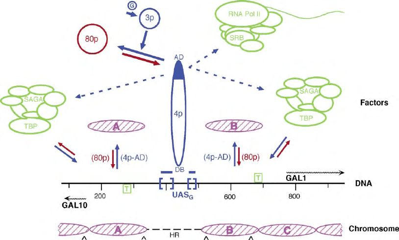
FIGURE 1 An outline of GAL1–10 regulation. The DNA sequence organization of the yeast GAL1–10 promoter region is shown in the center of
the diagram (“DNA”), with UAS
G
(blue brackets) and TATA boxes (green, boxed “T”) located to scale along the sequence (gray line). The numbers
refer to base pairs from an EcoRI site in the GAL10 gene. The transcription start sites are located at the origins of the two wavy arrows below
(GAL10) or above (GAL1) the line. The chromosomal structure of this region in the inactive state of gene expression is shown at the bottom
(“Chromosome”) with nucleosomes A, B and C (pink) located to scale on the sequence and the nonnucleosomal, highly accessible region in the
chromosome indicated by a dashed line (“HR”). The locations of intrinsic, sequence specific DNA bends are shown by “ ^ ”. In the topmost portion
of the Figure (“Factors”), various factors and complexes known or thought to be involved in regulating and implementing GAL1– 10 expression are
shown. The GAL-specific factors (“3p”, Gal3p; “4p”, Gal4p) are shown in blue and the reactions that occur in, or are characteristic of, the active
(galactose-induced, “G”) state are designated by solid blue arrows. Dotted blue arrows indicate the suggested recruitment of complexes by Gal4p
during gene activation, RNA polymerase II (“RNA pol II”), including SRB (Suppressor of RNA Polymerase B subunit) proteins and the SAGA (Spt-
Ada-Gen5-Acetyltransferase) complex. The activation domain (“AD”) of Gal4p is solid blue and the Gal4p DNA binding domain is identified
(“DB”). The negative GAL-specific factor, Gal80p (“80p”), is shown in red and the reactions that occur in, or are characteristic of, the inactive state
of expression are designated by red arrows. The association/dissociation of TBP with the GAL1 and GAL10 TATA and the disruption/redeposition
equilibria of promoter nucleosomes A and B (pink) are also designated by solid blue/red arrows. The involvement of Gal4p and Gal80p in the
promoter nucleosome disruption/redeposition equilibrium is indicated by their appearance in parentheses next to the appropriate arrow.
Nucleosome C, which also undergoes disruption/redeposition, is not shown in this upper portion of the figure.
430 YEAST GAL1–GAL10 SYSTEM
The (Three) States of GAL1–10
Gene Expression
THE ACTIVE (GALACTOSE-INDUCED)
S
TATE
In galactose, GAL1– 10 are transcribed at very high
levels, indicating that the activation mechanisms for
their expression are quite robust. Under these con-
ditions, Gal4p is bound, via its N-terminal DNA-
binding domain, to the UAS
G
elements that lie between
GAL1 and GAL10 while promoting transcription of
the genes through its C-terminal activation domain.
In this state, binding of TBP to the GAL1 and GAL10
TATA will be greatly aided by the apparent removal
(disruption) of the promoter nucleosomes A–C, a
process mediated directly or indirectly (through other
factors) by the Gal4p activation domain. Transcription
activation by Gal4p also involves the direct or indirect
recruitment of cellular complexes (Figure 1). The UAS
G
are . 200 bp from the GAL1 –10 transcription start sites
suggesting that the three-dimensional structure of Gal4p
and promoter chromatin architecture, both unknown,
may also impact on the activation process. This variety
of Gal4p activation depends upon the galactose-induced
release of Gal80p inhibition that is mediated through
Gal3p (and Gal1p?).
THE INACTIVE STATES
(REPRESSED OR POISED)
There are two distinct types of inactive states, repressed
(in glucose) or poised for expression (in nonfermentable
carbon sources like glycerol/lactate). In both inactive
states, Gal80p binds to the Gal4p activation domain,
masking its activity, and nucleosomes A –C are present
on the GAL1–10 promoter region, covering the TATA
and transcription start sites.
The presence of glucose results in an additional
negative feature, Gal4p absence from the UAS
G
. This
absence prevents GAL1–10 from being (rapidly) indu-
cible in glucose. The UAS
G
are still present in an open
chromosomal region and should thus be available
to Gal4p. Gal4p absence from the UAS
G
is thought to
result mainly from a decrease in Gal4p levels due to
decreased expression of the GAL4 gene, imposed by the
global catabolite repression apparatus. The decrease in
expression is fairly modest but Gal4p–UAS
G
binding
should be very sensitive to Gal4p levels due to its
highly cooperative nature (multiple UAS
G
/two Gal4p
per UAS
G
).
In carbon sources that are neither repressing nor
inducing, such as glycerol lactate, the GAL1–10 genes
are not expressed at all but they can be very rapidly
(within minutes) induced to full expression by galactose.
Rapid inducibility is due to the presence of Gal4p on
the UAS
G
in these carbon sources, strongly protecting
these elements as in galactose-grown cells, and to the
(low-level) presence of the signal transducer Gal3p.
Elevated Gal4p levels (GAL4 is most actively expressed
in these carbon sources) plus constitutive accessibility of
the UAS
G
elements probably account for the UAS
G
occupation by Gal4p in this state. Thus, although
inactive, GAL1–10 are poised for expression; only
Gal80p inhibition of Gal4p (and the presence of nucle-
osomes A– C) prevents transcription. A poised state
would probably have been a major advantage to wild
yeast growing on poor carbon sources, by allowing
them to utilize galactose even if it were only transiently
available.
Key Themes in
GAL1–10 Regulation
GAL4P:COMPLETELY SEPARABLE DNA
B
INDING AND TRANSCRIPTION
ACTIVATION FUNCTIONS
The ability of Gal4p to bind strongly to the UAS
G
in the
poised inactive state demonstrates that activator binding
and transcription activation are separable aspects of
GAL1–10 expression. As shown unambiguously by the
Ptashne lab, this reflects the independence of the DNA
binding and transcription activation functions of Gal4p.
(This feature found an important general application in
two-hybrid analysis.) These two functions of Gal4p are
also differentially controlled: DNA binding by Gal4p
levels, transcription activation by Gal80p.
HOW GAL4P ACTIVATES
TRANSCRIPTION
As shown by Ptashne and co-workers, Gal4p can
function throughout the eukaryotic kingdom, in
microbes, animals, and plants. Thus, it must utilize
basic and conserved mechanisms of transcriptional
activation and target universal components of the
transcription apparatus. TBP is a likely recruitment
target of the Gal4p activation domain and recent work
has suggested that the spt-ada-Gcn5-acetyltransferase
(SAGA) complex mediates this recruitment. Gal4p has
also been suggested to recruit RNA polymerase.
Disruption of the promoter nucleosomes (A–C,
Figure 1) in galactose is another explicit function of
the Gal4p activation domain; this event is not simply
an indirect consequence of transcription (as shown by
the Majors lab). Exposing the DNA in these nucleo-
some-covered promoter regions is necessary to
provide access for factors that initiate the transcription
YEAST GAL1–GAL10 SYSTEM 431
process like TBP, which cannot bind to DNA that is
nucleosome-covered. Surprisingly, the activation func-
tions of Gal4p do not appear to depend on specific
amino acids or protein structural motifs and sequence
variations in the activation domain are exceptionally
well tolerated as shown the labs of Johnston, Ptashne,
and others.
THE CENTRAL ROLE OF THE NEGATIVE
REGULATOR GAL80P
In many respects, Gal80p may be viewed as the key
regulator of GAL1–10 expression. It directly inhibits
the activator Gal4p in nongalactose carbon sources, it
responds to the Gal3p-dependent signal in galactose,
thus mediating the activation response, and it even
appears to temper GAL1– 10 expression levels in galac-
tose. The latter function may explain the surprising fact
that expression of this negative regulator is significantly
increased in galactose (up five- to tenfold), in a
Gal4p/UAS
G
-dependent transcription process. Gal80p
also mediates, directly or indirectly, redeposition of the
disrupted promoter nucleosomes A–C when conditions
(activation signals, cellular energy levels) are unfavor-
able, as shown in the Lohr lab, and its effects on GAL1–
10 expression in galactose, in particular, may reflect
this activity.
THE IMPORTANCE OF
PROTEIN –PROTEIN CONTACTS
IN
GAL1–10 REGULATION
GAL1–10 regulation is implemented mainly through
protein–protein interactions: Gal4p with Gal80p,
Gal80p with Gal3p, Gal4p with transcription factors
(TBP/SAGA/RNA pol II), and Gal4p/Gal80p with
nucleosomes. The latter two may also involve other
factors. The prominent role of protein–protein contacts
might account for the importance of regulator (Gal4p,
Gal80p, Gal3p) stoichiometries for proper regulation,
which in turn are reflected in the levels and carbon
source variations in regulatory gene expression.
THE ROLE OF CHROMOSOME
STRUCTURE IN GAL1–10 REGULATION
Chromatin structure is now seen as a major facet of
eukaryotic gene regulation; studies of GAL1–10
chromatin as early as the mid 1980s in the Lohr lab
provided indications of this feature. The advantages of
maintaining the major promoter elements in an
accessible chromosomal region, particularly for the
poised state of expression, have been discussed pre-
viously. As shown by the Grunstein lab, the promoter
nucleosomes A–C play regulatory roles in both the
inactive and active states of GAL1–10 expression.
These nucleosomes help inhibit transcription in non-
galactose carbon sources; nucleosome depletion allows
TATA-driven (Gal4p-independent) GAL1 expression,
even in glucose. The N-terminal tails of histone H4 are
needed for full levels of galactose-induced GAL1
expression and therefore play a positive role in this
process; this role involves the nucleosome B region. In
contrast, the removal of H3 histone tails results in an
elevated level of induced GAL1 expression, in an effect
that depends on the UAS
G
region; therefore the H3 tails
must normally function in a process that attenuates
induced expression. Occupancy of the TATA regions on
the GAL1–10 promoter probably involves a compe-
tition between TBP and the promoter region nucleo-
somes A and B/C (Figure 1). Galactose-inducing
conditions favor TBP binding and thus transcription,
as the nucleosomes are disrupted and the promoter
region is exposed by a Gal4p-dependent process(es);
under inactivating conditions, nucleosome binding to
the regions is favored, and transcription is disfavored,
by a process(es) that is dependent on Gal80p (Figure 1).
Therefore, nucleosome presence on the promoter region
(and thus transcriptional activity) is dependent on the
state of a disruption/redeposition equilibrium, which is
controlled by the gene-specific regulators Gal4p and
Gal80p. The specific roles of the histone tails men-
tioned above may be linked to this regulator-dependent
promoter region disruption/redeposition equilbrium.
Only the promoter nucleosomes A–C show this
behavior, not the GAL1 coding region nucleosomes,
which is consistent with the suggested regulatory role of
the promoter nucleosomes and their link to Gal4p/
Gal80p action.
SEE ALSO THE FOLLOWING ARTICLES
Chromatin: Physical Organization † Chromatin
Remodeling † RNA Polymerase II and Basal Transcrip-
tion Factors in Eukaryotes † RNA Polymerase II
Elongation Control in Eukaryotes † RNA Polymerase
II Structure in Eukaryotes † RNA Polymerase Reaction
in Bacteria † RNA Polymerase Structure, Bacterial † T7
RNA Polymerase
GLOSSARY
activator Proteins that enable or enhance transcription of genes.
gene-specific factors Proteins whose function is associated with
expression of a specific gene(s).
nucleosome The complex of histone proteins wrapping up 147 bp of
DNA that is the basic structural unit of eukaryotic chromosomes.
promoter DNA sequences that mediate gene expression.
432 YEAST GAL1–GAL10 SYSTEM
