Lennarz W.J., Lane M.D. (eds.) Encyclopedia of Biological Chemistry. Four-Volume Set . V. 4
Подождите немного. Документ загружается.

As would be expected, the interpretation of some of the
biomedical data changed with the accumulation of more
information. An example of this is the changing attitude
toward how the kidneys handle ketone bodies. As will be
discussed in detail later, these ketone bodies play several
metabolic roles and are essential for survival during
prolonged starvation.
Metabolic Alteration Related to
Starvation: Fed to Fasted to Fed
FED STATE
When food is presented to an average hungry human
who has undergone a mere 12 h overnight fast, he/she
can readily eat at a rate of 150 times the individual’s
resting metabolic rate. Only minor perturbations occur
in the concentrations of fuels in the blood but the
postprandial concentration of insulin, the major ana-
bolic hormone, can increase 50-fold in obese individ-
uals. It may take several hours to digest, absorb, oxidize,
and store the excessive caloric content of the food. Some
of the nutrients that are consumed in overabundance are
transiently stored as glycogen and intracellular amino
acids or protein during the early postabsorptive period.
However, the dominant storage form of energy in
the body is triglyceride in adipose tissue. A human can
consume about as much as 10 000–12 000 kcal per
day for many days. If this excessive caloric intake
continues over a long period of time, the adipose tissue
mass can become huge, accounting for as much as
500 000–1 000 000 kcal of energy present as triglycer-
ides. However, the maximum body fat content is usually
limited to about one-half of the total body mass. Lean
body mass also increases to support the fat mass. Thus, a
150 kg (330 lb) man or woman can have a triglyceride
(fat) mass of . 75 kg (165 lb). The stored fat, along with
some of the body protein and glycogen, is mobilized
during semi-starvation or total fasting, usually when
approximately 20–50 g of carbohydrate and 15–30 g
of protein are eaten. However, during total, prolonged
starvation among morbidly obese people, when amino
acids are continuously mobilized from vital organs, (e.g.
the heart), death may occur before the entire fat mass of
the body is depleted. Thus, grossly obese humans
subjected to prolonged starvation can die with body
fat remaining.
STARVED STATE
In going from the fed state to the starved state the body
shifts from storing fuels to mobilizing fuels. The factors
that control production and utilization of specific fuels
change rapidly during this transition. In contrast,
starving volunteers enter a near steady-state of
metabolism after . 2–3 weeks of starvation, where
day-to-day changes are minimal. During starvation,
catabolism is tightly regulated and the supply of fuels
from various depots adjusts to meet the body’s energy
requirements. Hepatic glycogen breakdown is brief
(only 600 Kcal are available as hepatic glycogen in a
70 kg human), while proteolysis is continuous to
supply gluconeogenic amino acid for energy and to
maintain acid–base balance. The triglyceride present
in adipose tissue is the major and persistent fuel
reserve that supports metabolic process during star-
vation; it can account for as much as 90–93%
of the body’s energy needs during periods of
prolonged starvation.
REFEEDING
During a period of semi-starvation, a constant aware-
ness of hunger dominates the thought processes of
humans. During refeeding after periods of starvation or
undernutrition, emaciated people may ingest as much
food as possible, sometimes vomiting because of
excessive intake. This craving for food persists until
weight gain occurs largely from the deposition of fat in
white adipose tissue. Eventually, weight gain plateaus
and weight loss may occur until the body weight of an
individual reaches an equilibrium that is at or near their
weight before semi-starvation. On the other hand, after
total starvation, the obese volunteers who become
outpatients experience mild abdominal pain and may
pass fecaliths. During the first 2–3 weeks of refeeding,
these individuals routinely retain fluid and become
edematous and are encouraged to maintain a low caloric
intake until diuresis develops. However, the success rate
for preventing the re-accumulation of body fat is low.
Some clinical investigators hypothesize that the body
possesses a “set-point” or has a body fat “lipostat.” This
is questionable because it is just as likely that individuals
eat more than they need to replace body fat, and gain
additional weight predominantly as adipose tissue.
Weight Loss, Body Composition,
and Energy Requirements
The measurement of body weight is usually obtained
accurately and easily. However, edema can complicate
the interpretation of this simple measurement. Measur-
ing body composition and energy requirements has
several shortcomings. FFM or “active tissue” (lean
body mass) is a conceptualized mass and difficult to
measure accurately. This is because there are differences
in the loss of mass among different organ systems in both
the lean and obese individuals during starvation. The
skeletal muscles and subcutaneous fat lose the greatest
STARVATION 101

quantity of weight. Among organs, the liver and spleen
lose at least 50% of their usual mass. The gastrointestinal
tract becomes atrophic; the diameter of the lumen of the
small intestine becomes reduced to . 50% of its normal
diameter, and the villae become flattened. Gut motility
also decreases. The heart decreases in size and the pulse
rate and blood pressure decrease. The loss of bone mass is
less than the loss of adipose tissue and skeletal muscle
during experimental periods of starvation.
The energy needs of the body are determined by the
sum of the metabolic requirements of various tissues
(e.g. brain, liver, muscle, heart, adipose tissue, spleen,
leucocytes, etc.) that depend on the mass and activity
of individual organs. During the resting state, there is a
50- to 100-fold variation per unit mass for energy
requirements among different tissue types (e.g. brain
and adipose tissue). The energy requirements of skeletal
muscles can change 12-fold in the transition from the
resting state to strenuous exercise.
The proposed “metabolic efficiency” during star-
vation has long been claimed in lean people, but it has
been difficult to demonstrate in starving, obese humans.
Studies by Owen and colleagues differ from those of
Leibel and associates in this regard. There is no question
that as body size decreases in lean and obese humans;
their metabolic requirements also decrease. Nonetheless,
metabolic adaptation to starvation, where the energy
requirements per unit of measurement decrease out of
proportion to changes in unit of measurement, remains a
perplexing issue. This is partly due to the method by
which the data are calculated or expressed. The
nonresting metabolic rate is more difficult to measure
and is more inaccurate than estimates of the resting
energy expenditure. Therefore, if there is an adaptive
metabolic efficiency induced by food deprivation, it
should be demonstrable by the (more) standardized
RMR measurement. However, this has not been
consistently demonstrated in all studies. Therefore, the
increase in efficiency may be more apparent than real.
Thus, there remains an inadequately defined relation-
ship between weight loss and metabolic rate. Contra-
dictory data have been presented as to whether there is a
decreased RMR per unit mass in adult men produced by
prolonged semi-starvation. However, the normal RMR
varies widely from low, normal or high values for
humans of identical weight, gender and age. Therefore,
it is not surprising that the literature on RMR for
humans, based on the standard of measurement, is
inconsistent. The differences among the various thyroid
hormones, T
4
,T
3
, and rT
3
, in lean and obese people
subjected to protracted food deprivation may influence
the RMR. It is well known that after about a week of
total starvation, when the diuretic phase of starvation is
completed, body weight loss was only . 0.32% per day.
Much later it was reported that the RMR of obese,
starving volunteers, based on oxygen consumption
per kg fat-free mass per day, and corrected for urinary
nitrogen loss during starvation, is constant. This was
truly disheartening for obese people who desperately
wanted to lose body fat. It also showed clinical
investigators how limited total starvation really is as a
method for weight reduction for morbidly obese
patients. Nonetheless, useful knowledge was gathered
about starvation from these noble volunteers.
The Nature and Quantity of Fuels
Oxidized during Starvation
Indirect calorimetry closely reflects the nature and
quantity of the fuels oxidized. The results obtained
using this method are influenced by dietary intake, total
metabolic requirements, and state of physical activity.
When lean individuals fast overnight, their nonprotein
respiratory quotient (npRQ) is . 0.84. This matches the
previous day’s intake of 12– 20% protein, 40 –45% fat,
and 40–45% carbohydrate. When obese people eat the
same balanced diet, but with a greater caloric intake to
match their greater metabolic needs, the npRQ is
significantly lower and decreases as body weight
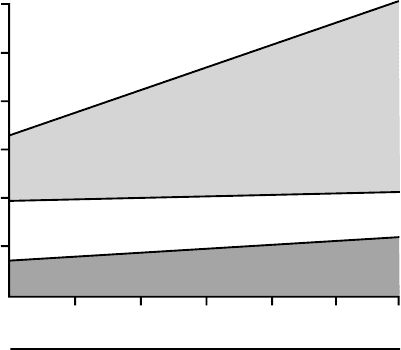
increases. Figure 2 shows the trend in the nature and
quantity of fuels oxidized after an overnight fast in lean
and obese humans. The data show that as weight and
body fat increase, the npRQ falls. The greater the RMR,
the greater the quantity of fat that is mobilized to meet
the energy requirements of fasting humans. Due to body
energy demands, obese humans shift into an accelerated
rate of fat mobilization. However, this tendency to
mobilize and oxidize stored fat becomes more obvious in
1.8
1.5
1.2
0.9
0.6
0.3
0
kcal/min
Fat
Carbohydrate
Protein
55 75 95 115 135 155 175
Weight (kg)
0.84 0.82 0.80 0.78 0.76 0.75 0.73
npRQ
FIGURE 2 The nature and quantity of fuels oxidized by humans
after a 12–14 h fast. Fat oxidation increases as body size increases.
102 STARVATION
both lean and obese people as fasting is prolonged. As
starvation is extended to 2–3 days and beyond, lipids
furnish 90–93% of the resting energy requirements in
both lean and obese humans. These changes in the
nature and quantities of fuels oxidized by tissues such
as muscle and brain, spare carbohydrate (glucose)
from oxidation; this provides the physiologic reasoning
for insulin-resistance or insensitivity when carbo-
hydrate oxidation must be curtailed for survival during
food deprivation.
The absolute minimal rates for the major fuel
utilization before death occurs have not been defined.
The minimum requirement for fat and protein oxidation
in grams/day/kg FFM has been approximated as follows:
. 2.98 ^ 0.21 g per kg per day FFM for fat;
. 0.52 ^ 0.10 g per kg per day FFM for protein.
Glucose is derived from glyceride-glycerol, amino acid,
and recycled lactate and pyruvate. About 1.91 ^ 1.04 g
per kg per day FFM are synthesized. However, most of
the glucose that is synthesized during starvation is
recycled from lactate and pyruvate (the Cori cycle),
glycerol, alanine, and glutamine. The quantity of
glucose that undergoes oxidation to CO
2
and H
2
Ois
primarily determined by the catabolism of glucose in the
central nervous system. This quantity is not closely
related to the FFM because the size of the brain is
unrelated to the FFM. The terminal oxidation of glucose
is . 40–45 g per day, which is derived primarily
from gluconeogenesis from amino acids (alanine and
glutamine) and glycerol.
In obese humans, during starvation there is no
decrease in the metabolic requirement per unit mass,
based on measurements of oxygen consumption. How-
ever, when the npRQ decreases below 0.7, the theoreti-
cal minimum for fat oxidation, there is a difficulty in
extrapolating the respiratory exchange rates of O
2
and
CO
2
and urinary nitrogen excretion into energy
expenditure. The production of CO
2
falls disproportio-
nately to O
2
consumption, creating a mysterious
situation characterized by a calculated npRQ of
0.62–0.65. This unusual finding cannot be eliminated
by correcting for ketonuria. However, it has been
postulated that some of fatty acid released from
triglyceride stores may undergo desaturation before
being recycled to storage depots. The process of
desaturation consumes oxygen and produces heat, but
releases no carbon dioxide. Desaturated-FFAs in the
blood have been identified in starving animals. If this
process occurs in humans, the respiratory quotient (RQ)
should rise before death, when unsaturated fatty acids
are released and oxidized for energy. The RQ does rise in
animals and humans during the pre-mortal period of
starvation. This phenomenon has not been fully
explained but is usually attributed to the mobilization
and oxidation of amino acids. It is just as likely that it is
due to oxidation of desaturated fatty acids.
Changes in the Concentration
of Substrates and Hormones
in the Blood
The concentration of glucose in the blood after an
overnight fast is . 4.5 mM; this value falls to . 3.6 mM
after 3 days of starvation. Thereafter, the concentration
of glucose in the blood reaches a plateau. Anaerobic
glycolysis in tissues such as muscle, brain, red blood
cells, and kidney medulla converts glucose into lactate
and pyruvate, and the liver extracts lactate and pyruvate
to produce glucose. The concentration of lactate in the
venous blood is less than 1.0 mM, while the concen-
tration of pyruvate is less than 0.1 mM and is constant in
the resting state. The concentration of FFA in the plasma
rises from . 0.6 mM, to . 1.4 mM during the first few
days of starvation. Thereafter, the concentrations of
these fuels remain elevated and relatively constant. The
rate of uptake and disposal of FFA is largely determined
by its concentration in the blood. Insulin regulates the
release of FFA from adipose tissue, primarily by
influencing lipolysis, and thus the availability of FFA.
The concentration of glycerol in the blood derived from
lipolysis is less than 0.1 mM after an overnight fast and
rises to 0.15 mM on the third day of starvation.
Thereafter, glycerol remains constant because uptake,
primarily by the liver, to synthesize glucose matches its
release by adipose tissue. The concentration of triglycer-
ides in the blood is less than 1.0 mM and remains
constant during fasting.
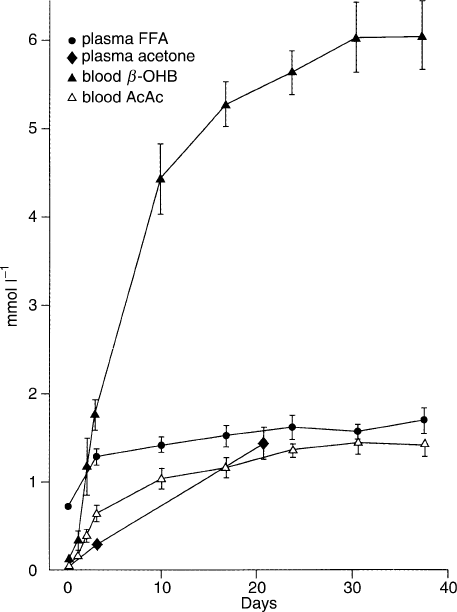
A characteristic of fasting in humans is the presence of
increased concentrations of ketone bodies in the blood
(Figure 3) and urine. These water-soluble, short-chained
compounds are synthesized primarily in the liver from
the acetyl CoA derived from fatty-acid oxidation and
serve as alternate fuels for tissues such as the brain.
Ketone bodies replace glucose as the dominant energy
source for the brain during starvation. Physical activity
augments catabolism during starvation and promotes
hyperketonemia. There are no other fuels in the blood
that can change as markedly as the concentration of
ketone bodies during starvation. This is partly due to the
low concentration of ketone bodies in blood during the
postprandial period, after a mixed meal containing
adequate carbohydrate. After an overnight fast, lean
people have blood AcAc
2
and
b
-OHB
2
concentrations
of . 0.05 mM, while this value is slightly higher in obese
individuals. Acetone is virtually absent after an over-
night fast, unless the individual is regularly consuming a
high-fat diet. There is an exponential rise in the con-
centration of AcAc
2
and
b
-OHB
2
in the blood during
starvation, until new steady levels develop. During the
first 3 days of fasting, the AcAc
2
concentration in the
blood increases to . 1.5 mM, while
b
-OHB
2
continues
STARVATION 103

to rise to . 4.5 mM after day ten of starvation. AcAc
2
plus
b
-OHB
2
reaches a plateau at . 6–8 mM after
18 days of total starvation (water, salt, and vitamins
were provided). The smell of acetone halitosis becomes
evident after . 2–3 days of starvation when the blood
concentration is . 0.25 mM. Acetone slowly increases
to . 0.35 mM after 21 days of starvation. A minimum
estimate of the change in the concentration of ketone
bodies in the blood from an overnight fast to 18 or more
days of total starvation is 75–160 fold (0.1–0.2 mM to
15.0–16.0 mM). Blood AcAc
2
and
b
-OHB
2
are anions
and the hydrogen produced during ketogenesis com-
bines with bicarbonate to form CO
2
and water. CO
2
is exhaled thus decreasing the concentration of
bicarbonate in the blood; this creates the typical anion
gap of ketonemia. Blood pH also falls appropriately
(7.4 to 7.3), and a mild metabolic acidosis of starvation
becomes evident.
The concentration of total amino acids in the plasma
declines slowly from an overnight fasting value of
. 4.6 mM to . 3.7 mM after 40 days of starvation.
However, there are four basic patterns of change among
the amino acids during total starvation, represented by
alanine, glycine, valine, and glutamine. Alanine rapidly
decreases to . 30% of its overnight concentration.
Glycine does the opposite; it rises nearly threefold,
before reaching a plateau. The concentration of valine
doubles in the plasma during the first 7–10 days of
starvation, and then slowly and progressively falls to a
value below its overnight fasting value. Venous gluta-
mine, the predominant plasma amino acid, remains
relatively stable during 5–6 weeks of fasting.
Blood urea nitrogen mimics the concentration of
alanine in the blood. The concentration of creatinine in
the blood slowly drifts downward, reflecting the
decrease in muscle mass.
Hormonal Changes
Insulin is the main hormone that controls the anabolic
processes that maintain fuel homeostasis in humans. Its
secretion is primarily regulated by the blood glucose
concentration, but the levels of amino acid, FFA
and ketone bodies also modulate insulin release by the
b
-cells of the pancreas. The influence of insulin on
the metabolism of the major organs is readily demon-
strated in adipose tissue, skeletal muscle, liver, white
blood cells, and other tissues. Insulin also inhibits
glucagon secretion, a significant counter-balance cata-
bolic hormone.
The concentration of insulin in the blood parallels the
changes in the levels of glucose. The fall in the
concentration of insulin diminishes some of its inhibi-
tory effects on peripheral proteolysis and lipolysis. In the
starved state, the concentration of insulin falls, resulting
in a decrease in the rate of uptake of glucose, amino
acids, and fatty acids in peripheral tissues and a
subsequent increase in the rates of gluconeogenesis,
lipolysis, and proteolysis. After an overnight fast, the
concentration of insulin in the portal blood is 2– 3 times
greater than the peripheral venous concentration. After
2 to 3 days of starvation, the portal-peripheral insulin
concentration gradient is small. During starvation, there
is still a high enough concentration of insulin in the
blood to limit the maximal rates of glycogenolysis,
gluconeogenesis, and ketogenesis. In addition, insulin
has at least two indirect effects on hepatic glucose and
ketone body production. Insulin decreases the delivery
of gluconeogenic precursors and FFA from the extra-
hepatic-tissue stores to the liver. As the concentration of
insulin in the mesenteric blood decreases, the inhibitory
effect of insulin on the
b
-cells of the pancreas curtails the
secretion of glucagon. Thus, the low concentration of
insulin in the blood promotes glucagon secretion.
Insulin has dual roles in controlling the release of
glucose and ketone bodies from the liver (and kidney
cortex), and the release of amino acid from muscle and
FFA from adipose tissue. Glucagon has the opposite
effect. It augments hepatic (and renal) gluconeogenesis
and ketogenesis, and promotes peripheral lipolysis and
proteolysis. A relatively low blood insulin concentration
FIGURE 3 Changes in the concentration of ketone bodies and free
fatty acids (FFA) during starvation.
104 STARVATION
and relatively high glucagon concentration creates a
blood insulin/glucagon ratio that promotes fuel avail-
ability. These pancreatic hormones serve collectively to
finely balance the fuel needs of various tissues. The
catabolic role of glucagon is augmented by catechol-
amines, glucocorticoids, and growth hormones. Fuel
homeostasis is critical for survival so there are multiple
levels of hormonal control of this process.
Appetite Regulation
During starvation there are a number of factors that
regulate the control of appetite. Ghrelin, an orexigenic
hormone, primarily secreted by the stomach and
duodenum, normally increases in the blood before
meals and falls after meals. Ghrelin is thought to
stimulate hunger, increase body weight, and decrease
metabolic rate. Ghrelin rises with semi-starvation but its
physiologic roles regarding hunger need further elucida-
tion. Adipose tissue produces a satiety hormone, leptin,
which suppresses appetite. The leptin concentration in
the blood falls in parallel with insulin during the first
three days of starvation. As body fat decreases during
starvation, the blood leptin concentration decreases. A
fall in the levels of leptin in the blood increases the
hypothalamic concentration of neuropeptide Y, greatly
stimulating appetite. However, the control of appetite is
not a simple process, but rather involves the interaction
of a number of neuroendocrine systems. There is no
single hormonal neuropeptide that controls hunger in a
starving human. Prolonged starvation causes exagger-
ated hunger, a psychosomatic state that dominates the
conscious mind.
Insulin plays an important role in the regulation of
appetite. Acting together with leptin, insulin circulates
at concentrations proportional to the body fat content.
Both hormones enter the CNS and bind to specific
receptors in neurons involved in controlling food intake.
The administration of leptin and insulin directly to the
hypothalamus decreases appetite and suppresses food
intake. It is probable that leptin is the more important
of the two hormones since leptin deficiency, not a lack
of insulin, results in obesity. The mechanism by which
these hormones influence the CNS is the subject of
intensive study. A detailed discussion of this area is
beyond the scope of this article. However, it should be
clear that during starvation, as the mass of adipose
tissue decreases and the secretion of insulin by the
pancreas is depressed due to a lack of food intake,
the appetite centers of the brain would be stimulated
by the lack of satiety signals such as leptin, insulin,
and ghrelin.
Adipose tissue secretes a number of other regula-
tory peptides, besides leptin, that control fuel depo-
sition. These include adiponectin, resistin, adipsin,
acetylation-stimulating protein, angiotensinogen, and
cytokines. The most abundant adipose specific hormone
is adiponectin. The concentration of adiponectin in the
blood is positively correlated with insulin sensitivity.
Adiponectin promotes glucose uptake in muscle and
fatty tissue. Its concentration in plasma is inversely
related to adipose tissue mass, especially abdominal fat
mass. The levels of adiponectin in the blood fall during
semi- and total starvation, when glucose intolerance has
been documented. Resistin is another protein secreted
into the blood by adipose tissue, but it has an effect that
is opposite to that of insulin. Its concentration in the
blood of humans after prolonged starvation has not been
defined. The behavior of adipose tissue cytokines in
humans during weight reduction caused by semi-
starvation is under investigation.
Biochemical Changes
during Starvation
Starvation induces a number of specific biochemical
changes that permit an individual to survive in the
absence of food for a prolonged period of time. These
include alterations in glucose synthesis, fatty acid
utilization by specific tissues, and the preservation of
nitrogen to insure the maintenance of lean body mass.
Starvation is a catabolic state in which fuel reserves are
mobilized to support metabolic needs. To understand
the metabolic adaptations to starvation, it is first
necessary to describe the available energy reserves of a
human as starvation begins. It has been estimated that a
70 kg human contains 144 000 kcal as fat (largely as
triglyceride), 24 000 kcal as protein (lean body mass),
and only 900 kcal as carbohydrate (liver and muscle
glycogen). The relatively low level of carbohydrate
reserve in humans necessitates a number of biochemical
adaptations to insure the appropriate energy supply to
specific tissues. For example, the brain uses . 600 kcal
of glucose each day; this means that the total carbo-
hydrate reserve is depleted in about one day of
starvation! It is clear that mechanisms must exist for
the use of fuels other than glucose (fatty acids and ketone
bodies) by tissues such as the muscle and the brain, to
insure survival during starvation. This process is called
“fuel sparing.”
CONTROL OF FATTY ACID RELEASE
DURING
STARVATION
Since fat is the major caloric reserve in humans, the
regulation of mobilization of FFA from adipose
tissue during starvation is a critical factor for survival.
The breakdown of triglyceride via lipolysis is under
the control of a number of hormones, including
STARVATION 105
epinephrine, glucagon, glucocorticoids, and insulin. The
concentration of insulin in the blood falls by . 50%
after 3 days of starvation. At the same time there is an
increase in the concentration of glucagon. This change in
the insulin to glucagon ratio is a critical factor in the
increase in lipolysis and the enhanced rate of hepatic and
renal gluconeogenesis that occurs during starvation.
Insulin is the major antilipolytic hormone and its
decrease is the major factor in insuring the increased
availability of the fatty acids needed for energy
metabolism during starvation.
Adipose tissue contains a hormone-sensitive lipase
that is sensitive to the state of its phosphorylation. An
increase in the concentration of cAMP in the tissue
(caused by a rise in epinephrine, glucagon and growth
hormone, and a drop in the level of insulin) activates
protein kinase A (PKA) that in turn phosphorylates,
and thus activates the hormone-sensitive lipase; this
results in the breakdown of triglyceride and generation
of FFA. Insulin counters this process, partly by
decreasing the levels of cAMP in the adipose tissue,
and by increasing the activity of a phosphoprotein
phosphatase that dephosphorylates the hormone-sensi-
tive lipase, thereby inactivating it. There is also
evidence that insulin increases the activity of a
phosphodiesterase in the adipocyte, causing a fall in
the concentration of cAMP in the tissue. The net result
of prolonged starvation is an enhanced rate of FFA
release from adipose tissue that is used as a fuel by a
number of tissues.
About 50% of the plasma FFA presented to the liver
during starvation is extracted and . 50–80% of the
extracted fatty acids undergo partial oxidation to
synthesize equal quantities of AcAc
2
and
b
-OHB
2
.
Most of the remaining quantity of FFA extracted by the
liver is converted to triglycerides and recycled to adipose
tissue as VLDL. It is interesting that such a large fraction
of the FFA released by lipolysis is re-esterified back to
triglyceride in adipose tissue or in the liver and other
tissues, and returned to the adipose tissue for the
resynthesis of triglyceride. Fatty acid recycling via this
so-called triglyceride–fatty acid cycle can account for as
much as 60% of the fatty acid released after 3 to 4 days
of starvation in humans. The synthesis of triglyceride
requires the generation of 3-phosphoglycerol, usually
from glucose. During starvation, when glucose is at a
premium, the 3-phosphoglycerol is generated from
pyruvate, lactate, and amino acids via an abbreviated
version of gluconeogenesis termed glyceroneogenesis.
The triglyceride/fatty acid cycle is a “futile cycle” that
uses energy for the synthesis of triglyceride (6 molecules
of ATP per molecule of triglyceride synthesized), so it
must have a role in preserving the FFA that was
released by adipose tissue to be used later as a fuel by
peripheral tissues.
PROTEOLYSIS AND AMINO ACID
METABOLISM
After a meal containing proteins, amino acids largely
escape hepatic extraction and are carried by the blood to
extrahepatic tissues. Insulin promotes the active trans-
port of amino acids into cells, primarily skeletal muscle.
Normally, amino acids are in a state of flux; they are
precursors for protein synthesis and then appear as free
amino acids after protein breakdown. Within the first
day of starvation, however, protein catabolism dom-
inates the metabolic flux. As starvation progresses,
relatively more proteolysis occurs and amino acids are
mobilized from protein depots. The dominant fate of the
carbon skeletons and the amino and amide groups
derived from the breakdown of amino acids in muscle, is
conversion to alanine and glutamine that is mobilized
from muscle and other depots and transported to liver
and kidney to be utilized to synthesize glucose, urea,
and ammonia.
Protein catabolism in the splanchnic tissues is
somewhat less responsive to insulin than it is in
skeletal muscles. In the periphery, the basal concen-
tration of insulin that is present during starvation,
limits proteolysis. Nonetheless, glucose must be con-
tinually synthesized from amino acids by the liver and
kidney cortex during starvation; amino acids from
muscle protein are a major source of carbon for
gluconeogenesis. The first proteins degraded during
starvation are the digestive enzymes secreted from the
stomach, pancreas, and small intestine. The liver also
loses various enzymes needed to process incoming
nutrients into plasma protein, e.g., albumin. There-
after, the largest protein mass of the body, striated
muscle, begins to be drained, not only of intracellular
proteins, such as enzymes, but also the contractile
elements. The disintegrating muscles can easily be seen
as skeletal muscle atrophy during prolonged starvation
in humans.
The control of proteolysis in muscle is a complex
process. Insulin retards proteolysis and enhances protein
synthesis. In addition, metabolic acidosis promotes
protein breakdown to insure the generation of
ammonia to titrate the acidity of the tubular urine.
The low plasma insulin concentration and mild
metabolic acidosis of starvation are conducive to
proteolysis. The best characterized pathway for protein
catabolism is an ATP-independent system of acid
proteases (cathepsin) and hydrolases contained in
cellular lysosomes. In addition, there are calcium-
dependent proteases, as well as cytosolic ATP-dependent
and independent pathways. The most important
muscle proteolytic system employs the ubiquitin protea-
some pathways.
Amino acids generated from proteolysis undergo
deamination and/or deamidation before entering the
106
STARVATION
tricarboxylic acid (TCA) cycle to participate in further
transformations to alanine or glutamine which efflux
from cells. Alanine plus glutamine account for . 80% of
the amino acids released from muscle after prolonged
starvation, and are the principal vehicles for transport-
ing nitrogen from peripheral tissues to liver and kidney.
These two amino acids together account for only 10% of
the composition of skeletal muscle protein, but they
provide the majority of the carbon for the synthesis of
glucose by the liver and kidney during starvation. Thus,
there is a net conversion of other amino acids into
alanine and glutamine in the muscle. Alanine is the
major nitrogenous compound released from muscle and
extracted by the liver for gluconeogenesis and ureagen-
esis. There is a “glucose– alanine” cycle between muscle
and liver. Glutamine is the other amino acid released in
high levels by muscle, but it is extracted from the blood
by the kidney cortex to produce glucose and ammonia.
During the catabolism of muscle, branched-chain amino
acids provide the amino groups to
a
-ketoglutarate to
form glutamate via glutamate dehydrogenase; glutamine
is derived from glutamate and ammonia by the action of
glutamine synthase.
The TCA cycle is the key metabolic pathway for
energy production: acetyl CoA is oxidized to carbon
dioxide. During starvation, the TCA cycle has another
fundamental role in metabolism. Amino acids are
catabolized to 4- (aspartate, asparagines, phenylalanine,
tyrosine, methionine, isoleucine, and valine) and 5-
(glutamine, glutamate, histidine, proline, and arginine)
carbon intermediates that enter the TCA cycle by a
process termed anaplerosis. The TCA cycle cannot serve
as a carbon sink; therefore, the carbon atoms that enter
the TCA cycle must leave the TCA cycle by a process
called cataplerosis. Anaplerosis and cataplerosis are
reciprocal reactions that must be balanced. These
reactions have been reviewed in detail elsewhere in
this volume.
During starvation, more fuels are released from
storage depots or synthesized in liver and kidney than
are oxidized to generate energy. About 60% of the fuels
that enter the bloodstream are recycled, e.g., FFA,
glucose and amino acids, especially alanine and
glutamine.
KETOGENESIS
The liver not only stores glucose as glycogen, it also
converts fuels such as FFA, amino acids, lactate,
pyruvate, and glycerol to glucose and ketone bodies; it
also detoxifies the ammonia from amino acid breakdown
by converting it to urea. After an overnight fast, hepatic
glycogenolysis, gluconeogenesis, and ketogenesis pro-
vide . 50% of the total energy-yielding fuels for the body
in the resting state. Lipolysis of triglyceride in adipose
tissue supplies FFA and glycerol; these substrates become
precursors for ketone body (fatty acids) and glucose
(glycerol) syntheses. Amino acids released primarily by
skeletal muscles complement glycerol as gluconeogenic
precursors. As fasting is prolonged, the kidney cortex
also contributes to fuel homeostasis by conserving sub-
strates and sharing the gluconeogenic role with the liver.
Ketone bodies are the only fuels synthesized in the
body that do not recycle. Unlike FFA, amino acids and
glucose, ketone bodies are either oxidized or excreted in
the urine and/or the breath (acetone). A small amount of
acetone can be converted to glucose.
b
-OHB
2
and
AcAc
2
are synthesized in the liver (and kidney)
mitochondria by the following reactions. Two molecules
of acetyl CoA condense head-to-tail to form acetoacetyl
CoA; this reaction is catalyzed by acetoacetyl CoA
thiolase. Another molecule of acetyl CoA is joined by
b
-hydroxy-
b
-methylglutaryl CoA (HMG CoA) synthase
to form HMG CoA, also generating a hydronium ion
(H
þ
). HMG CoA lyase cleaves HMG CoA into AcAc
2
and acetyl CoA. AcAc
2
is then reduced to
b
-OHB
2
by
b
-hydroxybutyrate dehydrogenase. Acetone and CO
2
are formed from the degeneration of AcAc
2
. Ketone
bodies are synthesized from the acetyl CoA that is
derived from the
b
-oxidation of fatty acids in the
mitochondria. A small quantity can be synthesized
from ketogenic amino acids during starvation. In
addition, acetoacetyl CoA can be formed from FFA
and cleaved to AcAc
2
in the kidneys. The hepatic
production of
b
-OHB
2
and AcAc
2
are about equal.
During hyperketonemia, the concentration of
b
-OHB
2
in the blood is . 3 times greater than AcAc
2
. This is due
to the preferential removal of AcAc
2
or conversion of
AcAc
2
to
b
-OHB
2
by skeletal muscle. The brain
removes
b
-OHB
2
and AcAc
2
in a concentration-
dependent manner.
The metabolism of ketone bodies during starvation is
a critical element in the control of fuel homeostasis in
humans. The demonstration that
b
-OHB
2
and AcAc
2
could serve as major fuels for the metabolism of the
brain during starvation provided the information needed
to evaluate the roles of fatty acid oxidation, amino acid
mobilization, glucose conservation, and urinary nitro-
gen excretion during prolonged starvation. From this
research it became clear that the abundance of FFA
stored in humans provides a substantial reserve for the
synthesis of ketone bodies. The metabolism of ketones
by the brain during starvation greatly limits the need to
use amino acids to make glucose to support the
metabolism of this tissue. This is an important example
of “fuel sparing.”
There are few synthetic processes that are quantitat-
ively as large as the daily rate of ketogenesis during
starvation. After an overnight fast, hepatic ketogenesis
amounts to . 10 g per day. After 2–3 days of starvation
the liver synthesizes over 100–150 g per day of ketone
bodies. Over this period, the average resting human
STARVATION 107

oxidizes a minimum of . 3 g of fat per kg FFM per day.
Thus, a large, but not obese adult man, weighing 80 kg,
with a body composition of 80% FFM (64 kg) and 20%
fat mass (16 kg) oxidizes a minimum of . 192 g of fat
per day. About 60 g of the FFA derived from the lipid
stores undergo
b
-oxidation in the liver to yield an
estimated 113 g per day of ketone bodies. Ninety
percent of these water-soluble fuels undergo terminal
oxidation, primarily by the brain and muscle. Most of
the remainder is excreted in the urine. It is interesting to
note that about half of the molecular weight of AcAc
2
and
b
-OHB
2
is oxygen. Thus, if 10–12 g of ketone
bodies are excreted in the urine, only 5–6 g of the
carbon skeleton is derived from stored triglycerides.
Since
b
-OHB
2
and AcAc
2
are excreted with near
equimolar quantities of NH
4
þ
, ketosis is an energetically
cheap way to excrete nitrogen (the synthesis of urea
requires 4 molecules of ATP per molecule of urea).
Finally, for each gram of nitrogen lost in the urine
. 3.57 g of glucose is synthesized.
GLUCONEOGENESIS
The de novo synthesis of glucose from nonhexose
precursors (gluconeogenesis) occurs in the liver, kidney
cortex, and perhaps to a minor extent in the small
intestine. The precursor molecules for gluconeogenesis,
and the organs involved in this process, change as
starvation progresses. After an overnight fast, the liver is
the primary, if not the sole, organ that makes a net
contribution to glucose in the blood. The kidney extracts
and releases glucose after an overnight fast but its overall
contribution is minimal. After 3 days of starvation, renal
gluconeogenesis increases and by 10–18 days of total
starvation the kidney contributes . 50% of the glucose
added to the blood. However, during prolonged
starvation, total glucose added to the blood from
gluconeogenesis is only . 50% of what it was after an
overnight fast when gluconeogenesis is greatly comple-
mented by hepatic glycogenolysis. In addition, during
prolonged starvation only half of this glucose contrib-
uted to the blood is oxidized to CO
2
and H
2
O. This is
the result of the “sparing” of glucose by tissues that use
more abundant fuels such as ketone bodies.
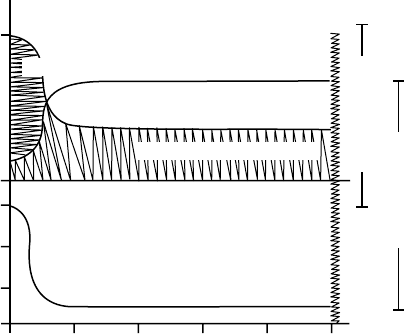
A schematic representation of the quantities of ketone
bodies and glucose contributed to the blood by the liver
and kidneys is shown in Figure 4. In general, glycogen-
olysis decreases as gluconeogenesis increases, and glucose
production decreases as ketogenesis increases. An
interesting aspect of gluconeogenesis during starvation
is the relationship between renal gluconeogenesis and
ammoniagenesis. Glutamine produced by the muscle is
used by the kidney cortex as the major source of carbon
for gluconeogenesis. The amino and amide groups are
removed from glutamine to form
a
-ketoglutarate,
which then enters the TCA cycle and is converted to
glucose via renal gluconeogenesis. The kidney also has
the ability to synthesize glucose from lactate, pyruvate,
glycerol, amino acids and other precursors, but only
glutamine has been closely associated with net renal
gluconeogenesis.
The decreased rate of glucose production during
starvation is linked to the increased ketone body
production. Hyperketonemia is accompanied by keto-
nuria and ammoniagenesis. Renal NH
4
production is
coupled to renal gluconeogenesis, while the release of
NH
3
by the kidney is linked to hepatic urea production.
In summary, gluconeogenesis, ketogenesis, ammonia-
genesis, acid–base balance, and ureagenesis are all
tightly interdependent during starvation.
Urinary Nitrogen Excretion
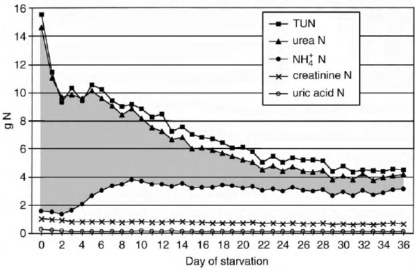
Total, daily urinary excretion undergoes an asymptotic,
progressive decrease during starvation (Figure 5).
Humans that consume a diet with 100 g protein
(16.0 g nitrogen) excrete 15.5 g of nitrogen in the
urine. Most of the nitrogen (85%) is excreted as urea.
Ammonium, uric acid, and creatinine account for most
of the remaining urinary nitrogen. Starvation initially
induces an acute decrease in total nitrogen excretion.
After 3–6 days of starvation, total urinary nitrogen
decreases gradually, so that after 36 days of starvation
Renal gluconeogenesis
Hepatic ketogenesis
Renal ketogenesis
0 3 6 9 12 15+
Hepatic glycogenolysis
Hepatic gluconeogenesis
Days of starvation
0.86
0
1.0
mmol/min/1.73m
2
=550 cal/min/1.73m
2
=550 cal/min/1.73m
2
FIGURE 4 Schematic representation of fuel delivery by the liver and
kidney during starvation. Hepatic gluconeogenesis, glycogenolysis and
ketogenesis, and renal gluconeogenesis are expressed as mmol/min
and converted to calories per 1.73m
2
body surface area. Hepatic
glycogenolysis is rapidly dissipated during the first 1–3 days of fasting.
The liver and kidney share the role of gluconeogenesis as
starvation progresses. Hepatic ketogenesis becomes a dominant
mechanism for supplying fuels to the blood after liver glycogenolysis
is curtailed. Renal ketogenesis may contribute to ketonuria, but not
to ketonemia.
108 STARVATION

the nitrogen excretion is . 4.6 g per day. The biggest
decrease is in the exponential decay in urea nitrogen
excretion. The excretion of ammonium ion (NH
4
þ
)
parallels the excretion of AcAc
2
and
b
-OHB
2
;it
peaks at . 120–140 mmol per day at 6– 9 days of
starvation. After 15–18 days of drinking only water,
NH
4
þ
overtakes urea as the dominant urinary nitrogen
compound. Urinary NH
4
þ
gradually decreases as star-
vation is prolonged. NH
4
þ
, AcAc
2
,and
b
-OHB
2
urinary
excretion rates are sensitive to small quantities of
ingested glucose. In early studies of metabolism during
starvation, subjects were allowed to drink beverages that
contained carbohydrates. This distorted both urea and
NH
4
þ
urinary excretion. The excretion of creatinine
decays in a linear manner that is dependent upon
diminishing body muscle mass that occurs as starvation
progresses. The output of uric acid in the urine drops
acutely during the first 4 days of starvation as ketonuria
rises. Thereafter uric acid nitrogen excretion plateaus at
. 2 mmol per day (120 mg per day).
The rate of urea excretion provided the first clue that
the concept that brain metabolized only glucose for
energy had to be modified. Research in the first half of
the twentieth century established that for each gram of
urinary nitrogen excreted . 3.5 g of glucose could be
synthesized from amino acids. Excluding the glycerol
that is present in triglycerides, only trivial quantities of
glucose could be derived from triglyceride. The brain
requires . 100–125 g of glucose or equivalent energy
sources daily. Plasma FFAs cannot pass the blood–brain
barrier to provide a source of energy. In 1967 it was
shown that AcAc
2
and
b
-OHB
2
were the primary
fuels for brain metabolism during starvation. Therefore,
gluconeogenesis is clearly important during starvation,
but the quantity of glucose formed is less than the
quantity needed to provide energy for the brain.
However, it should be pointed out that tissues such as
the red blood cells, kidney medulla, and lens of the eye
use glycolysis for energy production. The lactate and
pyruvate generated by these tissues are recycled to the
liver for the production of glucose.
Fuel Consumption
during Starvation
While the liver and kidney release glucose into the blood
during starvation, only the liver makes a net contri-
bution of ketone bodies. However, more urinary
excretion of AcAc
2
and
b
-OHB
2
occurs than is
extracted across the renal vascular beds, suggesting
that the kidneys synthesize some of the ketone bodies
excreted in the urine. Ketonuria augments ammoniagen-
esis. The deamination of glutamine to form
a
-ketoglu-
tarate generates two molecules of NH
4
þ
, which are
released into the tubular urine to maintain electroneu-
trality and acid–base balance. As mentioned above, the
a
-ketoglutarate produced in this process enters the TCA
cycle via anaplerosis, and is subsequently converted to
glucose that is released into the blood. The kidney can
also convert lactate, pyruvate, other amino acids, and
perhaps glycerol into glucose. Clearly the kidney shares
the role of glucose production with the liver, but the
kidneys make a net contribution to blood only after
several days of starvation.
FIGURE 5 The total quantity and various components of urinary nitrogen excretion during prolonged starvation when five men and five women
volunteers were given water, salt, and vitamins. TUN is an abbreviation for total urinary nitrogen. The nitrogen contents of urea, ammonium,
creatinine, and uric acid are displayed.
STARVATION 109
Early in starvation, the small intestine may provide
gluconeogenic amino acids to the splanchnic (portal)
system, but by 3 days of fasting there is no easily
detected release of alanine or glutamine into the portal
blood in fasting humans.
After . 3 days of starvation skeletal muscles derive
. 50% of their energy requirements from ketone bodies
and, as fasting progresses, muscle preferentially extracts
a small quantity of AcAc
2
and may release
b
-OHB
2
.At
this time in starvation, FFA provides the great majority
of fuels for muscle.
After an overnight fast the brain derives its energy
from glucose, consuming . 100 –125 g per day. As
starvation progresses and blood glucose concentration
decreases and the concentration of AcAc
2
and
b
-OHB
2
increases, ketone bodies become the major fuels for
brain metabolism. These water-soluble fuels have access
to neurons and can supply . 2/3 of the total energy
requirements during prolonged starvation.
Concluding Statement
Humans are capable of surviving long periods
without food, but there are major limitations in this
ability. When about one-half of lean body tissue is
consumed to provide energy, death occurs. Morbidly
obese people subjected to prolonged starvation can
deplete the vital organs of structural proteins and
die fat. The adaptations that permit humans to survive
in the absence of food provide a fascinating look
at the underlying biochemical adaptations to star-
vation. Much of what we know about the biological
response to long-term starvation came from research
with human volunteers who deserve our heartfelt
thanks.
SEE ALSO THE FOLLOWING ARTICLES
A-Kinase Anchoring Proteins † Amino Acid Meta-
bolism † Fat Mobilization: Perilipin and Hormone-
Sensitive Lipase † Fatty Acid Oxidation † Glycogen
Metabolism † Insulin- and Glucagon-Secreting Cells of
the Pancreas † Insulin Receptor Family † Ketogenesis †
Leptin † Pyruvate Carboxylation, Transamination and
Gluconeogenesis † Tricarboxylic Acid Cycle
GLOSSARY
fuel homeostasis The steady-state maintenance of fuels.
gluconeogenesis The synthesis of glucose from nonhexose precursors.
glycogenolysis The breakdown of glycogen.
ketogenesis The synthesis of acetoacetate and
b
-hydroxybutyrate
and the generation of acetone.
starvation The deprivation of any or all of the elements for nutrition.
FURTHER READING
Balasse, E. O. (1979). Kinetics of ketone body metabolism in fasting
humans. Metabolism 28(1), 41 –50.
Cahill, G. F. Jr. (1970). Starvation in man. N. Engl. J. Med. 282,
668–675.
Felig, P., and Bergman, M. (1995). The endocrine pancreas: Diabetes
mellitus. In Endocrinology and Metabolism (P. Felig, J. D. Baxter
and L. A. Frohman, eds.) 3rd edition, pp. 1107–1250. McGraw-
Hill, New York.
Keys, A. J., Brozek, J., Henschel, A., Mickelsen, O., and Taylor, H. L.
(1950). The Biology of Human Starvation, Vols I and II, University
of Minnesota Press, Minneapolis.
Leibel, R. M., Rosenbaum, M., and Hirsch, J. (1995). Changes in
energy expenditure resulting from altered body weight. N. Engl.
J. Med. 332, 621–628.
Owen, O. E., and Reichard, G. F. Jr. (1971). Human forearm metabo-
lism during progressive starvation. J. Clin. Invest. 50, 1536– 1545.
Owen, O. E., Morgan, A. P., Kemp, H. G., Sullivan, J. M., Herrera,
M. G., and Cahill, G. F. Jr. (1967). Brain metabolism during fasting.
J. Clin. Invest. 46, 1589–1595.
Owen, O. E., Licht, J. H., and Sapir, D. G. (1981). Renal function and
effects of partial rehydration during diabetic ketoacidosis. Diabetes
30, 510–518.
Owen, O. E., Smalley, K. J., D’Alessio, D. A., Mozzoli, M. A., Knerr,
A. N., Kendrick, Z. V., Kavle, E. C., Donohoe, M., Tappy, L., and
Boden, G. (1990). Resting metabolic rate and body composition of
achondroplastic dwarfs. Medicine 69(1), 56–67.
Owen, O. E., Kalhan, S. C., and Hanson, R. W. (2002). The key role of
anaplerosis and cataplerosis for citric acid cycle function. J. Biol.
Chem. 277(34), 30409–30412.
BIOGRAPHY
Richard W. Hanson is Professor of biochemistry at Case Western
Reserve University School of Medicine in Cleveland, Ohio. For many
years he has been an Associate Editor of the Journal of Biological
Chemistry and was the President of the American Society of
Biochemistry and Molecular Biology. His research interests include
the dietary and hormonal control of gene transcription and the
regulation of gluconeogenesis and glyceroneogenesis in mammals.
Oliver E. Owen was previously Professor of medicine and Co-Principal
Investigator and Program Director, General Clinical Research Center,
Temple University School of Medicine and Hospital, and formerly
Professor and Chair of medicine, Southern Illinois University School
of Medicine.
110 STARVATION
