Lennarz W.J., Lane M.D. (eds.) Encyclopedia of Biological Chemistry. Four-Volume Set . V. 4
Подождите немного. Документ загружается.


Steroid/Thyroid Hormone Receptors
Ramesh Narayanan and Nancy L. Weigel
Baylor College of Medicine, Houston, Texas, USA
Steroid and thyroid hormones are important in regulating a
wide variety of normal physiological processes, including
development, metabolism, and reproduction. The receptors
for these hormones are members of the nuclear receptor
superfamily of ligand-activated transcription factors. For the
most part, each hormone interacts with a unique receptor
although the receptor may have multiple forms derived from
the same gene by various mechanisms. Exceptions include
estradiol, which activates two receptors derived from
independent genes, estrogen receptor
a
(ER
a
) and the
recently discovered estrogen receptor
b
(ER
b
), thyroid
hormone receptors (TR
a
and TR
b
), and retinoic acids,
which activate the retinoic acid receptors (
a
,
b
, and
g
). In
response to their cognate hormones, nuclear receptors bind to
specific DNA sequences altering transcription. In addition to
their best-characterized actions as DNA-binding transcription
factors, the receptors also influence transcription and result-
ing cell function through direct interactions with other
transcription factors, as well as through alterations in cell
signaling. These functions and the structures of the receptors
are described in this article.
Overview of Nuclear Receptor
Ligands and Mechanism of Action
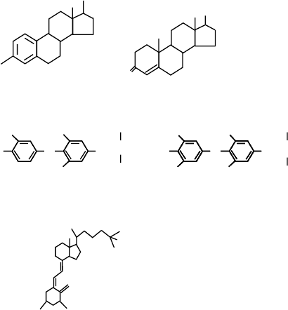
Figure 1 shows the structures of some of the ligands for
members of the nuclear receptor family. Estradiol is the
primary ligand for both estrogen receptors, ER
a
and
ER
b
. Testosterone, which is closely related to estradiol,
is one of the two major ligands for the androgen
receptor (AR). The ligands for the thyroid hormone
receptor (T
3
and T
4
) and the vitamin D receptor
(1,25(OH)
2
D
3
) are also shown. The five major classes
of steroids are synthesized from pregnenolone, which is
derived from cholesterol through the actions of the
cholesterol side-chain cleavage enzyme (P450scc). The
synthesis of the hormones is complex, with a number
of alternate pathways leading to the same hormone.
Testosterone and estradiol are derived from 17
a
-
hydroxy pregnenolone. Testosterone, the major circu-
lating androgen, is synthesized in the testes of males
and in the ovaries of females. It is important for
development of the male reproductive tract, fertility,
and secondary male characteristics. Estradiol, the
major circulating estrogen, is produced in the ovaries
of females. Estradiol is important in the female
reproductive tract, playing roles in breast and uterine
development, fertility, and also in bone and other
tissues. Progesterone is synthesized directly from
pregnenolone, and the major site of synthesis in
females is the ovary. Progesterone, acting through the
progesterone receptor, plays important roles in the
breast and uterus and in the maintenance of pregnancy.
Progesterone is a precursor of corticosterone and
aldosterone, which are synthesized in the adrenal
glands. The mineralocorticoid, aldosterone, is import-
ant for salt retention in the kidney. The glucocorticoid,
cortisol, is produced in the adrenals from 17
a
-hydroxy
pregnenolone and is important for regulation of
carbohydrate metabolism; it also plays a role in
suppressing immune responses. The secosteroid,
1,25(OH)
2
D
3
, is derived from cholesterol through a
UV-catalyzed reaction in the epidermis followed by
sequential hydroxylations in the liver and kidney. The
action of 1,25(OH)
2
D
3
is important for calcium
homeostasis and also plays a role in the differentiation
of a variety of tissues. Thyroid hormones are produced
from tyrosines and iodide in the thyroid gland. Thyroid
hormones have multiple actions in regulating metab-
olism, typically increasing oxidation rates. The
hormones are transported through the blood to their
sites of action.
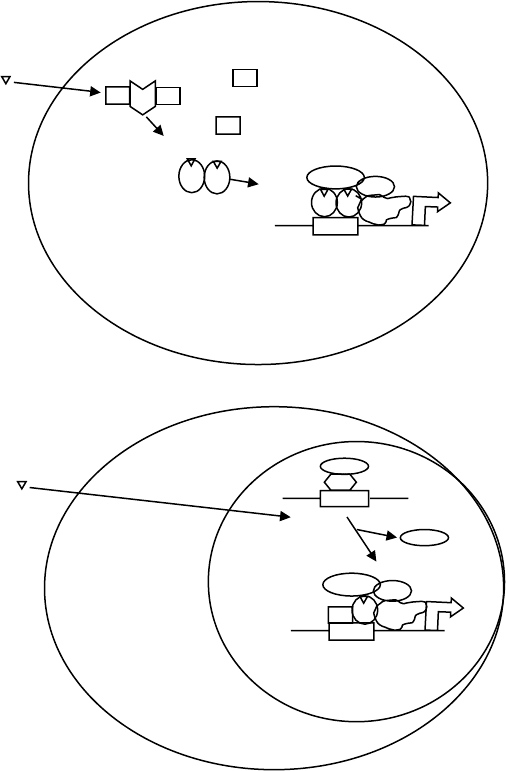
Figure 2 depicts the general mechanism of action of
nuclear receptors. The ligands are all lipophilic
compounds, which enter the cells by passive diffusion.
They bind to their cognate intracellular receptors
located either in the cytoplasm or the nucleus.
The receptors can be divided into two classes – those
that do not bind DNA in the absence of hormone
(Figure 2A) and those that bind to DNA in the absence
of hormone (Figure 2B). The regulation of their
activities differs somewhat. Classical steroid receptors
such as AR, glucocorticoid receptor (GR), progesterone
receptor (PR), mineralocorticoid receptor (MR), and
Encyclopedia of Biological Chemistry, Volume 4. q 2004, Elsevier Inc. All Rights Reserved. 111
ER belong to the first class; they are maintained in
an inactive conformation capable of ligand binding
by a complex of chaperone proteins including
hsp90 and p23. Whether these complexes are nuclear
or cytoplasmic depends upon the receptor. Upon ligand
binding, the receptor changes its conformation, no
longer binding to heat-shock proteins, homodimerizes
and, if the unliganded receptor is localized in the
cytoplasm, translocates to the nucleus. There, the
receptor binds to DNA containing specific sequences
called hormone response elements (HREs) that are most
often found in the 5
0
flanking region of target genes.
The receptor recruits components of the basal tran-
scription machinery, as well as proteins termed
coactivators that perform a variety of functions includ-
ing histone acetylation that enhance transcription.
Unlike the receptors for classical steroids, thyroid
receptors (TRs) are not bound to heat-shock proteins
in the absence of hormone and, instead, are bound
to the DNA as a heterodimer with the retinoid X
receptor (RXR), another member of the steroid/thyroid
hormone receptor superfamily (Figure 2B). In the
absence of ligand, TR binds corepressors, which in
turn bind histone deacetylases resulting in lower levels
of histone acetylation and repression of target gene
transcription. Hormone binding releases corepressors
and promotes binding of coactivators, and subsequent
transcription follows.
The Steroid/Thyroid Hormone
Receptor Superfamily
The steroid/thyroid receptors are the largest family of
ligand-activated transcription factors. In addition to
the well-characterized steroid, thyroid, retinoid,
and vitamin D receptors, the family contains receptors
for numerous lipophilic metabolites and xenobiotics as
well as receptors, termed orphans, for which ligands
have not yet been identified.
RECEPTOR STRUCTURE
Despite some evolutionary and functional differences,
the steroid receptor family members have many simi-
larities especially in their structure. As shown in Figure 3,
there are multiple domains in the receptors, with all
receptors containing domains A–E and only a subset
containing the additional F domain.
The N Terminus
The N terminus or the A/B domain of the receptor is the
least conserved domain among the family members. This
region is the most variable in length ranging from a few
amino acids to more than 500. This region has an
activation function, AF-1, which contributes to the
transcriptional activity of the receptor through binding
of coactivators.
The DNA-Binding Domain (DBD)
The DNA-binding domain, region C, is important for
the binding of receptor to the DNA and is the most
highly conserved domain. This region has two type-2
zinc finger motifs, which are responsible for DNA
recognition and dimerization (Figure 3B). Each finger is
composed of four cysteines that coordinate with one zinc
atom. Amino acids in this region also participate in
receptor dimerization.
The Hinge Region
Downstream of the DBD is the hinge region (D), which
contains a nuclear localization signal. This is a short
lysine-rich region, with a high homology to the simian
virus 40 T antigen nuclear localization signal. Additional
functions of this region are receptor specific.
The Ligand-Binding Domain
The ligand-binding domain (E) is essential for the
binding of ligand. The primary interaction site for the
hsp complex is also in this domain. Also located in this
region is the second activation function domain, AF-2,
NH
2
Estradiol
Testosterone
T
4
1,25-dihydroxyvitamin D
3
OH
OH
OH
O
OHOH
OH
O
OH
I
I
I
CH
2
CH
COOH
O
OH
I
I
I
CH
2
CH
NH
2
COOH
I
III
II
T
3
FIGURE 1 Structure of steroid and thyroid hormones. Estradiol is
the ligand for estrogen receptor, testosterone is the ligand for androgen
receptor, T
3
-3,5,3-L-triiodothyronine, T
4
-thyroxine are ligands for
thyroid receptor, and 1,25-dihydroxyvitamin D
3
is the ligand for the
vitamin D receptor.
112 STEROID/THYROID HORMONE RECEPTORS
which is responsible for ligand-mediated transcription
of target genes. The relative importance of AF-2 and
AF-1 in inducing transcription is receptor- and cell-
type specific. The structures of the hormone-binding
domains of several receptors have been determined
using X-ray diffraction. The hormone-binding
domain consists of a series of 12
a
-helices. Binding of
hormone causes a substantial conformational change in
the receptor exposing AF-2 for interactions with
coactivators. This domain also contains the strongest
dimerization interface in most steroid receptors.
The function of the F domain, located at the C
terminus of some receptors such as the ER is not
well defined.
RECEPTOR BINDING TO DNA
All of the receptors bind to their cognate HREs as
dimers. The consensus binding sequence for AR, PR,
GR, and MR, shown in Figure 3C, contains two half-
sites separated by three nucleotides with the sites
oriented to form a palindrome. ER recognizes a related
pair of half-sites with the same spacing and orientation.
Each monomer binds to a half-site. The class-II
receptors, including VDR and TR, bind to pairs of
half-sites whose sequences are identical to the ER half-
site, but whose orientation (direct or inverted repeats)
and spacing (0–6 nucleotides) determines the specificity
of binding. TR and VDR each heterodimerize with
HSP
HSP
HSP
HSP
SR
SR
SR
SR
TR
RXR
TR
CoR
CoR
SR
CoA
CoA
Steroid
hormone
GTF
HRE
mRNA
A
B
Thyroid
hormone
CoA
CoA
HRE
GTF
mRNA
HRE
FIGURE 2 Mechanism of steroid (A) and thyroid (B) hormone action. The two classes are distinguished by whether they are associated with heat-
shock proteins (A) like classical steroid receptors or are bound to DNA in the absence of hormone (B) like thyroid receptor. In both cases, binding of
agonist causes dissociation of proteins that repress activity and promotes a conformation that induces recruitment of coactivators stimulating
transcription of the target gene. SR, steroid receptor; HRE, hormone response element; GTF, general transcription factor; RXR, retinoid X receptor;
TR, thyroid receptor; CoA, coactivator; CoR, corepressor.
STEROID/THYROID HORMONE RECEPTORS 113
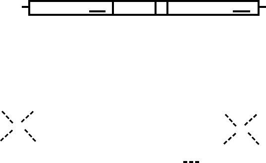
RXR and bind to the 3
0
end (half of the HRE), whereas
RXR binds to the 5
0
end (half of the response element).
Although these sequences represent the consensus
binding sites, natural sequences may differ significantly
and promoters may contain combinations of HREs as
well as individual half-sites all of which contribute to
the final activity.
Steroid Receptor Coregulators
When the receptor binds to the DNA, it recruits
proteins in the basal transcription machinery such as
TFIIB and RNA polymerase II. The receptors also bind
additional proteins or protein complexes that modu-
late receptor activity; these are termed coactivators
and corepressors. Coactivators are defined as proteins
that interact with the receptors and increase their
ability to transactivate the target gene. The mechanism
by which individual coactivators achieve this can vary.
More than 100 candidate coactivators have been
identified. While some coactivators function only
with steroid receptors and a small subset of other
transcription factors, others are used by many
transcription factors. The best characterized of the
steroid receptor coactivators (SRCs) is the p160 family
of coactivators: SRC-1, SRC-2 (GRIP1, TIF2), and
SRC-3 (Rac3, AIB1). These bind to the receptor
recruiting additional coactivators including CBP
(CREB-binding protein), p/CAF (CBP-associated fac-
tor) and CARM-1 (coactivator-associated arginine
methyltransferase). CBP/p300, p/CAF and some of
the p160 proteins are histone acetyl transferases
(HATs) and their binding increases local histone
acetylation. Other coactivators include the DRIP/
TRAP (D receptor interacting protein/thyroid hormone
receptor-associated protein) complex. Many coactiva-
tors interact with AF-2 located in the LBD. In other
cases, coactivators interact with the AF-1 region and
some interact with both domains. Interactions with
N
C
AF-1
A/B C E/FD
AF-2
GR
777
A
B
C
5′ AGAACAnnnTGTTCT 3′
3′ TCTTGTnnnACAAGA 5′
GRE
5′ AGGTCAnnnTGACCT 3′
3′ TCCAGTnnnACTGGA 5′
ERE
1
Zn
Cys
Cys
Cys
Cys
Lys
Leu
Lys
Val
Ser
Asp
Glu
Ala
Ser
Cys
Cys
His
Tyr
Cys
Val
Leu
Thr
Cys
Ser
Lys
Val Phe Phe Lys Arg Ala Val Glu Cys
Tyr
Leu
Zn
Cys
Cys
Cys
Cys
Ile
Ile
Asp
Lys
Ile
Arg
Arg
Lys
Cys
Pro
Ala
Lys
Ala
Cys
Arg
Glu
Arg
Tyr
Arg
Lys
Gln
Ala CysMet
5′ AGGTCAn
X
AGGTCA 3′
3′ TCCAGTn
X
TCCAGT 5′
VDRE, TRE
FIGURE 3 Receptor structure and DNA binding elements. Panel (A) shows the common structural features of nuclear receptors. The A/B region
contains AF-1, a region important for transcriptional activation. C is the DNA-binding domain, the most conserved region in the nuclear receptors.
D contains a nuclear localization sequence. E contains the hormone-binding domain and second activation function AF-2. Some receptors also
contain a C terminal extension, termed the F domain, whose physiological function is not well described. Panel (B) shows the sequence of the DNA-
binding domain of GR. Panel (C) shows sequences of consensus hormone response elements. The consensus sequence for a GRE (binds GR, AR, PR)
and an ERE (binds ER) are shown. Vitamin D receptor and the thyroid hormone receptors bind to direct repeats separated by three and four
nucleotides, respectively. Other receptors bind to direct or inverted repeats with a spacing of 0–6 (n
x
indicates that the half-site may be separated by
0–6 nucleotides). GRE, glucocorticoid response element; ERE, estrogen response element; VDRE, vitamin D response element; TRE, thyroid
response element; GR, glucocorticoid receptor; AF, activation function.
114 STEROID/THYROID HORMONE RECEPTORS
AF-2 are typically mediated by LXXLL (L ¼ Leucine
and X ¼ any amino acid) motifs in the coactivator.
Another class of proteins, termed corepressor, reduces
the activation of target gene transcription through
interaction with the receptors. The best-characterized
nuclear receptor corepressors are nuclear receptor
corepressor (NcoR) and silencing mediator of retinoid
and thyroid (SMRT) receptors. These proteins bind
histone deacetylase complexes and also interact with
class II receptors in the absence of hormone resulting in
local reductions in histone acetylation. Corepressors do
not bind to unliganded steroid receptors. However,
steroid receptor antagonists cause changes in the
conformation of the hormone-binding domain that
induce binding of the corepressors.
Steroid Receptor Agonists
and Antagonists
Although steroid receptor family members are import-
ant for normal physiological processes, there are a
number of instances in which it is desirable to block
the actions of selected steroid receptors. These include
breast cancer (ER) and prostate cancer (AR). Thus,
although natural antagonists of steroid receptor action
have not been identified, much effort has been
devoted to identifying compounds that will antagon-
ize hormone action. These compounds compete with
the natural ligand for binding to the hormone-binding
domain of the receptors. Although some antagonists
block dissociation from heat-shock protein complexes
or destabilize the receptor, most of the antagonists
promote dissociation from heat-shock proteins and
cause the receptors to bind to DNA. However, the
conformation induced by the antagonist differs from
that induced by agonist. This prevents recruitment of
coactivators to AF-2 and, instead, promotes recruit-
ment of corepressors. In some cases, it is desirable to
maintain the activity of a receptor in some tissues
while inhibiting activity in other tissues. The most
common example of this is the need for tissue-specific
regulation of ER activity. Estradiol is important for
maintaining bone mass and postmenopausal women
frequently develop osteoporosis. However, estradiol
can promote uterine cancer and may also be detri-
mental in breast. Thus, a great deal of effort has been
devoted to developing selective estrogen receptor
modulators (SERMs) which have tissue-specific agon-
istic and antagonistic activities. Tamoxifen (a SERM)
has been used in the treatment of breast cancer, but
increases the risk of uterine cancer. A newer SERM,
raloxifene, is an antagonist in both breast and uterus,
but acts as an agonist in bone.
Crosstalk between Nuclear
Receptors and Cell
Signaling Pathways
PHOSPHORYLATION OF RECEPTORS
AND
COACTIVATORS
The nuclear receptors and their coactivators are phos-
phoproteins; phosphorylation regulates various func-
tions of these proteins. In some cases, enhanced cell
signaling is sufficient to induce the transcriptional
activity of the receptor. The ability to be activated by
cell signaling pathways alone is receptor specific,
although changes in cell signaling modulate the activity
of all of the receptors. Estrogen receptors are activated
both by growth factor pathways and by activation of
protein kinase A. Other receptors, such as GR, require
hormone for activity.
FUNCTIONAL INTERACTIONS
BETWEEN
NUCLEAR RECEPTORS
AND
SIGNAL-REGULATED
TRANSCRIPTION FACTORS
In addition to altering transcription through direct
binding to DNA, nuclear receptors alter transcription
through interactions with other transcription factors. In
some cases these protein–protein interactions prevent
binding of the transcription factor to its DNA target
site. In other cases, the receptor binds to the factors
on their DNA target sites influencing (either þ or 2 )
the transcription of a target gene. Many of the
anti-inflammatory actions of GR are a result of these
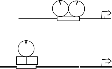
protein–protein interactions. Figure 4 shows the
ER ER
mRNA
ER
mRNA
Fos Jun
A
B
AP-1 RE
ERE
FIGURE 4 Mechanisms of transcriptional activation by nuclear
receptors. Panel (A) shows the classical pathway for transcriptional
activation with a steroid receptor dimer binding directly to a hormone
response element. Panel (B) shows an alternative mode of activation. In
this case, a receptor such as the estrogen receptor binds to another
transcription factor and influences transcription through its inter-
actions with the transcription factor and recruitment of coregulator
proteins to the complex. ER, estrogen receptor; AP-1 RE, AP-1
response element; ERE, estrogen response element.
STEROID/THYROID HORMONE RECEPTORS 115
comparison between two of these pathways for ER. In
the upper panel is the classical DNA-binding-dependent
induction of transcription. We next depict the ability of
ER to induce transcription through interactions with
AP-1 complexes. In this instance, both estradiol as well
as SERMs will stimulate the activity of AP-1.
NUCLEAR RECEPTOR STIMULATION
OF
CELL SIGNALING PATHWAYS
Both of the pathways above can be considered genomic
pathways in that the nuclear receptor acts through
altering transcription. Nuclear receptors can also act
through stimulating kinase activity, although the final
downstream target may be a change in transcription.
These actions are rapid (minutes) and are termed non-
genomic. The pathways are less well characterized,
but there is evidence that activation of nuclear receptors
can lead to downstream activation of mitogen-activated
protein kinase (MAPK) In some cases, this is through
activation of Src kinase and in others through generation
of a ligand for a growth factor receptor. There are
numerous other examples of these rapid actions.
Induction by estradiol of nitric oxide synthase activity
in endothelial cells is a rapid response that does not
require transcription. Thus, steroid/thyroid hormones
alter cellular activities through multiple mechanisms.
SEE ALSO THE FOLLOWING ARTICLES
A-Kinase Anchoring Proteins † Mitogen-Activated
Protein Kinase Family † Thyroid-Stimulating Hormone/
Luteinizing Hormone/Follicle-Stimulating Hormone
Receptors
GLOSSARY
agonists Natural or synthetic ligands that bind to the hormone-
binding domain of the receptor stimulating activity.
antagonists Ligands that compete with agonists for binding to the
hormone-binding domain, but do not cause activation of the
receptor.
nuclear receptors Ligand-activated transcription factors character-
ized by conserved zinc-finger motifs in their DNA-binding
domains and smaller conserved regions in the hormone-binding
domain.
phosphorylation A posttranslational modification of an amino acid
in which a phosphate group is added by a kinase.
FURTHER READING
Falkenstein, E., Tillmann, H. C., Christ, M., Feuring, M., and
Wehling, M. (2000). Multiple actions of steroid hormones—a
focus on rapid, nongenomic effects. Pharmacol. Rev. 52(4),
513–556.
McDonnell, D. P., Connor, C. E., Wijayaratne, A., Chang, C. Y., and
Norris, J. D. (2002). Definition of the molecular and cellular
mechanisms underlying the tissue selective agonist/antagonist
activities of selective estrogen receptor modulators. Recent Prog.
Horm. Res. 57, 295– 316.
McKenna, N. J., Lanz, R. B., and O’Malley, B. W. (1999). Nuclear
receptor coregulators: Cellular and molecular biology. Endocr.
Rev. 20(3), 321 –344.
Tsai, M. J., and O’Malley, B. W. (1994). Molecular mechanisms of
action of steroid/thyroid receptor superfamily members. Annu.
Rev. Biochem. 63, 451–486.
Weigel, N. L., and Zhang, Y. (1998). Ligand-independent activation of
steroid hormone receptors. J. Mol. Med. 76(7), 469–479.
Whitfield, G. K., Jurutka, P. W., Haussler, C. A., and Haussler, M. R.
(1999). Steroid hormone receptors: Evolution, ligands, and
molecular basis of biologic function. J. Cell Biochem.
32–33(suppl.), 110– 122.
BIOGRAPHY
Nancy Weigel is a Professor of Molecular and Cellular Biology at
Baylor College of Medicine. Her interests are in steroid receptor action
and crosstalk with cell signaling pathways as well as the role of steroid
receptors in prostate cancer. She received her Ph.D. from Johns
Hopkins University and received her postdoctoral training at Baylor
College of Medicine.
Ramesh Narayanan is a postdoctoral fellow in the Department of
Molecular and Cellular Biology at Baylor College of Medicine. He
studies steroid receptor action and crosstalk with cell signaling
pathways. He received his Ph.D. from the University of Madras, India.
116 STEROID/THYROID HORMONE RECEPTORS

Store-Operated Membrane
Channels: Calcium
Indu S. Ambudkar
National Institute Of Dental and Craniofacial Research, Bethesda, Maryland, USA
Ca
21
entry via plasma membrane Ca
21
influx channels
regulates a wide array of physiological functions such as
neurotransmission, muscle contraction, secretion, and gene
expression. A number of different types of Ca
21
channels have
been identified in excitable and nonexcitable cells, including
voltage-gated Ca
21
channels, primarily found in neuronal and
various types of muscle cells; receptor-operated channels that
are activated by extracellular ligand; and second-messenger-
activated channels that are activated by intracellular “ligands”
such as cGMP. Over the last decade considerable interest has
been focused on store-operated Ca
21
channels (SOCCs),
which mediate store-operated Ca
21
entry (SOCE, also referred
to as capacitative Ca
21
entry, CCE). SOCE is activated
following stimulation of cell-surface receptors that lead to
phosphatidylinositol bisphosphate (PIP
2
) hydrolysis, gener-
ation of diacylglycerol (DAG) and inositol-1, 4, 5-trispho-
sphate (IP
3
), and IP
3
-mediated release of Ca
21
from internal
Ca
21
stores via the inositol trisphosphate receptor (IP
3
R). The
concept of SOCE was proposed by Putney in 1986 according to
which depletion of Ca
21
in intracellular Ca
21
store(s) acts as a
trigger for activation of plasma membrane Ca
21
influx. Ca
21
entering the cells via this pathway not only achieves refilling of
the intracellular Ca
21
stores but also provides a sustained
elevation of cytosolic [Ca
21
] ([Ca
21
]
i
) that is critical in the
regulation of a variety of cellular functions. Despite the large
number of studies that have been directed towards SOCE, the
molecular composition of these channels as well as the
mechanisms that activate or inactivate them have not yet
been elucidated.
Characteristics of SOCE
Store-operated Ca
2þ
entry (SOCE) was originally
identified in nonexcitable cells, although it has now
been shown to be present in excitable cells as well. Thus,
our knowledge of the characteristics of SOCE is
primarily based on studies with nonexcitable cells that
span over two decades. Early studies using radioactive
Ca
2þ
and subsequent studies using Ca
2þ
-sensitive
fluorescent probes together demonstrate that neuro-
transmitter stimulation of cells leads to a biphasic
increase in cytosolic Ca
2þ
. An immediate increase that
is not substantially altered by the removal of external
Ca
2þ
suggesting that it is due to internal Ca
2þ
release,
primarily mediated by IP
3
. This is followed by a rela-
tively sustained elevation of Ca
2þ
, completely dependent
on the presence of external Ca
2þ
, that is due to influx of
Ca
2þ
from the external medium. Although other Ca
2þ
influx pathways might contribute to this sustained
[Ca
2þ
]
i
elevation, SOCE accounts for a major part, or
all, of this Ca
2þ
influx. Several key studies lead to the
conclusion that this Ca
2þ
influx is triggered by the
depletion of Ca
2þ
in the internal Ca
2þ
store. (1) Ca
2þ
influx is not activated by IP
3
or its metabolites. (2) Ca
2þ
influx remains active even after the receptor-coupled
signaling is stopped by addition of the antagonist.
(3) Ca
2þ
influx is inactivated after Ca
2þ
is reintroduced
into the cell and allowed to refill the internal Ca
2þ
store.
(4) The same type of Ca
2þ
influx is activated by treating
cells with the SERCA inhibitors such as Tg which induce
a rapid and specific block of Ca
2þ
uptake into the ER
and unmask a “leak” of Ca
2þ
from the ER.
The first channel activity associated with SOCE was
measured in RBL cells by Penner and co-workers in
1992. This channel CRAC has been extensively studied
and has also been found in lymphocytes and megakar-
yotes. CRAC is characterized by a high Ca
2þ
/Na
þ
permeability ratio (. 500), as well as a relatively rapid
Ca
2þ
-dependent feedback inhibition. The channel dis-
plays strong anomalous mole fraction behavior
suggesting that under normal physiological conditions
external Ca
2þ
blocks the entry of Na
þ
via the channel,
thus Ca
2þ
is the favored ion to permeate this channel.
Since 1992, store-operated Ca
2þ
influx channels have
been measured in many different cell types, including cell
lines and primary cell cultures. It is now clear that,
although store-operated Ca
2þ
channels are all activated
by the same, presently unknown, mechanism associated
with internal Ca
2þ
store depletion they are not
homogeneous. They display distinct biophysical proper-
ties, e.g., selectivity to Ca
2þ
, which suggest possible
molecular diversity in their composition as well as
differences in their modulation. An interesting question
Encyclopedia of Biological Chemistry, Volume 4. q 2004, Elsevier Inc. All Rights Reserved. 117
that arises from such data is whether these distinct
properties of store-operated Ca
2þ
channels (SOCCs)
reflect their cell-specific physiological functions.
Mechanism of SOCE
Although agonist-stimulation of Ca
2þ
influx was first
recognized in secretory cells almost three decades ago by
Douglas and Poisner, the molecular mechanisms that
activate or inactivate this Ca
2þ
influx have not yet been
established. Unraveling this mechanism has been a
major challenge in the field of Ca
2þ
signaling. Since
SOCE was first identified, several mechanisms have been
proposed to describe how it is activated. The earliest
model proposed that Ca
2þ
in the internal store
negatively regulates Ca
2þ
influx. When the store Ca
2þ
content is decreased, Ca
2þ
influx is activated and
external Ca
2þ
is somehow directly accumulated into
the ER, bypassing the cytosol. This model was primarily
based on experiments which revealed that during
refilling of the internal Ca
2þ
store (e.g., after agonist
stimulation and addition of antagonist) by Ca
2þ
entry
via the SOCE pathway, there is no detectable increase in
[Ca
2þ
]
i
. This model was disproved by studies in which
external Ca
2þ
was substituted by divalent cations such
as Mn
2þ
, which enter the cell via SOCE but are not
pumped into the ER by the SERCA. These studies lead to
the proposal that Ca
2þ
first enters the cytosol from
where it is rapidly taken up into the ER lumen by the
SERCA activity, and thus does not produce any
substantial increase in [Ca
2þ
]
i
. This results in refilling
of the internal Ca
2þ
stores which leads to inactivation of
SOCE. Thus, there is reciprocal regulation of the ER
Ca
2þ
store and plasma membrane Ca
2þ
channels.
Later models addressed the nature of the signal that
conveys the status of the internal Ca
2þ
store to either
activate or inactivate SOCE in the plasma membrane.
While a number of different models have been proposed
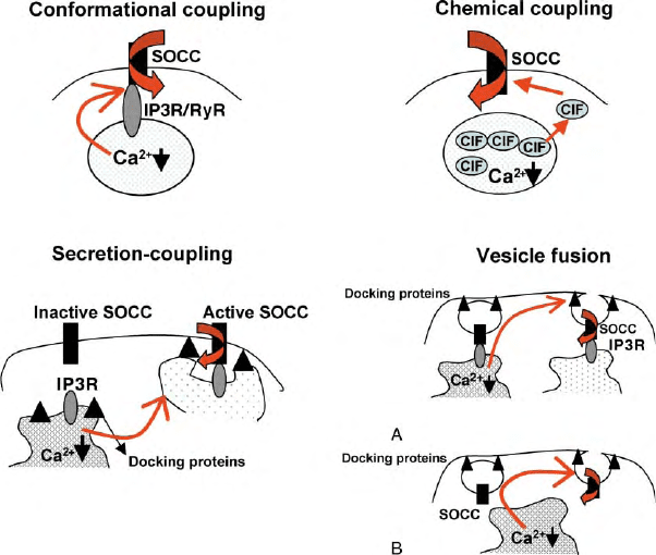
in an effort to explain this “ER–PM coupling,” three
major mechanisms have garnered the most attention;
(1) conformational coupling, (2) a diffusible factor, and
(3) regulated recruitment of channels by fusion of intra-
cellular vesicles (Figure 1). Conclusive data are presently
lacking to either support or rule out any of these
proposed mechanisms for SOCE activation. A major
hurdle in these efforts has been the lack of knowledge
regarding the molecular identity of the SOCC channel.
Molecular Candidates for SOCC
The relatively recent discovery of mammalian homol-
ogues of the Drosophila Trp (transient receptor poten-
tial) gene has propelled the field of SOCE in a new
direction. TRP proteins form a large functionally diverse
FIGURE 1 Proposed models for activation of SOCE. All components are labeled in the figures. See text for description. Two mechanisms are
shown for the vesicle fusion model. (A) “Kiss and Run” model predicts that SOCC-containing vesicles might be docked to the ER and therefore
sense depletion which initiates fusion to PM. Ion channel is retrieved during inactivation. (B) SOCC-containing vesicles are present in the subplasma
membrane region and sense depletion of store that leads to vesicle fusion and channel insertion into the PM. Inactivation and retrieval could be
independent events.
118 STORE-OPERATED MEMBRANE CHANNELS: CALCIUM
superfamily of ion channel proteins and are found in all
excitable and nonexcitable tissues. Members of the
TRPC subfamily and some members of the TRPV
subfamily form channels that are activated in response
to receptor-coupled Ca
2þ
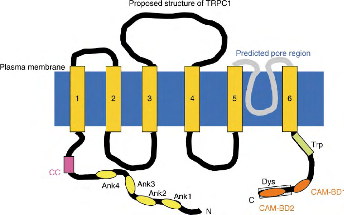
signaling events (Figure 2
shows the proposed structure of TRPC1). TRP channels
appear to fall into two general classes; nonstore
operated such as TRPC3, TRPC6, TRPC7, which are
activated by agonists and exogenous addition of
diacylglycerol (DAG), but not by thapsigargin. Thus, it
is suggested that this family forms channels that are
activatedbyDAGgeneratedinresponsetoPIP
2
hydrolysis. The other group which includes TRPC1,
TRPC4, TRPC5, TRPV6 has been shown to form
store-operated channels. In this case, studies include
heterologous expression, knockdown of endogenous
proteins, and site-directed mutagenesis. However, there
are exceptions. For example, TRPC3 has been shown to
form SOCC and also to be regulated by the internal
Ca
2þ
store via interaction with inositol trisphosphate
receptor (IP
3
R). Further, TRPCs have also been shown
to interact to form heteromultimeric channels which
display a wide range of biophysical characteristics.
Thus, it is possible that TRPCs could be involved in the
formation of a diverse range of SOCCs. Although
further work is required to conclusively establish that
TRP proteins are molecular components of SOCCs,
presently they are the only viable candidates for these
channels. Further, TRPs provide useful tools to test the
validity of the models proposed for SOCC activation.
Signaling from ER to
the Plasma Membrane
CONFORMATIONAL COUPLING
This hypothesis was originally proposed by Irvine in
1990 and was based on functional analogy between
IP
3
R and ryanodine receptors (RyR) in muscle cells. RyR
are Ca
2þ
release channels in the muscle sarcoplasmic
reticulum (SR) and have been suggested to physically
couple to the L-type Ca
2þ
channels in the T-tubule
plasma membrane. During excitation–contraction
coupling, Ca
2þ
inflow via the plasma membrane
channels regulates Ca
2þ
-induced Ca
2þ
release via RyR
in the SR. Although in the case of SOCE, the flow of
information can be predicted to occur in the reverse
direction, i.e., from ER to the plasma membrane, the
homology between IP
3
R and RyR channeled the
hypothesis that regulation of SOCC could be mediated
via a direct physical association between the IP
3
R and
the SOCC in the plasma membrane. The model proposes
that there are preformed IP
3
R–SOCC complexes and
that store depletion is detected by the IP
3
R, leading to a
conformational change that results in activation of
SOCC. A caveat to the IP
3
R requirement in the
regulation of SOCC is its activation by SERCA
inhibitors or by depletion of Ca
2þ
stores by loading
the cytosolic or ER with Ca
2þ
buffers. To explain this
discrepancy, it was proposed that only a certain pool of
IP
3
R’s interacts with SOCC, and that these are localized
FIGURE 2 Proposed structure of transient receptor potential canonical 1 (TRPC1): The structure includes six-transmembrane domains (1– 6),
extracellular as well as intracellular domains (black lines) and a pore-domain between the fifth and sixth TMs (gray). The N terminus of the protein
contains ankyrin repeats (ank) as well as a coil–coil domain (CC). The C terminus has the conserved TRP motif (Trp, EWKFAR), calmodulin-
binding domains (CAMBD), and a dystrophin domain (Dys).
STORE-OPERATED MEMBRANE CHANNELS: CALCIUM 119
in ER membranes situated in close proximity to the PM.
This also implied that this pool of IP
3
R is not involved in
internal Ca
2þ
release, but only in SOCC regulation.
Currently, there are conflicting data regarding the role of
IP
3
R in the regulation of SOCC. Studies using a gene
knockout approach have shown that IP
3
Rs are not
required for thapsigargin-stimulated Ca
2þ
entry,
although they are clearly required for IP
3
-mediated
internal Ca
2þ
release. Other studies suggest that RyR,
which are present in several nonmuscle cell types, can
also couple with SOCC and regulate its function. Thus,
it is possible that RyR could replace IP
3
Rs in cells where
IP
3
R expression has been down-regulated or eliminated.
However, further studies will be required to rule out or
provide conclusive evidence for the conformational
coupling hypothesis.
SECRETION-LIKE COUPLING
VESICLE
FUSION
The activation of SOCE is a relatively slow process.
A lag time of about 10 s has been detected between
internal Ca
2þ
release and Ca
2þ
influx. Thus, it has been
proposed that vesicle trafficking and fusion events could
be involved in activation of SOCE. Two possible
processes could occur. The first is a variation of the
conformational coupling model and suggests that the
ER–PM interaction is a dynamic, reversible, process and
that the ER membrane moves towards and docks with
the PM upon stimulation. The docking enables proteins
in the PM and ER to interact, thus resulting in activation
of SOCC. Although the ER protein is considered to be
IP
3
R or RyR, other proteins could also be involved in the
ER–PM signaling. Since the ER and PM are apposed to
each other at the site of interaction, there is no particular
requirement that the ER or PM protein should have very
long cytosolic domains. Support for the secretion-
coupling hypothesis has been mainly provided by studies
using reagents to disrupt the cytoskeleton and alter the
spatial arrangement of cellular organelles. Further,
TRPC1–IP
3
R interaction was shown to be disrupted
by reagents that induce cortical actin formation.
However, several other studies have refuted this model
as a possible activation mechanism for SOCC. The
second mechanism that can be suggested involves vesicle
trafficking and exocytotic insertion of the channel
proteins. Here again, there are data to both support
and refute the model. Experiments have shown that
disruption of the SNARE proteins involved in exo-
cytosis, inhibits activation of SOCE. However, in other
studies, such maneuvers did not affect SOCC activation.
An important point that needs to be considered when
assessing the possible mechanisms for activation is
whether the different SOCCs that have been detected
in various cell types are activated by the same
mechanism or does internal Ca
2þ
store-depletion induce
a variety of cellular signals which can then activate
different channel types. For example, if different TRP
channels are involved in the SOCCs in the different cell
types, can that account for differences in their regu-
lation? Voltage-gated Ca
2þ
channels represent a family
of proteins that are activated by various thresholds of
membrane potential. They are also regulated differently,
exhibit distinct characteristics and carry out specific
physiological functions. Analogous to this, we might
have to consider SOCC channels as a family of channels
that sense the same fundamental signal, but are
regulated by subtly distinct mechanisms. What these
mechanisms are, presents a challenging question for
future studies in this field.
METABOLIC COUPLING
Another hypothesis that has received sporadic attention
is that an as yet unknown diffusible factor, referred to as
CIF, is either released from the ER with Ca
2þ
or is
generated during this process. CIF can reach the PM
SOCC channels and either activate it directly or bind to
a regulatory protein and enable channel activation.
Evidence in support of this shows that extracts from
stimulated cells can increase Ca
2þ
influx in unstimulated
cells. However, these findings have not held up for all
types of cells. Other metabolites that have been shown to
regulate SOCC are of the cytochrome P450 epoxygenase
pathway. Modifiers of the lipoxygenase pathway has
been shown to affect I
CRAC
in RBL cells. A role for
arachidonic acid has also been suggested. Thus, further
studies are needed to establish whether CIF is involved in
SOCC activation. It should be noted that the require-
ment for CIF and secretion-like coupling need not be
mutually exclusive, since dynamic trafficking of ER to
the PM would decrease the diffusion restraints for CIF.
In addition, reassembly of the cortical actin can also play
a role in the access to the PM and diffusion of CIF.
Interestingly, the status of the actin is controlled by the
PIP
2
levels in the plasma membrane. Thus, the hydro-
lysis of PIP
2
not only initiates Ca
2þ
signaling but also
remodeling of the actin cytoskeleton in order to facilitate
the regulation of cellular function. In fact, modulation of
PIP
2
metabolism, i.e., inhibition of PI-3 kinase has been
shown to alter SOCE in some cells. Thus, it is becoming
exceedingly evident that regulation SOCE is a highly
orchestrated process with several orders of complexity
that might be determined by the particular SOCC that is
present, and the specific physiological function that it
contributes to, in any given type of cell.
Ca
21
Signaling Microdomains
Recent studies have highlighted spatio-temporal aspects
of Ca
2þ
signaling in cells. It has been demonstrated that
agonist-stimulated Ca
2þ
influx occurs within specific
120
STORE-OPERATED MEMBRANE CHANNELS: CALCIUM
