Lennarz W.J., Lane M.D. (eds.) Encyclopedia of Biological Chemistry. Four-Volume Set . V. 4
Подождите немного. Документ загружается.

lamps, particularly those of Edgerton and colleagues.
However, construction of the first ruby laser used in
biological studies by Bunkenberg and DeVault opened
up a whole new time domain, not only because of the
monochromaticity of the laser (which greatly simplified
the filter leakages) but also because of the duration
of the light pulse (in the microsecond region), which
for the time was extraordinarily short. From this study
came the totally unexpected observation that the
photochemical oxidation of cytochrome c by photo-
activated chlorophyll of Chromatium was temperature
independent, not only from room temperature to liquid
nitrogen temperature, but eventually at liquid helium
temperatures, extending the time range of the spectro-
scopic method to one of the primary or near primary
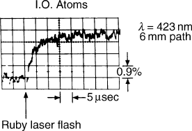
results and/or reactions of photosynthesis (Figure 4),
and also introducing the concept of electron tunneling
in biochemical reactions, a concept pursued by Les
Dutton and Harry Gray.
The history of spectroscopy is checkered with
unexpected results and meaningful interpretations on
the nature of electron transfer reactions. In fact, the study
of reactions obtained from photolysis activation of CO
compounds in the presence of O
2
caused ligand exchange
to occur, especially in the case of CO. Together with
Carlo Saronio, Chance discovered a number of inter-
mediate steps involving higher oxidation states of iron
mirroring those obtained with peroxidase and H
2
O
2
, but
much faster and more complex. This was because copper
oxidation was involved in the active site of CO, enabling
donation of four electrons sequentially for the reduction
of oxygen to water without significant amounts of free
radical intermediates. This contrasted with the photo-
activation of porphyrins in the absence of electron
donors, resulting in the creation of singlet oxygen, a
process used extensively in photodynamic therapies.
FLUOROCHROMES OF TISSUES
Spectroscopy of biological fluorochromes, such as
NADH and flavoprotein compounds that exhibit
strong fluorescence in mitochondria, led Chance and
co-workers to study them extensively in vivo by a simple
method in which excitation was obtained by the very
strong 366 and 436 lines of the mercury arc. This yielded
emission spectra characteristic of the chromophores in a
variety of functional states, characterizing electron
transfer through the citric acid cycle as reductant and
the cytochrome chain as oxidants, giving essential
features of the redox state of the mitochondrial matrix.
Interpretations of the importance of metabolic control
and thermodynamic principles of the system were
derived from these, particularly by R. L. Veech.
HEMOGLOBIN AND CYTOCHROME
It is perhaps ironic that the cell biologists and
etymologists of the Molteno Institute focused on the
cytochromes of hemoglobin-free cells and tissues and
that the “real” physiology of oxygen delivery to
tissues by hemoglobin and its utilization by CO had to
wait for more sophisticated methods. Even now, the
observation of cytochrome oxidase absorption bands
in the presence of physiological concentrations of oxy
and deoxy-hemoglobin is fraught with controversy, to
the point that no reliable spectroscopic distinction of
the copper component of cytochrome oxidase and the
overlapping spectra of oxy-hemoglobin has been
obtained. In fact, nearly all studies find that the so-
called cytochrome oxidase Cu changes track those of
HbO
2
due to the similarity of their spectroscopic
absorption bands in the near infrared (NIR) region,
and the great predominance of hemoglobin absorption
over that of cytochrome oxidase.
NADH AS AN OXIMETER
It has been shown that the fluorescence of NADH and
flavoprotein, when used in a ratiometric manner, can
exhibit a reasonable immunity to changes of hemoglobin
concentration, and has afforded standards for the
independent changes of hemoglobin and NADH in
transient hypoxia and re-oxidation. Such independence
of the measures of cytochrome and hemoglobin has
never been clearly demonstrated for the copper com-
ponent of cytochrome oxidase, due to the overlapping
spectra of oxyhemoglobin.
An alternate approach was based upon the fluor-
escence of the newly discovered NADH and flavoprotein
components of the mitochondrial respiratory chain,
particularly by using the ratio of these two fluoro-
chromes. Fortunately, NADH fluoresced in the reduced
state while flavoprotein fluoresced in the oxidized state,
so their ratio was a measure of the redox state of
mitochondria, a signal that was found to be only
marginally affected by changes in the oxygenation of
hemoglobin in model systems. Using this criterion on the
FIGURE 4 Typical recording of the oxidation of cytochrome c of
photosynthetic bacteria at low temperatures as activated by a light
flash of the ruby laser. Remarkably this reaction rate was affected very
little by the transition from room temperature to liquid nitrogen
temperature due to electron tunneling.
SPECTROPHOTOMETRIC ASSAYS 71
very strong signal of NADH only, it was possible to
show that the fluorescence of NADH was unchanged
until the oxy/deoxy transition of hemoglobin was almost
complete in a system in which functional activity was
measured by the photoaction potential of an animal
model, removing any doubt about the higher oxygen
affinity of cytochrome in vivo as compared to hemo-
globin (, 20 Torr). This was a very important milestone
for physiologists, who know that the deoxygenation
of hemoglobin is very high and the critical pO
2
of
mitochondrial function is compromised. Thus, the
calibration of tissue oximeters in the region of intra-
venous saturation of hemoglobin (i.e., 20–30%) must
be precisely measured to indicate critical tissue hypoxia
in vivo. This indeed was subsequently validated by
measurements of tissue energetics through phosphorus
nuclear magnetic resonance (
31
P NMR), particularly by
measurements of the phosphocreatine:phosphate ratio.
NIR Spectroscopy
Jobsis used Kramer’s technique to measure in the
infrared, and developed a technology for the measure-
ment of absorption of the copper component of
cytochrome oxidase in the region of 830 nm based
upon studies of the cat model and the heads of neonates,
which he termed “transcranial spectroscopy.” He
further developed algorithms based upon fluorocar-
bon-perfused cat brain to give optical pathlengths that
were believed to be transferable to the neonate brain and
allow a deconvolution of blood volume and saturation
changes from those of cytochrome oxidase signals using
the full-length light algorithm. Delpy and co-workers
avidly followed the lead of Jobsis and developed a close
correlation between the decreased concentration of
oxyhemoglobin and the so-called copper signal in a
number of models, suggesting that the mitochondria in
tissues contained a low-affinity cytochrome oxidase and
responded to pO
2
(in a slice) in a way similar to that of
hemoglobin. However, isolation of rat brain mitochon-
dria failed to support this contention. Furthermore, the
freeze-trapped hypoxic brain failed to show the absorp-
tion band of reduced cytochrome c in mild hypoxic
stress that caused deoxygenated hemoglobin. In fact, the
absorption band of reduced cytochrome c was not
observed until the band of oxyhemoglobin was no
longer detectable, according to the work of Bashford.
While attempts were made to detect the copper
absorption band of hemoglobin in the NIR region,
animal studies showed that the fluorescence of NADH
and the flavoprotein could be used to detect anoxia in
the presence of hemoglobin, particularly when the ratio
of the fluorescent oxidized flavoprotein and the fluor-
escent reduced NADH were employed; this value was
relatively insensitive to the hemoglobin concentration.
In fact, further demonstrations showed that the fluor-
escence of NADH was unaffected by the deoxygenation
of hemoglobin in animal model brain. The NADH
fluorescence increased in hypoxia only when the
hemoglobin was already almost completely deoxyge-
nated. This observation suggests that measurements of
the critical pO
2
in hypoxia require accurate measure-
ment of extreme values of hemoglobin desaturation, at
the critical pO
2
for mitochondrial function.
NIR SPECTROSCOPY OF BRAIN AND THE
BOLD EFFECT MEASURED BY MRI
Much interest in the NIR method is based upon Ogawa’s
finding that changes in deoxyhemoglobin concentration
(changes of the paramagnetic species of deoxyhemog-
lobin) enhanced water relaxation in the brain. This
opened up the field of study of the activation phenom-
enon in the human brain, in which changes in
deoxyhemoglobin levels are measured by NMR and by
NIR tissue spectroscopy. The MRI changes are precisely
imaged, while the NIR images, although crude, are
measures of the rapidity of the changes. But in addition
to incremental changes of deoxyhemoglobin, NIR could
measure the saturation value of hemoglobin, which, for
reasons involving Beer’s law, originates mainly from the
arteriolar/capillary/venolar bed. This feature, namely,
the value of local oxygen extraction due to incremental
changes of mitochondrial functional activity (i.e.,
localized activation), is not measured by MRI. The
two techniques are now widely accepted as indicative of
localized brain activation and have afforded the basis for
in-depth studies of visual and sensory motor function.
But, most importantly, NIR gives an excellent rendition
of prefrontal cortex (PFC) signals without the difficulty
of the large water content of the ocular system
encountered with NMR (Figure 5). The use of activation
images is appropriate to the NIR system, where baseline
values may be somewhat variable and difficult to
calibrate. The incremental changes of blood volume
measured as changes of total hemoglobin, together with
the aforementioned oxygen extraction measure, i.e.,
desaturation of hemoglobin, can be directly related
to local metabolic activity, opening up a new field of
NIR study of the semi-quantitative nature of the
hemoglobin signals.
NIR Imaging
While the above-mentioned studies used dual-wave-
length technology stemming from that of Glenn
Millikan, a completely new concept was introduced by
the discovery that photon migration through tissues can
72
SPECTROPHOTOMETRIC ASSAYS
be modeled by the diffusion equation. Multiple sources
and detectors give very reasonable two- and three-
dimensional imaging, and the propagation of light
through tissue can be quantified by pulse time and
phase and amplitude measurements, much as has been
the case with measurement of fluorescence.
PHOTON MIGRATION IN TISSUES
The discovery that photons migrating through tissue
followed the diffusion equation and that the tracks could
be simulated by Monte Carlo methods, together with the
adaptation of time-correlated single photon counting
(TCSCP) to the task of measuring propagation times in
tissues, opened up an entire field of NIR spectroscopy
and imaging. This grew to be a field of medical science in
a manner similar to NMR, but differing in the fact that
the necessary equipment is not nearly as expensive, so
the proliferation of the technique in research labora-
tories could be much more rapid. Because the profit
margins did not match those of NMR, commercial
production of NIR imagers has been restricted to two or
three companies.
TISSUE OPTICAL PROPERTIES
Three techniques are outstanding in the measurement of
tissue optical properties. The first, and still the foremost,
is the pulse time method, in which photon delay is
caused by scattering and photon attenuation is caused by
absorbers such as hemoglobin, water, and lipid. Because
TRS (time-resolved spectroscopy) immediately decon-
volutes scattering and absorption by time domain
analysis, it is a preferred method. Similarly variable
frequency modulated light will unravel by Fourier
transformation exactly the same quantities as those
obtained by TRS. However, the difficulty of stabilizing
the phase shifts of electronic systems of variable
frequency, together with the limitations of detection
response to high-frequency radio waves, has limited this
system to approximately 400 MHz. Nevertheless, many
instruments have been made in the frequency range of 50
to 200 MHz that are used for quantifying absorption
and scattering in multi-wavelength systems and that are
capable of measuring hemoglobin saturation with
significant precision. Such systems have adopted some
cell phone components and are therefore compact and
cheap. The most reliable and most used system
modulates the light at very low frequencies in either, or
time shares the light sources in a multiplex system, and
appears to be the preferred system for many appli-
cations. The deconvolution of scattering and absorption
can be obtained if sufficient data are taken at various
source detector separations and optical wavelengths to
include the scattering variations.
CANCER DETECTION
In cancer detection, scanning the breast for example, an
activation signal based upon angiogenesis and hyperme-
tabolism is given, causing more blood volume and more
deoxygenated hemoglobin to be present. While this
criterion may not be applicable to all cancers, it has
given remarkably good scores in one breast cancer study.
MUSCLE STUDIES
A series of studies has been based upon activation
measurements in which (e.g., in both muscle and brain),
functional activation causes hemodynamic changes due
to mobilization of blood flow and saturation changes
due to varying metabolic activity (Figure 5). This has
been used in muscle not only to evaluate exercise
capability but also to quantify disability due to occlusive
disease in the limbs.
BRAIN FUNCTIONAL ACTIVATION
In brain studies, whereas NMR and the optical method
both measure hemodynamic activation due to functional
activity, the NIR method is unique in that it measures
changes of hemoglobin saturation caused by varying
degrees of oxygen extraction from the capillary blood
vessels of the brain. Thus, the convenience and economy
of the optical method lends itself to studies of minimally
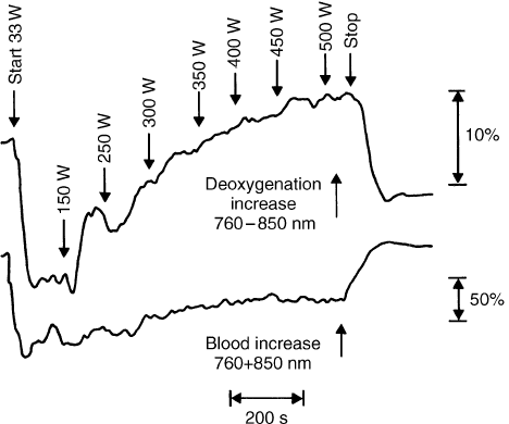
FIGURE 5 Evidence of activation of metabolism measured in the NIR
(near infrared) by the dual wavelength method of 760–850 nm
(equibestic wavelengths) which show progressive deoxygenation of
the quadriceps muscle of a trained athlete during bicycle exercise up to
the remarkable level 500 watts, at which nearly complete deoxygena-
tion of hemoglobin can occur. As measured separately by the sum of
signals at these two wavelengths. With appropriate coefficient, the
increase of blood flow in the muscle also occurs giving a larger absor-
bance signal. At this level of exercise, it is probable that myoglobin is not
deoxygenated as indicated by separate experiments with animal models.
SPECTROPHOTOMETRIC ASSAYS 73
perturbed human subjects, be they adults or neonates
(Figure 6).
BRAIN STUDIES:THE NEURONAL SIGNAL
The early studies of David Hill, Tasaki, and Richard
Keynes on transparency changes of axons upon
stimulation suggest that a functional optical signal
could be obtained in animal and human brain. While
the studies of Salzberg did indeed verify the scattering
changes in isolated preparations, studies using NIR to
seek similar changes in the human brain suggest that
only very small and somewhat irreproducible signals can
be obtained. For example, the early experiments of
Gabriele Gratton have not been duplicated either by
himself or by others working in the field (Villringer and
Franceschini, and Wolf), in part due to instrumental
difficulties, and in part due to the very small size of the
signal. The rise time of the signals appears to be in
the range of 100 ms and the amplitude as small as one
part in 10
4
or even smaller. However, the relatively
robust signals obtained from the hemodynamic and
metabolic activations discussed previously show the
feasibility of measurements of functional activity in
the human brain, particularly in the prefrontal region,
and open up a reliable and economical method for
human brain studies that predict a vibrant future of the
optical methods in human studies.
Summary
The story of the development of optics in biochemistry
and biophysics does not end here. In fact, some might
say this is just the beginning of the transferability of
optical tomography and optical biopsy to small animals
on the one hand and human beings on the other. Perhaps
spurred by the interest in online methods for evaluating
the growth and recession of cancers under the influence
of appropriate drugs in small animals, transferability
of these principles to human subjects is becoming
important. The optical method is taking its place along
with MRI, PET, ultrasound, CT, and X-ray mammo-
graphy as methods for studying pathologies in the
human body.
SEE ALSO THE FOLLOWING ARTICLES
Cytochrome bc
1
Complex (Respiratory Chain Com-
plex III) † Cytochrome c
GLOSSARY
Brown converter An electrical chopper developed by the Leeds and
Northrup Company that had very little contact potential variation.
cytochrome oxidase The terminal enzyme of most oxygen-using
systems.
dual-wavelength spectrophotometer A spectrophotometer with time
shared to adjacent wavelengths in order to minimize the effect of
scattering changes upon absorbance changes, because scattering
varies very slowly with wavelength, while cytochrome absorption
varies sensitivity with wavelength.
DuBridge electrometer (DU) A remarkable use of the suppressor grid
of the pentode to control electron flow with very high input
impedance, affording the basis of the Beckman pH meter and
spectrophotometer.
electron tunneling The transfer of electrons between two proteins at a
distance without collision of their active sites.
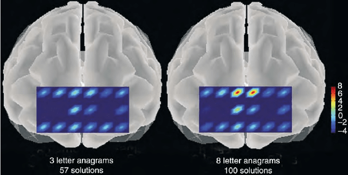
FIGURE 6 Illustrating the use of the NIR CW (continuous wave) dual wavelength system in localizing the particular voxels in the human
forebrain at which brain functional activity is indicated to be increased by the oxygenation changes with respect to the baseline level of 3 letter
anagrams. Each colored symbol represents the average of over 100 tests of a particular individual giving an average value of 8 mM oxygenation
under this condition of maximal stress. The region observed is the Broadman’s 9 and 10 or ear to ear and hairline to eyebrow region of the
projection of the prefrontal cortex. This separation was 4 cm in order to ensure signaling from the prefrontal cortex to accentuate the absorbance
changes due to the prefrontal cortex and minimize those which might be associated with tissue layers at smaller depths.
74 SPECTROPHOTOMETRIC ASSAYS
EXAFS X-ray absorption spectroscopy used to obtain high resolution
structures of Fe and Cu enzymes.
flavoprotein A widely spread pigment, but in this article the
prosthetic group of ketoglutarate and pyruvate dehydrogenase.
mercury arc A very useful light source for biological studies, which
gives light at exactly the correct wavelength for hemoglobin and in
some cases cytochrome studies.
Molteno Institute A famous institute directed by David Keilin
through the 30s, 40s and 50s. It became a Mecca for those working
with cell respiration.
near infrared (NIR) imaging The use of the wavelengths in the red
region just at the verge of invisibility between 700 and 900 nm to
better penetrate tissue.
photochemical action spectrum The effect of light upon a biological
system often used to activate a carbon monoxide inhibited
cytochrome.
photomultiplier A highly sensitive light detector used in many
spectrophotometers.
photon migration The phenomenon of photon diffusion through
tissues used in great detail recently to image subsurface objects.
ruby laser One of the early forms of the laser, emitting in the red
region.
scattered light Light that does not proceed directly through tissue.
spectrophotometer A device that measures the absorbance of
materials as a function of wavelength or, in some cases, energy.
time resolve spectroscopy (TRS) Spectroscopy using sharp pulses of
light to distinguish the scattering from the absorption of tissues.
X-ray absorption spectroscopy (EXAFS) Spectroscopy used to obtain
high-resolution structures of Fe and Cu enzyme.
FURTHER READING
Carafoli, E. (2003). Historical review: Mitochrondria and calcium:
Ups and downs of an unusual relationship. Trends Biochem. Sci.
28, 175–181.
Chance, B. (ed.) (1989). Photon Migration in Tissues. Plenum Press,
New York, NY.
Chance, B. (1991). The optical method. Ann. Rev. Biophys. Biophys.
Chem. 20, 1 –18.
Keilin, D. (1966). The History of Cell Respiration and Cytochrome.
Cambridge University Press, Cambridge, UK.
Slater, E. (ed.) (1966). Flavins and Flavoproteins.Elsevier,
Amsterdam.
BIOGRAPHY
Britton Chance is Eldridge Reeves Johnson University Professor
Emeritus of Biochemistry and Biophysics and Physical Chemistry and
Radiologic Physics at the University of Pennsylvania School of
Medicine. His current research interests focus on optical spectroscopic
methods for the study of brain cognition, breast cancer detection, and
physiological function. He has received numerous honors and awards
and honorary Ph.D. and M.D. and is noted for his contributions in
basic science research and technological development.
SPECTROPHOTOMETRIC ASSAYS 75

Sphingolipid Biosynthesis
Martina Leipelt and Alfred H. Merrill, Jr.
Georgia Institute of Technology, Atlanta, Georgia, USA
Sphingolipids are a complex family of compounds that perform
diverse structural and regulatory functions for eukaryotes and
some prokaryotes and viruses. They share a common structural
feature, a sphingoid base backbone that is synthesized de novo
from serine and a fatty acyl-coenzyme A, then converted into
ceramides, phosphosphingolipids, glycosphingolipids, and
other species, including protein adducts. Several diseases result
from disruption of de novo sphingolipid biosynthesis by
environmental factors or hereditary defects, but modulation
of sphingolipid biosynthesis is also being explored as a means to
control other diseases, including sphingolipid storage diseases
and cancer.
Structures and Nomenclature
Sphingolipids can be divided into several major cat-
egories: the sphingoid bases and their simple derivatives,
ceramides, and more complex sphingolipids (Figure 1).
The International Union of Pure and Applied Chemists
(IUPAC) has recommended a systematic nomenclature
for sphingolipids. The root name “sphingosin,” in
reference to the sphinx, was given by J. L. W.
Thudichum in 1884 “in commemoration of the many
enigmas which it presented to the inquirer.”
SPHINGOID BASES
The structure of sphingosine, the major sphingoid base
of mammals, is (2S, 3R, 4E)-2-aminooctadec-4-ene-1,
3-diol (it is also called
D-erythro-sphingosine and
E-sphing-4-enine) (Figure 1). This is only one of many
sphingoid bases found in nature, which vary in alkyl
chain length and branching, the number and positions of
double bonds, the presence of additional hydroxyl
groups, and other features. The structural variation
has functional significance; for example, sphingoid bases
in skin have additional hydroxyls at position 4 and/or 6
that can interact with neighboring molecules, thereby
strengthening the permeability barrier of skin.
Sphingoid bases function as intra- and extracellular
signals and second messengers in the form of free
sphingoid bases, sphingoid base 1-phosphates (Figure 1),
and possibly other species. Nonetheless, sphingoid bases
are present in cells primarily as the backbones of more
complex sphingolipids.
CERAMIDES
Ceramides are fatty acid derivatives of sphingoid bases
(Figure 1). The fatty acids are typically saturated or
mono-unsaturated with chain lengths from 14 to 26
carbon atoms (or even longer in the special case of skin),
and sometimes have a hydroxyl group on the
a
-or
v
-
carbon atom. These structural features favor the
segregation of ceramides and some complex sphingoli-
pids into specialized regions of the membrane (called
“rafts” and “caveolae”) that participate in cell signaling,
nutrient transport, and other functions.
Ceramides also serve as second messengers that
regulate cell growth, senescence, and programmed cell
death (apoptosis). Their biologic activity depends on the
type of sphingoid base and fatty acid; for example,
dihydroceramides (i.e., without the 4,5-double bond of
the sphingosine backbone) (Figure 1) are less potent than
ceramides as inducers of apoptosis, whereas phytocer-
amides (i.e., with 4-hydroxysphinganine or “phyto-
sphingosine” as the backbone) are more potent.
MORE COMPLEX PHOSPHO- AND
GLYCO-SPHINGOLIPIDS
The major phosphosphingolipids of mammals are
sphingomyelins (ceramide phosphocholines) (Figure 1),
whereas insects contain mainly ceramide phosphoetha-
nolamines and fungi have phytoceramidephosphoinosi-
tols and inositol phosphates. Some aquatic organisms
also contain sphingolipids in which the phosphate has
been replaced by a phosphono- or arsenate group.
Glycosphingolipids are classified on the basis of
carbohydrate composition: (1) neutral glycosphingoli-
pids contain one or more uncharged sugars such as
glucose (abbreviated Glc, hence, glucosylceramide is
GlcCer), galactose (Gal), N-acetylglucosamine
(GlcNAc), N-acetylgalactosamine (GalNAc), and fucose
(Fuc); and (2) acidic glycosphingolipids contain ionized
functional groups (phosphate or sulfate) attached to
neutral sugars, or charged sugar residues such as sialic
acid (N-acetylneuraminic acid). The latter are called
gangliosides, and the number of sialic acid residues is
usually denoted with a subscript letter (i.e., mono-, di- or
tri-) plus a number reflecting the subspecies within that
Encyclopedia of Biological Chemistry, Volume 4. q 2004, Elsevier Inc. All Rights Reserved. 76
category (see examples in Figure 1). For a few glyco-
sphingolipids, historically assigned names as antigens
and blood group structures are still in common usage
(e.g., Lexis x and sialyl Lewis x).
PROTEIN ADDUCTS
Some sphingolipids are covalently attached to protein,
e.g.,
v
-hydroxy-ceramides and -GlcCers are attached to
surface proteins of skin and inositolphosphoceramides
are used as membrane anchors for some fungal proteins,
in a manner somewhat analogous to the glycosylpho-
sphatidylinositol (GPI) anchors that are attached to
proteins in other eukaryotes.
De novo Synthesis of the
Ceramide Backbone
Sphingolipid biosynthesis is widespread among eukary-
otic cells, and it appears that new synthesis (i.e., de novo)
is relied upon more than reutilization of sphingolipids
from exogenous sources, such as food. The biosynthetic
pathway for such a diverse family of compounds
(conservatively estimated to be in the tens of thousands)
is obviously complex; however, its fundamental features
can be summarized in Figures 2 and 3.
SERINE PALMITOYLTRANSFERASE
Serine palmitoyltransferase (SPT) catalyzes the initial
step of the pathway which, for many organisms, is
the condensation of serine and palmitoyl-CoA to form
3-ketosphinganine (Figure 2). However, for organisms
that produce sphingoid bases with other alkyl chain
lengths (such as the C14 species of insects), the first
enzyme of the pathway utilizes a different cosubstrate
(dodecanoyl-CoA, in this example) and could be
renamed “serine dodecanoyltransferase.”
SPT is a pyridoxal 5
0
phosphate-dependent enzyme
comprised of two gene products (termed SPTLC1 and
SPTLC2 for humans, and LCB1 and LCB2 for yeast); a
third has also been identified in yeast, but does not
appear to have a homologue in mammals. In
most organisms, SPT is associated mainly with the
HO
O
O
OH
OH
O
O
O
OH
O
OH
H
O
O
OH
HO
AcNH
4
HO
O
OH
HO
OH
H
OH
O
HNAc
OH
HO
2
C
HO
HO
H
H
Cer
Glucose
Galactose
N-acetyl galactosamine
Galactose
N-acetyl neuraminic acid
G
M3
G
M2
G
M1
Lactosylceramide (LacCer)
Glucosylceramide (GlcCer)
3
1
4
3
1
2
1
11´
O---_---
Ceramide (N-acylsphingosine)
OH
NH
D-
erythro
-sphingosine
O
P(O
2
–
)O-choline
Phosphosphingolipids:
Sphingomyelin
Glycosphingolipids
(examples):
Gangliosides:
Examples of other sphingoid
base backbones:
OH
NH
2
4-hydroxysphinganine
(phytosphingosine)
OH
OH
NH
2
D-
erythro
-sphinganine
(dihydrosphingosine)
OH
HO
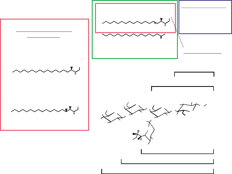
FIGURE 1 Structures of representative sphingolipids. Shown are several examples of sphingoid bases (sphingosine, sphinganine and
4-hydroxysphinganine, boxed in red), ceramide (in green), sphingomyelin (blue), and neutral (GlcCer and LacCer) and acidic (gangliosides G
M1
,
G
M2
, and G
M3
) glycosphingolipids.
SPHINGOLIPID BIOSYNTHESIS 77
endoplasmic reticulum, as are the other enzymes of
ceramide biosynthesis. SPT activity is affected by a wide
range of factors: sphingosine 1-phosphate, endotoxin
and cytokines, heat shock, UVB irradiation, cytotoxic
drugs (including many cancer chemotherapeutic drugs),
retinoic acid, and a number of small molecule inhibitors
produced by microorganisms (one of which, ISP1 or
myriocin, is often used to block de novo sphingolipid
synthesis by cells in culture). The mechanisms of SPT
regulation are not fully understood, but include (for
example) both acute modulation by heat shock and
increased expression of SPT mRNA by cytokines.
Mutations in human SPTLC1 cause hereditary sensory
neuropathy type I (HSN1), which is the most common
hereditary disorder of peripheral sensory neurons.
CERAMIDE SYNTHASE
3-Ketosphinganine is rapidly converted to sphinganine
by an NADPH-dependent reductase, then ceramide
syntase(s) acylate sphinganine to dihydroceramides
using fatty acyl-CoA’s varying in length from C16 to
C30 (and usually saturated or mono-unsaturated)
(Figure 2). Ceramide synthase is actually a family of
enzymes, each of which appears to arise from a different
gene and to utilize a particular subset of fatty acyl-CoA’s
(e.g., TRH4 utilizes palmitoyl-CoA whereas UOG1 uses
stearoyl-CoA).
Ceramide synthase is activated by a number of stimuli,
including cancer chemotherapeutic drugs and irra-
diation, and the increased production of ceramide is
thought to mediate the toxicity of these treatments.
Ceramide synthase is also the target of a number of
mycotoxins (fumonisins), which are produced by fungi
that grow on corn and, when consumed, result in
spectrum of diseases that are important to agriculture
(equine leukoencephalomalacia and porcine pulmonary
edema) as well as human cancer and possibly birthdefects.
DIHYDROCERAMIDE DESATURASE
Insertion of double bond(s) into the sphingoid base
backbones occurs mainly after formation of dihydrocer-
amide(s) (Figure 2). For mammals, introduction of the
Sphinganine
Dihydro-
ceramide
Palmitoyl-CoA
3-keto-sphinganine
3-ketosphinganine
reductase
NH
H
CH
2
OH
O
HO
H
(-)OOC
CH
2
OH
NH
3
(+)
O
SCoA
O
H
CH
2
OH
NH
3
(+)
H
CH
2
OH
HO
NH
3
(+)
NH
H
CH
2
OH
O
HO
CH
3
(CH
2
)
10
CH
2
Ceramide
(Dihydro)ceramide
synthase
Dihydroceramide
desaturase
Serine
+
Serine palmitoyltransferase
CH
3
(CH
2
)
10
CH
2
CH
3
(CH
2
)
10
CH
2
CH
3
(CH
2
)
9-19*
CH
2
CH
3
(CH
2
)
9-19*
CH
2
CH
3
(CH
2
)
10
CH
2
CH
3
(CH
2
)
10
CH
2
NADPH
Fatty acyl-CoA
NAD[P]H
Sphingomyelin
Glucosylceramide
(Galactosylceramide)
Sphingomyelin synthase
Glc(Gal)ceramide synthase(s)
Dihydrosphingomyelin
Dihydroglucosylceramide
(DHGalactosylceramide)
Sphingomyelin synthase
Glc(Gal)ceramide synthase(s
)
Sphingosine
H
CH
2
OH
HO
NH
3
(+)
CH
3
(CH
2
)
10
CH
2
Ceramidase
Sphingosine
kinase
Sphingosine
1-phosphate
H
CH
2
OPO
3
2–
NH
3
(+)
CH
3
(CH
2
)
10
CH
2
HO
Sphingosine
kinase
Sphinganine
1-phosphate
H
CH
2
OPO
3
2–
NH
3
(+)
CH
3
(CH
2
)
10
CH
2
HO
FIGURE 2 The de novo biosynthetic pathway for sphingoid bases, ceramide, (dihydro)sphingomyelins and (dihydro)glucosylceramides. The
color coding distinguishes the enzyme names (in red) and the metabolites (in blue).
78 SPHINGOLIPID BIOSYNTHESIS
4,5-double bond is catalyzed by two pyridine nucleo-
tide-dependent desaturases (DES1 and DES2), one of
which may also be responsible for addition of the 4-
hydroxyl-group of phytoceramides.
Synthesis of More
Complex Sphingolipids
Ceramides in their various forms (i.e., ceramides,
dihydroceramides, phytoceramides, etc.) are at a key
branch point of complex sphingolipid biosynthesis
where these intermediates are partitioned into either
phosphosphingolipids or glycosphingolipids. For cells
that produce more than one category of glycolipid (for
example, mammalian epithelial cells, which have both
GlcCer and GalCer), the glycolipid arm can have
multiple branches. The fate of a given intermediate is
governed by the relative activities and selectivity of the
enzymes at this branch point as well as by the subcellular
localization of the participants.
SPHINGOMYELIN AND OTHER
PHOSPHOSPHINGOLIPIDS
Sphingomyelins are synthesized by transfer of phosphor-
ylcholine from phosphatidylcholine to ceramides
(Figure 2). This reversible reaction links glycerolipid
and sphingolipid metabolism and signaling, because
ceramides and diacylglycerols both function as metabolic
intermediates and as intracellular second messengers.
This may explain why cells produce dihydroceramides as
the initial products of de novo sphingolipid biosynthesis
since that allows a relatively innocuous intermediate to
accumulate if later steps in the pathway slow.
Relatively little is known about the biochemistry of
sphingomyelin synthase, including whether the activities
in the Golgi apparatus and plasma membranes represent
a single enzyme, or several different enzymes (two
mammalian sphingomyelin synthase genes have been
identified, SMS1 and SMS2). The regulation of sphingo-
myelin biosynthesis is also intriguing – with changes in
development, neoplasia, and other normal and abnormal
cell states.
Ceramide phosphorylethanolamines are synthesized
from phosphatidylethanolamine and ceramides in a
reaction analogous to sphingomyelin synthase (i.e.,
transesterification with phosphatidylethanolamine),
and once formed can be methylated to sphingomyelins in
some species. Inositolphosphoceramides are also formed
by transesterification (from phosphatidylinositols).
GLYCOSPHINGOLIPIDS
A pathway that is responsible for the biosynthesis
of hundreds (to thousands) of different glycosphingoli-
pids is obviously complex, but these compounds
are nonetheless produced using surprisingly few
LacCer
G
M3
G
D3
G
T3
G
D2
G
D1b
G
T1b
GlcCerCer
G
A2
G
A1
G
M1b
G
D1a
G
M1a
G
M2
G
T2
G
T1c
G
Q1c
GalNAcT
GalT II
SAT IV
SAT V,
SAT X
SAT IIISAT IISAT I
GalT I
GlcT
G
D1c
G
D1a
G
T1a
G
T1a
G
Q1b
G
Q1ba
G
P1c
G
P1ca
0-series a-series b-series c-series
FIGURE 3 A representation of the combinatorial nature of glycosphingolipid biosynthesis. Shown are the reactions leading to the major
ganglioside series and the enzymes involved. The abbreviations refer to ceramide (Cer), glucosylceramide (GlcCer), lactosylceramide (LacCer) and
the different categories of gangliosides designated by “G” and subscripts for the number of sialic acids (M,D,T and Q representing 1,2,3 and 4,
respectively) and other structural features. Abbreviations: GalNAcT, N-acetylgalactosaminetransferase; GalT, galactosyltransferase; GlcT,
glucosyltransferase; and, SAT (sialyltransferase) with the Roman numerals reflecting the subtypes. (Modified from Kolter, T., Proia, R. L., and
Sandhoff, K. (2002). Combinatorial ganglioside biosynthesis. J. Biol. Chem. 277, 25859–25862.)
SPHINGOLIPID BIOSYNTHESIS 79
glycosyltransferases. Efficiency is achieved by a “combi-
natorial” biosynthetic pathway that directs precursors
and intermediates toward the desired products by
modulating the activities of key combinations of
enzymes (see Figure 3 for an illustration).
The addition of the carbohydrate headgroups is
catalyzed by glycosyltransferases that transfer a specific
sugar from the appropriate sugar nucleotide (e.g., UDP-
Glc, UDP-Gal, etc.) to ceramide or the nonreducing end
of the growing carbohydrate chain attached to ceramide.
GlcCer and GalCer are synthesized by UDP-Glc(or
Gal):ceramide glucosyltransferases, hence, a major
determinate of the types of glycosphingolipids made by
a given cell type will be whether it expresses one or both
of these genes. Factors that regulate these enzymes
include cell type, the nature of the ceramide substrate
(ceramides with
a
-hydroxy fatty acids are mainly utilized
for GalCer synthesis), and exposure of the cells to
agonists such as endotoxin and acute phase response
mediators. A number of inhibitors of these glycosyl-
transferases are being tested for efficacy in sphingolipid
storage diseases (caused by inherited defects in glyco-
sphingolipid hydrolyases), based on the rationale that
slowing biosynthesis may counterbalance these defects.
Additional glycosyltransferases are responsible for
subsequent addition of sugars to make dihexosylcera-
mides, trihexosylceramides, etc. as well as for addition of
neutral sugars to gangliosides (Figure 3). Likewise,
gangliosides are synthesized by the stepwise transfer of
neutral sugars and sialic acids. In general, the enzymes
responsible for these reactions are located in the lumen of
the Golgi apparatus, and the region corresponds to the
order in which the sugars are added. For example, the
sialyltransferase catalyzing the synthesis of a simple
ganglioside (ganglioside G
M3
) is in the cis-Golgi, whereas
enzymes involved in terminal steps of more complex
gangliosides are located in the more distal trans-Golgi
network.
Regulation of complex glycosphingolipid biosyn-
thesis involves both transcriptional and posttranscrip-
tional factors. For example, developmentally regulated,
tissue selective variations in ganglioside amounts and
types in mammalian tissues are under transcriptional
control, but the activities of glycosyltransferases can be
fine tuned by posttranslational modification.
The biosynthesis of sulfatides (i.e., sulfated glyco-
sphingolipids such as 3
0
-sulfo-GalCer) is catalyzed
by sulfotransferases (in this example: 3
0
-phosphoadeny-
lylsulfate:GalCer 3
0
-sulfotransferase), which utilize
the activated sulfate donor 3
0
-phosphoadenosine-
5
0
-phosphosulfate.
OTHER SPECIES
Although once thought to be only intermediates of sphin-
golipid turnover, lysosphingolipids such as sphingosine
1-phosphate and sphingosylphosphocholine (lysosphin-
gomyelin) are now known to be synthesized as
important signaling molecules. Sphingosine 1-phosphate
formation requires the release of sphingosine from
ceramide (note that sphingosine is not a direct inter-
mediate of de novo sphingolipid biosynthesis but first
appears in ceramide) by ceramidase(s) followed by
transfer of phosphate from ATP by sphingosine kinase(s)
(Figure 2). Less is known about the origin of sphingo-
sylphosphocholine, although it is plausible that this
could be made by a phospholipase A
2
-type cleavage of
sphingomyelin, the transfer of phosphocholine to
sphingosine, or both.
Sphingolipidomics
The large number and structural complexity of sphin-
golipids has made quantitative analysis of all of the
molecular species technically difficult, and heretofore
impossible with small samples such as cells in culture.
However, it is now feasible to map the sphingolipid
“metabolome” due to the relatively recent availability of
tandem mass spectrometers of multiple configurations
(e.g., tandem quadrupole, time-of-flight, and ion traps
as well as hybrids of these technologies) and modes of
ionization (such as electrospray and matrix-assisted
laser-desorption ionization (MALDI)), especially
when combined with high-performance liquid
chromatography. When complemented by the tools of
genomics and proteomics, the new field of “sphingoli-
pidomics” will finally be able to answer the many
riddles of how these molecules are made, and for
what functions.
SEE ALSO THE FOLLOWING ARTICLES
Glycolipid-Dependent Adhesion Processes † Lipid
Bilayer Structure † Lysophospholipid Receptors †
Protein Palmitoylation † Sphingolipid Catabolism
GLOSSARY
ceramide An N-acyl-derivative of sphingosine that is both a
metabolic intermediate and a cell signaling molecule. In some
cases, the term is applied generically to any N-acyl-sphingoid base.
glycosphingolipid A compound with a carbohydrate bound to a
sphingoid base (and most often, attached to position 1 of an
N-acyl-sphingoid base).
glycosyltransferase An enzyme that transfers a carbohydrate from a
donor (usually a UDP-sugar) to an acceptor which, in the case of
sphingolipids, is either ceramide or a carbohydrate chain attached
to ceramide.
phosphosphingolipid A compound with a phosphate or phosphodie-
ster linked headgroup attached to a sphingoid base (or more often,
to position 1 of an N-acyl-sphingoid base).
80 SPHINGOLIPID BIOSYNTHESIS
