Lennarz W.J., Lane M.D. (eds.) Encyclopedia of Biological Chemistry. Four-Volume Set . V. 4
Подождите немного. Документ загружается.


GTPase Activating Proteins
The intrinsic hydrolysis rate of the majority of small
GTPases is surprisingly slow, and sufficiently slow to be
of little consequence in signaling cascades. Signals would
not only be transduced too slowly, but the “on” state
would persist for an excessive period of time. GEFs act to
accelerate small GTPase activation, whereas GAPs act to
accelerate the small GTPase hydrolysis and therefore
make the signaling more transient (Figure 1).
In comparison to the vast number of Ras and Rho
GEFs, the number of GAPs which act upon small
GTPases is significantly smaller, fewer than ten for all
the Ras family GAPs. There are structurally distinct
GAPs for Ras and Rho family GTPases. Similar to the
GEFs, these GAPs also typically contain additional
sequences beyond the GAP catalytic domain. These
sequences are involved in regulation, although much less
is known about GAP regulation. Additionally, GAPs
may possess functions, such as the effector function
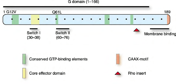
described for some Ras GAPs. Finally, the mutated Ras
proteins found in human cancers contain single amino
acid substitutions at glycine 12 (G12) or glutamine 61
(Q61), which render these mutants insensitive to GAP
stimulation (Figure 5). These GTPase-deficient mutants
are persistently GTP-bound in the absence of upstream
stimulation and consequently cause deregulated effector
activation. The experimental introduction of analogous
mutations at residues corresponding to G12 and Q61
also renders other small GTPases GAP-insensitive and
constitutively activated. Some wild-type small GTPases,
however, possess naturally occurring sequence variation
at these two residues, and are persistently GTP-bound
proteins (e.g., RhoE/Rnd3). Thus, while GDP/GTP
regulation is the major mode of small GTPase regu-
lation, for some GTPases, regulation by other mechan-
isms (e.g., gene transcription, membrane association) is
also important.
Other Small GTPase Regulatory Mechanisms
Guanine Nucleotide Dissociation Inhibitors (GDIs)
GDIs have been identified for Rho and Rab proteins.
Two distinct negative regulatory functions have been
ascribed to GDIs. First, they bind to and mask the
isoprenoid lipid modification in the carboxyl terminus of
the small GTPase, thus preventing the association of the
small GTPase with membranes (Figure 4). Second, their
binding perturbs GAP and GEF regulation, preventing
small GTPase activation. Three Rho GDIs and one Rab
GDI have been described and can recognize multiple
family members. The mechanisms of regulation of GDI
activity are unclear, but may involve phosphorylation.
SMALL GTPASE STRUCTURE
Small GTPases and Conserved
Structural Elements
The structure of many forms of Ras proteins has been
determined and has established the general rules and
requirements for all small GTPases. H-Ras was the
first small GTPase to have its structure solved, and
FIGURE 5 Small GTPase functional domains. Members of the small GTPase family share sequence similarities that define distinct functional
domains. The residue numbers corresponding to H-Ras are shown. The G domain is comprised of a set of four conserved sequence elements
involved in GTP-binding it alone is sufficient for the guanine nucleotide binding and GTPase activity, and is structurally similar among small
GTPases. Mutated forms of Ras proteins are found in human cancers and possess single amino acid substitutions (e.g., G12V or Q61L) that render
the protein insensitive to GAP stimulation and result in constitutively activated proteins. Similar mutations in other GTPases also result in
constitutively activated proteins. Ras and Rho membrane association is facilitated by COOH-terminal sequences that include the CAAX motif as
well as sequences upstream of this sequence. Effector binding involves the core effector domain (residues 32–40) and residues that change in
conformation in the GDP- and GTP-bound states (switches I and II). Rho GTPases possess additional sequences (Rho insert) not found in other
small GTPases. Some small GTPases contain additional NH
2
- or COOH-terminal sequence extensions.
SMALL GTPases 51

subsequent structures of related proteins have demon-
strated a conserved overall canonical structural fold
(designated the G domain) shared with all GTPases, but
with variations in features for different small GTPase-
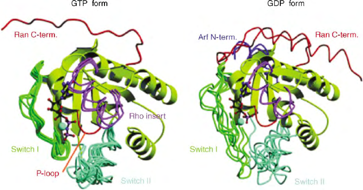
family members (Figure 6).
All small GTPases possess a set of conserved sequence
elements shared with other GTPases which represent the
GDP/GTP nucleotide-binding pocket and GTP hydro-
lytic machinery (some, however, are GTPase deficient
due to key residue changes), since the binding and
hydrolysis of guanine nucleotides is the uniting element
throughout small GTPases. A significant observation
from Ras structural studies was the discovery of
structural differences between the GDP- and GTP-
bound forms, which are localized in two regions, termed
switch I (residues 32–38) and switch II (residues
59–67). Similar switch-I and switch-II conformation
changes have also been identified for the GDP- and GTP-
bound states of other small GTPases.
Switch I and switch II show sequence divergence
between families, although their loop-like structures are
conserved. In addition to reflecting the nucleotide-bound
state of the small GTPase, the switch regions are also
involved in the interaction of small GTPases with
effectors as well as GAPs and GEFs. The divergent
sequence composition of these two regions contributes
to the specificity of different small GTPases for these
different binding partners.
Members of the Rho-family small GTPases possess a
unique structural feature absent on all other small
GTPases. The Rho family contains an insertion of 13
amino acids, called the insert region, positioned between
Ras residues 122 and 123, which forms a surface-
exposed loop (Figure 5). The primary sequence and
secondary conformation of this insert region varies
within the Rho family. Although the conformation of the
insert region is not influenced by nucleotide binding,
there is evidence that it is involved in effector interaction
and activation.
Finally, other small GTPases possess additional
amino- or carboxyl-terminal extensions important for
function. For example, Arf and Sar1 proteins contain an
amino-terminal extension necessary for insertion into
and interaction with the membrane, whereas Ran has an
elongated carboxyl-terminus that is crucial for its
function in nuclear transport.
Diverse Biology and Function
of Small GTPase
Despite their strong structural and biochemical
similarities, small GTPases facilitate a remarkably
divergent spectrum of cellular functions. This diversity
is accomplished by the input signals that regulate the
distinct GAPs and GEFs to modulate GDP/GTP
cycling, and by the unique set of effectors that are
recognized by the GTP-bound forms of small GTPases
within and between families, resulting in many
different cellular responses.
RAS PROTEINS AS SIGNALING
NODES AND REGULATORS OF
CELL PROLIFERATION
The frequent mutation of Ras proteins in human cancers
has made these small GTPases the most intensely studied
FIGURE 6 Canonical GTP conformation of small GTPases. Superimposition of a selection of Ras superfamily proteins on the G domain show the
similar conformation changes in the switch-I and switch-II regions in the GDP- and GTP-bound forms. Rho GTPases possess additional sequences
(Rho insert) not present in any other Ras superfamily proteins. Some small GTPases possess additional NH
2
- and COOH-terminal sequences: the
COOH-terminus of Ran is in red, the Rho insert in magenta and the Arf NH
2
-terminal helix in blue.
52 SMALL GTPases

and best characterized. Ras proteins serve as signaling
nodes, where a wide diversity of extracellular signals
such as growth factors (epidermal growth factor,
platelet-derived growth factor), hormones (insulin),
cytokines (interleukin-1), and the extracellular matrix
proteins (via integrins) converge on and cause activation
of Ras. Activated Ras in turn interacts with and
regulates the activities of downstream effectors with
highly divergent biochemical functions. Recent reviews
have provided detailed discussions of Ras effector
utilization. Therefore, we have highlighted various
themes.
The Raf serine/threonine kinases are important
effectors of Ras and facilitate activation of the ERK
mitogen-activated protein kinase cascade (Figure 7). Ras
promotes Raf activation by promoting the association of
the normally cytosolic Raf with the plasma membrane,
where a complex set of events, which includes protein
phosphorylation, activates Raf kinase function. Acti-
vated Raf then phosphorylates and activates the MEK1
and MEK2 dual specificity protein kinases, which
phosphorylate and activate ERK1 and ERK2.
Activated ERK translocates to the nucleus and phos-
phorylates a variety of targets that include Ets family
transcription factors. The Raf/MEK/ERK kinase cascade
contributes significantly to the growth-regulatory func-
tions of Ras.
The second best-characterized class of Ras effectors is
the phosphatidylinositol 3-kinases (PI3K), a family of
lipid kinases (Figure 7). A major activity of PI3K is the
conversion of membrane-associated phosphatidyl-
inositol 4,5-bisphosphate (PIP
2
) to phosphatidylinositol
3,4,5-trisphosphate (PIP
3
). PIP
3
in turn can regulate the
activity of a diverse range of targets, including activation
of the Akt serine/threonine kinase and Rac GEFs, via
PIP
3
interaction with pleckstrin homology domains
present in these proteins.
One significant theme that has emerged is that a
number of Ras effectors are GEFs that link Ras with
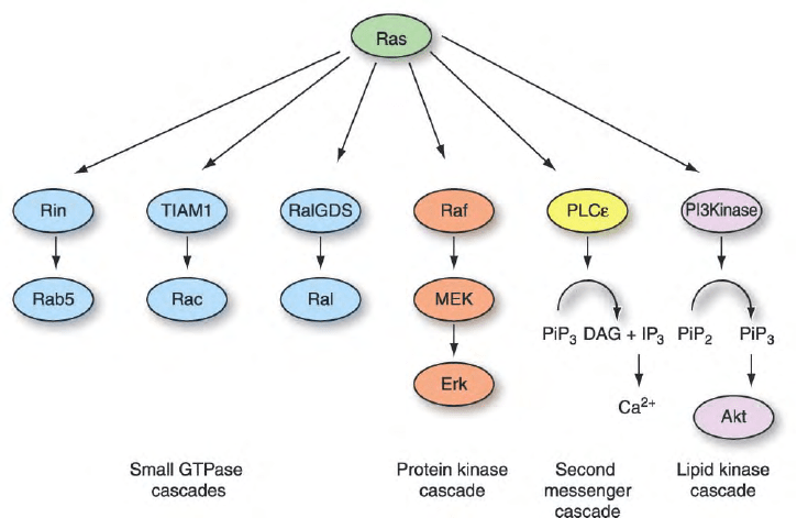
other small GTPases (Figure 7). First, there is a family of
Ral GEFs that binds activated Ras and promotes
activation of RalA and RalB, members of the Ras family
of small GTPases. Second, Rin1 functions as a GEF for
Rab5, thus linking Ras activity with regulation of
vesicular trafficking. Third, Ras binds and activates
Tiam1, which functions as a GEF for Rac. Fourth, Ras
binds phospholipase C epsilon, which also contains a
CDC25 homology domain that may activate the R-Ras
small GTPase.
While Ras GTPases function as positive regulators of
cell proliferation, some small GTPases appear to possess
opposing functions and may function as tumor suppres-
sors rather than oncogene proteins. This includes Rerg,
NOEY2/ARH1, Rig, and DBC2. Thus, while these
proteins share significant sequence and biochemical
functions with Ras, clearly their effector functions are
quite distinct. Additionally, whereas mutational acti-
vation of Ras proteins is associated with oncogenesis, it
is the loss of gene expression of these proteins that
accounts for their loss of function in tumor cells.
FIGURE 7 Ras effectors mediate diverse signaling outcomes. Activated, GTP-bound Ras can bind and regulate functionally diverse downstream
effector targets. These include activation of a protein kinase cascade (Raf), activation of the PI3K lipid kinase, and activation of PLCE and the
production of second messengers (DAG and calcium). Several Ras effectors are GEFs that activate other members of the Ras superfamily, including
Rab5 (Rin1), Ral (RalGEFs), and Rac (Tiam1). Thus, Ras functions as a signaling node, where diverse signals converge and cause its activation
(Figure 4), and once activated, results in activating of diverse downstream signaling pathways.
SMALL GTPases 53
RHO-FAMILY PROTEINS AND ACTIN
CYTOSKELETAL ORGANIZATION,CELL
MORPHOLOGY, AND CELL MOVEMENT
Similar to Ras proteins, Rho-family small GTPases also
function as signaling nodes activated by diverse extra-
cellular signals. Perhaps the best-characterized cellular
function of these proteins is their regulation of actin
cytoskeletal organization, which in turn influences cell
morphology, cell–matrix and cell–cell interactions, and
cell movement. For example, RhoA causes actin stress
fiber formation, Rac causes actin accumulation at the
leading edge of a motile cell and lamellipodia formation,
whereas Cdc42 promotes actin microspikes and filopo-
dia formation. These changes in actin organization can
influence cell shape, cell motility, and cell–cell
interactions.
SMALL GTPASES AND
MEMBRANE TRAFFICKING
Rab proteins constitute the largest subfamily of small
GTPases, with more than 60 mammalian members. Rab
proteins are involved in the modulation of specific steps
in eukaryotic membrane trafficking in the secretory and
endocytic pathways. Arf and Sar small GTPases are also
involved in the regulation of cytoplasmic vesicular
trafficking. By contrast, Ran is a regulator of nucleocy-
toplasmic transport. The regulation of the GDP/GTP
cycle is also critical for the functions of these small
GTPases in their distinct transport function.
SEE ALSO THE FOLLOWING ARTICLES
ARF Family † G
12
/G
13
Family † Phosphatidylinositol
Bisphosphate and Trisphosphate † Rab Family † Ran
GTPase † Ras Family † Rho GTPases and Actin Cyto-
skeleton Dynamics
GLOSSARY
cytoskeleton A dynamic network of filaments, microfilaments,
microtubules, and intermediate filaments.
effector proteins Proteins that bind preferentially to the GTP-bound
form of small GTPases and facilitate the biological response of
small GTPases.
GTPase High-affinity GDP-, GTP-binding, and GTP-hydrolyzing
proteins that act as molecular switches and timers that cycle
between inactive GDP-bound and active GTP-bound states.
GTPase activating protein Negative regulatory protein that accel-
erates the intrinsic GTP hydrolysis activity of small GTPases.
guanine nucleotide exchange factors Regulatory protein that
accelerates the intrinsic GDP/GTP exchange activity of small
GTPases.
guanine nucleotides Amine nucleotide bases composed of a guanine
moiety attached to one (guanosine monophosphate, GMP), two
(guanosine diphosphate, GDP), or three (guanosine triphosphate,
GTP) phosphate groups.
isoprenylation Post-translational, covalent modification of proteins
at carboxyl-terminal cysteine residues, by either C15 farnesyl or
C20 geranylgeranyl isoprenoid lipids.
kinase An enzyme with phosphorylating activity on either proteins
or lipids.
superfamily, family, and subfamily Small GTPases are classified into
hierarchical phylogenies on the basis of structural, sequence, and
functional similarity between members. Members of the Ras
family share , 50% amino acid identity with the four
Ras proteins whereas members of the Rho, Rab, and other Ras
superfamily GTPases share , 25–30% amino acid identity with
Ras proteins.
FURTHER READING
Der, C. J. (2002). Rho family proteins. In Encyclopedic Reference of
Cancer (M. Schwab, ed.) pp. 799– 804. Springer, Heidelberg,
Germany.
Hall, A. (ed.) (2000). GTPases. Oxford University Press, New York.
Pruitt, W. M., and Der, C. J. (2002). Ras proteins. In Encyclopedia of
Cancer (J. R. Bertino, ed.) 2nd edition, Vol 4, pp. 41–48. Academic
Press, Orlando, FL.
Seabra, M. C., Mules, E. H., and Hume, A. N. (2002). Rab GTPases,
intracellular traffic and disease. Trends Mol. Med. 8, 23–30.
Vetter, I. R., and Wittinghofer, A. (2001). The guanine nucleotide-
binding switch in three dimensions. Science 294, 1299 –1304.
BIOGRAPHY
Adam Shutes graduated from Oxford University before receiving
his Ph.D. from University College London in 2001. He is currently
a postdoctoral Fellow at the Lineberger Comprehensive Cancer Center,
UNC, where his interests lie in small GTPases and their regulation.
Channing Der is a Professor of Pharmacology at the University of
North Carolina at Chapel Hill, where his research is focused on Ras-
related proteins and their roles in cancer. He graduated from University
of California, Los Angeles, before receiving his Ph.D. from University
of California, Irvine, in 1981.
54 SMALL GTPases

Somatostatin Receptors
Agnes Schonbrunn
The University of Texas Health Science Center, Houston, Texas, USA
Somatostatin receptors (sst receptors) comprise a family of five
distinct plasma membrane receptors that bind the neuroendo-
crine peptides somatostatin and cortistatin. These receptors
exhibit the seven-transmembrane domain structure typical of
G protein-coupled receptors (GPCRs) and signal primarily
through pertussis toxin-sensitive G proteins. The different sst
receptor subtypes are found in specific endocrine and exocrine
tissues, including the pituitary and the pancreas, in addition to
being widely distributed in the central and peripheral nervous
systems and the gastrointestinal tract. They have important
physiological roles in inhibiting hormone secretion, particu-
larly the secretion of growth hormone, insulin, glucagon, and
gastrin, inhibiting exocrine secretion by the pancreas and
stomach, and modulating neuronal excitability and smooth
muscle contraction. Additionally, many neuroendocrine
tumors express high levels of somatostatin receptors, and,
because of their ability to inhibit tumor cell secretion as well as
proliferation, somatostatin analogues are now used to target
these receptors for both cancer therapy and diagnosis.
Physiological Somatostatin
Receptor Ligands
Somatostatin (SS), originally called somatotropin-release
inhibiting factor or SRIF, was discovered accidentally
over 30 years ago while investigators were hunting for the
brain peptide responsible for stimulating the release of
growth hormone, or somatotropin. Surprisingly, they
found that extracts of the hypothalamus, a specialized
brain region that regulates pituitary hormone secretion,
inhibited rather than stimulated the secretion of growth
hormone. Using this inhibition as an assay, R. Guillemin,
A. Schally, and their co-workers purified the active factor
from hypothalamic extracts and identified the 14-amino-
acid form of somatostatin, a discovery that contributed to
their receiving the Nobel Prize in 1977 for studies of
hormone production in the brain.
SS is now known to exist in two biologically active
isoforms, 14 (SS14) and 28 (SS28) amino acids in length,
which are produced by alternate proteolytic processing
from a common precursor of 116 amino acids. These
cyclic peptides are synthesized by specific endocrine,
gastrointestinal, immune, and neuronal cells, as well as
some tumors, and act either locally as paracrine,
autocrine, or neuronal modulators or through the
bloodstream as hormones.
The cDNA for the related peptide cortistatin was
discovered much later, in 1996, in the process of
characterizing region-specific rat brain mRNAs. Corti-
statin was named for its predominant expression in the
brain cortex and its ability to depress cortical neuronal
activity. Cortistatin is also synthesized as a precursor and
is proteolytically processed into two biologically active
products: a short (rat CST-14 and human CST-17) and
an amino-terminally extended (CST-29) form. Although
cortistatin is produced from a different gene than
somatostatin, the two peptides have 10 of their 14
carboxy-terminal amino acids in common (Figure 1).
Cortistatins are produced primarily by central nervous
system neurons but have also been found in immune
cells, lymphoid tissues, bone marrow, and the pancreas.
Because all forms of somatostatin and cortistatin bind
to the different somatostatin receptor subtypes (sst’s)
with similar high affinities, it is not surprising that the
peptides share many functional properties. However,
they also produce some distinct biological effects. For
example, intracerebroventricular injection of cortistatin
in rats increases slow-wave sleep but not REM sleep,
whereas somatostatin injection increases REM sleep.
Thus, the identification of a human cortistatin selective
G protein-coupled receptor (MrgX2, or Mas-related
gene X2) fulfills prior predictions for the existence of
distinct cortistatin-specific receptors. However, the
rodent orthologue of this receptor has not been identified,
and the relative roles of somatostatin receptors and
cortistatin receptors in mediating the physiological
actions of these peptides remain to be determined.
Biochemical and Molecular
Characterization of
Somatostatin Receptors
SOMATOSTATIN RECEPTOR SUBTYPES
Five somatostatin receptor genes, located on different
chromosomes, have been identified and named sst1 to
Encyclopedia of Biological Chemistry, Volume 4. q 2004, Elsevier Inc. All Rights Reserved. 55

sst5 in the order of their discovery (Table I). The coding
regions of sst1, sst3, sst4, and sst5 all occur within a
single exon. However, at least in some mouse and rat
tissues, the mRNA for sst2 undergoes alternative
splicing at the 3
0
end, producing two protein products.
The sst2A variant is the product of the unspliced mRNA,
whereas the sst2B variant contains an alternative exon
that encodes a different and somewhat shorter receptor
carboxy terminus. Only the sst2A variant has been
detected in normal human tissues.
Somatostatin receptors belong to the G protein-
coupled receptor (GPCR) family and are predicted to
contain seven
a
-helical transmembrane domains. Recep-
tors for somatostatin have been cloned from a variety of
mammals as well as from several nonmammalian
species, including a number of fish. The human sst
receptors exhibit between 40 and 57% amino acid
identity with each other, and sequence identity among
the rat, mouse, and human orthologues of each receptor
subtype is even higher. The greatest sequence similarity
between sst receptor subtypes occurs within the trans-
membrane domains; these domains, therefore, are
thought to be involved in ligand binding. Sequence
differences in the intra- and extracellular domains
are presumably responsible for the unique signaling
and trafficking properties of individual sst receptor
subtypes.
Based on sequence similarity, sst receptors have been
subdivided into two groups: sst1 and sst4 receptors form
the SRIF2 subgroup, and sst2, sst3, and sst5 constitute
the SRIF1 subgroup. In addition to a closer phylogenetic
relationship (Figure 2), members of each group share
several functional properties, such as their affinity for
short synthetic somatostatin analogues, including
octreotide and lanreotide, and sensitivity to agonist-
induced receptor internalization (Table I).
The GPCRs most closely related to the sst receptor
family are GPR7 and GPR8, which encode receptors for
two paralogous brain peptides, neuropeptide B (NPB)
and neuropeptide W (NPW) (Figure 2). The next most
closely related family is the opioid receptor family,
which also shares substantial sequence identity with the
cortistatin receptor MrgX2 (Figure 2).
SOMATOSTATIN RECEPTOR STRUCTURE
Somatostatin receptors contain many of the structural
features characteristic of GPCRs, including consensus
sites for Asn-linked glycosylation within the amino
terminus and multiple Ser and Thr phosphorylation sites
in the intracellular loops and carboxy-terminal tail
(Table I). All sst receptor subtypes except sst3 also
contain a conserved Cys residue in the carboxy-terminal
region, which provides a site for receptor palmitoyla-
tion. Lipid modification has yet to be demonstrated
biochemically for any of the sst subtypes. However, sst1,
sst2, sst3, and sst5 are known to be glycosylated from
Ala Gly Cys
s
s
s
s
Cortistatin-17
Somatostatin-14
Lys Asn Phe
Phe
Tr p
Lys
Thr
Phe
ThrSerCys
Asp Arg Met Pro Cys Arg Asn Phe
Phe
Tr p
Lys
Thr
Phe
SerSerCysLys
FIGURE 1 Structure of mammalian somatostatin-14 and human
cortistatin-17. The amino acids in the red circles are required for high-
affinity binding to somatostatin receptors. The amino acid differences
between the two peptides are shown by the green circles.
TABLE I
Properties of Human Somatostatin Receptor Subtypes
Property sst1 sst2A/B sst3 sst4 sst5
Chromosomal localization 14q13 17q24 22q13.2 20p11.2 16p13.3
Reference sequence
a
NM 001049 NM 001050 NM 001051 NM 001052 NM 001053
Amino acids 391 369/356 418 388 364
Asn glycosylation sites 3 4213
Receptor glycosylation þþþ2 þ
Cys palmitoylation site þþ2 þþ
Receptor phosphorylation þþþ2 nd
b
Octreotide/lanreotide binding affinity Low High Moderate Low Moderate
SS-stimulated receptor internalization Slow Fast Fast Slow Fast
a
Accessible at http://www.ncbi.nlm.nih.gov/.
b
nd, not determined.
56 SOMATOSTATIN RECEPTORS

the observation that treatment of these receptors with
peptide-N-glycosidase F (PNGaseF), an enzyme that
catalyzes the cleavage of N-glycosidically linked carbo-
hydrate chains from Asn, causes a large decrease in their
apparent molecular weights. Such a shift in molecular
weight was not observed for sst4, either because it is not
glycosylated or because glycosylation at its single
consensus site does not significantly affect its apparent
molecular weight.
Receptor phosphorylation, a covalent modification
known to be important for GPCR regulation and
signaling, has been examined biochemically for some
but not all sst receptor subtypes. Somatostatin stimu-
lates the incorporation of
32
PO
4
into sst1, sst2A, and
sst3 receptors, and protein kinase C activation also
increases phosphorylation of sst1 and sst2A. However,
phosphorylation of sst4 was not detected following
agonist stimulation, and sst5 phosphorylation has not
been investigated biochemically.
Pharmacology of Somatostatin
Receptor Subtypes
The native somatostatin peptides exhibit little
selectivity among the different receptor subtypes and
have a very short lifetime in plasma (half-life , 3 min).
Thus, analogues with increased metabolic stability
and greater receptor specificity have been developed
for therapy.
The earliest pharmaceuticals to target somatostatin
receptors were short peptides, 6 to 8 amino acids in
length, containing amino acids 7 to 10 of SS14, the
region required for receptor binding (Figure 1). These
agonists, including octreotide (SMS201-995), lanreotide
(BIM 23014), and seglitide (MK-678), also contain
D-amino acid substitutions and carboxy- or amino-
terminal modifications to increase metabolic stability.
These truncated peptides bind selectively to the SRIF1
group of receptors, exhibiting subnanomolar affinity for
sst2 and nanomolar affinities for sst3 and sst5 (Table I).
Further, octreotide does not bind to the MrgX2
cortistatin receptor. Both octreotide and lanreotide are
used clinically to treat hormone-secreting pituitary
adenomas and gastroenteropancreatic (GEP) tumors
and comprise the only FDA-approved pharmaceuticals
targeting sst receptors for therapy.
In an effort to generate receptor subtype specific
analogues that are resistant to proteolytic degradation,
S. Rohrer and co-workers at Merck screened combina-
torial libraries to successfully identify the first highly
selective, high-affinity, nonpeptide agonists for all sst
subtypes. Such reagents offer the possibility of oral
bioavailability, although they are not presently approved
for clinical use. Nonetheless, they supply valuable tools
for identifying the physiological roles of individual sst
receptor subtypes.
The recognition that many tumors express several
different sst receptors concomitantly has stimulated a
search for stable somatostatin analogues that can bind to
multiple somatostatin receptors. The hexapeptide ana-
logue SOM230 shows high affinity for four of the five
somatostatin receptor subtypes (sst1, sst2, sst3, and
sst5), whereas the nonapeptide analogue KE108 appears
to be truly universal since it binds to all five sst subtypes
with nanomolar affinity. Because of their excellent
metabolic stability, these broad-spectrum agonists have
significant therapeutic potential.
The first somatostatin receptor antagonist, CYN-
154806, described in 1996, showed high selectivity for
the sst2 receptor. Since then, antagonists have been
identified for all five somatostatin receptor subtypes,
although none are used clinically.
In addition to their therapeutic applications, ana-
logues of somatostatin have been developed to localize
and stage sst receptor-positive tumors in situ.The
radiolabeled derivatives used for tumor diagnosis con-
tain a chelating group covalently attached to a stabilized
somatostatin peptide, such that its receptor-binding
properties are not compromised. The chelating group
is then bound to a short-lived radioisotope, such as the
gamma emitter
111
In, and injected into patients. After
24 h, accumulation of the radiolabel can be visualized in
tumors expressing high levels of sst receptors by gamma
camera scintigraphy, thus permitting localization of the
tumors and their metastases. The analogue in current
NPB/WR-1 (GPR7)
NPB/WR-2 (GPR8)
CSTR (MrgX2)
sst1
sst4
sst2
sst5
sst3
KOR-3
KOR-1
MOR-1
DOR-1
FIGURE 2 Sequence relationships between somatostatin receptors
and closely related peptide receptors. The dendrogram was generated
with the Clustal W program using MacVector’s default parameters to
align the peptide sequences. Human sequences were used in all cases
and were obtained either from SwissProt or RefSeq. Somatostatin
receptors: hsst1, P30872; hsst2A, P30874; hsst3, P32745; hsst4,
P31391; hsst5, P35346. Cortistatin receptor: MrgX2, NP_473371.
Opioid receptors: KOR-1, P41145; MOR-1, P35372; DOR-1,
P41143; and KOR-3, P41146. Neuropeptide B/W receptors:
NPBWR-1 or GPR7, P48145; and NPBWR-2 or GPR8, P48146. The
plot shows that hsst1 and hsst4 form one subgroup and hsst2, hsst3,
and hsst5 form a second subgroup within the somatostatin receptor
family and that the cortistatin receptor is not a member of the sst
receptor family.
SOMATOSTATIN RECEPTORS 57
clinical use is an indium-labeled octreotide derivative,
[
111
In-DTPA-D-Phe
1
,Tyr
2
]octreotide (OctreoScan),
and hence is selective for the SRIF1 group of sst
receptors, showing the most sensitive detection of sst2-
expressing tumors. Unfortunately, this method cannot
be used to localize tumors expressing sst1 or sst4
receptors, as appropriate somatostatin analogues are
not yet available.
Somatostatin Receptor Signaling
Somatostatin receptors regulate a number of diverse
signaling effectors, including adenylyl cyclase, phospho-
lipases C and A2, calcium and potassium channels,
protein and lipid kinases, and tyrosine and serine/threo-
nine phosphatases. All sst receptors inhibit adenylyl
cyclase via pertussis toxin-sensitive G proteins and
thus decrease intracellular cyclic adenosine monophos-
phate (cAMP). However, other signal transduction
pathways modulated by somatostatin receptors
vary both with the receptor subtype and with the
target cell.
The mechanism by which somatostatin receptors
inhibit secretion in endocrine cells and neurons is
understood in some detail. In addition to reducing intra-
cellular cAMP levels, several sst receptors have been
shown to reduce intracellular calcium levels in excitatory
cells, also via pertussis toxin-sensitive G proteins. The
reduction in cytosolic calcium can result either from
a stimulation of various potassium channels, which
hyperpolarize the cell membrane and thereby decrease
influx through voltage-dependent calcium channels, or
from direct calcium channel inhibition. The decrease
in intracellular cAMP and calcium concentrations
together contribute to the inhibitory action of soma-
tostatin on secretion: when either signaling pathway is
blocked, the magnitude of somatostatin’s inhibitory
effect is reduced.
Somatostatin stimulates contraction of intestinal
smooth muscle cells by inhibiting adenylyl cyclase
and activating phospholipase C-
b
3 via the
a
-and
bg
-subunits, respectively, of pertussis toxin-sensitive
G proteins. The activated phospholipase catalyzes the
hydrolysis of the membrane lipid phosphatidylinositol
4,5-bisphosphate to form the second messengers inositol
trisphosphate (IP
3
) and diacylglycerol (DG). The bind-
ing of IP
3
to receptors on the sarcoplasmic reticulum
results in the release of calcium from intracellular stores,
producing a rise in cytosolic calcium concentrations.
The released calcium forms a complex with the protein
calmodulin, and this complex then activates myosin
light-chain kinase (MLCK) to phosphorylate the light
chain of myosin, leading to smooth muscle contraction.
Because protein kinase A (PKA) decreases the sensitivity
of MLCK to calcium, somatostatin inhibition of cAMP
formation facilitates the calcium effect by reducing the
activity of PKA. These pathways are also activated by
somatostatin in aortic vascular smooth muscle cells.
Interestingly, somatostatin does not activate phospho-
lipase C-
b
3 in pituitary cells, even though the level of
this enzyme in the pituitary appears to be similar to that
in smooth muscle cells. The explanation for the signaling
differences in these tissues remains to be elucidated, but
could be due to differences either in the sst subtypes or in
the signaling machinery present.
In endothelial cells, somatostatin inhibits cell
migration, stress fiber assembly, and cytoskeletal reor-
ganization produced by thrombin and other stimulators.
In humans, these effects are mediated by the sst1
receptor and have been implicated in somatostatin’s
antiangiogenic actions. Although the molecular steps
involved have not yet been identified, the mecha-
nism includes an unusual pertussis toxin-independent
inhibition of Rho, a low molecular mass GTPase
that plays a central role in regulating cytoskeletal
organization.
The mechanisms by which somatostatin inhibits
cell proliferation and stimulates apoptosis are also
poorly understood and appear to vary in the different
cell types in which they have been examined. In most
(though not all) cells, somatostatin activates the
mitogen-activated protein kinase (MAPK) pathway
and increases extracellular signal-related kinase
(ERK)1/2 phosphorylation by a pertussis toxin-
sensitive mechanism. However, this activation is often
observed whether somatostatin inhibits or stimulates
cell proliferation. Thus, it is likely that some of the
other effectors activated by the sst receptors contribute
to the final biological response. For example, in
pancreatic acinar cells, which express sst2 receptors
endogenously, somatostatin-induced growth arrest
involves enhanced expression of the cyclin-dependent
kinase inhibitor p27Kip and results from inhibition of
the phosphatidylinositol-3-kinase (PI3K) pathway. In
contrast, in sst2 transfected CHO cells, somatostatin
induction of p27Kip appears to be dependent on
stimulation rather than inhibition of PI3K, in that
PI3K inhibitors block the effect of somatostatin
analogues on ERK2 phosphorylation, which in turn is
required for p27Kip up-regulation. These and other
potential signaling pathways are under intense inves-
tigation in order to elucidate the clinically important
actions of somatostatin to inhibit cell proliferation and
stimulate apoptosis.
In summary, most but not all signaling by sst
receptors involves the pertussis toxin-sensitive G
i
/G
o
family. However, depending on the sst receptor
subtype and the cellular environment, a spectrum of
nonoverlapping signaling pathways can be activated.
The link between the different effectors regulated by sst
58
SOMATOSTATIN RECEPTORS
receptors and particular biological responses is under-
stood for some but by no means all of somatostatin’s
actions.
Somatostatin Receptor Regulation
Alterations in sst receptor responsiveness during
somatostatin exposure vary dramatically between tis-
sues, depending both on the response being measured
and on the nature of the target cell. In some instances,
desensitization occurs within minutes following
initiation of somatostatin treatment, whereas in others
no desensitization is detected even after years of
somatostatin analogue exposure. For example, in chick
sympathetic neurons, somatostatin inhibition of N-type
Ca currents desensitizes with a half-life of 3 min. In
contrast, desensitization to pharmacologic doses of the
somatostatin analogue octreotide does not occur in
months or years of therapy for pituitary tumors. The
molecular basis for such dramatic differences in somato-
statin receptor regulation remains poorly understood.
Studies of the different sst receptor subtypes in
transfected cells indicate receptor-specific differences in
their regulation. As has been shown for many GPCRs,
hormone treatment leads to the rapid phosphorylation
of several sst receptor subtypes (Table I). Interestingly,
however, this phosphorylation appears to have different
consequences for the different sst receptors. For
example, in CHO cells, although both sst1 and sst2
receptors are phosphorylated and desensitized within
minutes following somatostatin exposure, only the sst2
receptor is rapidly internalized.
The cellular environment also plays an important role
in the observed differences in the regulation of
somatostatin responsiveness. Interestingly, the sst2
receptor, which desensitizes within minutes of somato-
statin exposure in cultured cells but is resistant to
desensitization in human tumors, was recently shown to
be phosphorylated in situ in a human tumor. Hence, at
least this first step in the desensitization process appears
to be intact in human tumor tissue. As the other
molecular components of sst receptor desensitization
are characterized, it will be interesting to determine
whether their functions are altered in the desensitiza-
tion-resistant tumors.
Summary
The five known somatostatin receptors play important
physiological roles in the regulation of the endocrine,
neuronal, gastrointestinal, and immune systems. In
addition, they are recognized to be important targets
in the diagnosis and therapy of a number of neuroendo-
crine tumors.
Although a great deal remains to be learned about
many of somatostatin’s functions, studies with sst1,
sst2, and sst5 knockout mouse models, all of which
are viable, have begun to delineate the biological
importance of the individual receptor subtypes. Future
focus on these receptors in their normal cellular
environments can take advantage of the many new
receptor-specific agonists and antagonists that have
become available and will undoubtedly provide much-
needed insight into the function of this physiologically
and therapeutically important G protein-coupled
receptor family.
SEE ALSO THE FOLLOWING ARTICLES
Adenylyl Cyclases † Mitogen-Activated Protein Kinase
Family † Phospholipase A
2
† Phospholipase C † Rho
GTPases and Actin Cytoskeleton Dynamics † Voltage-
Sensitive Na
þ
Channels
GLOSSARY
acromegaly A condition caused by the excess secretion of pituitary
growth hormone after maturity, usually by a pituitary tumor.
The disease is characterized by enlargement of the extremities,
including the nose, jaws, fingers, and toes, as well as certain
internal organs.
cortistatins The short (rat CST-14 and human CST-17) and long
(CST-29) biologically active products produced by proteolytic
processing of the precursor peptide encoded by the cortistatin gene.
G protein One of a family of related heterotrimeric proteins that
bind GTP and GDP. The heterotrimeric forms, which are inactive,
become activated at the plasma membrane by agonist occupied
G protein-coupled receptors (GPCRs) that stimulate the binding
of GTP to the G protein
a
-subunit and cause dissociation of the
a
-subunit from the
bg
-subunit complex. When activated, the
G protein subunits regulate downstream effectors, such as ion
channels and enzymes that generate second messengers. G proteins
become inactivated by hydrolyzing GTP to GDP, which then permits
the reassociation of the
a
-subunit with the
bg
-subunit complex.
somatostatins (SSs) The 14-amino-acid (SS-14) and 28-amino-acid
(SS-28) peptides produced by alternative proteolytic processing
from a single 92-amino-acid precursor called prosomatostatin.
FURTHER READING
Alliance for Cellular Signaling (2004). The Signaling Gateway. http://
www.signaling-gateway.org/. ID numbers: A002204 for sst1,
A002205 for sst2, A002206 for sst3, A002207 for sst4, and
A000075 for sst5. Accessed May 2004.
Hofland, L. J., and Lamberts, S. W. (2003). The pathophysiological
consequences of somatostatin receptor internalization and resist-
ance. Endocr. Rev. 24, 28– 47.
Kreienkamp, H. J. (1999). Molecular biology of the receptors for
somatostatin and cortistatin. Results Probl. Cell. Differ. 26,
215–237.
Patel, Y. C. (1999). Somatostatin and its receptor family. Front.
Neuroendocrinol. 20, 157–198.
Reubi, J. C. (2003). Peptide receptors as molecular targets for cancer
diagnosis and therapy. Endocr. Rev. 24, 389–427.
SOMATOSTATIN RECEPTORS 59
Schonbrunn, A. (1999). Somatostatin receptors: present knowledge
and future directions. Ann. Oncol. 10, S17–S21.
Schonbrunn, A. (2001). Somatostatin. In Endocrinology (L. J. Degroot
and J. L. Jameson, eds.) 4th edition, pp. 427–437. Saunders,
New York.
Selmer, I., Schindler, M., Allen, J. P., Humphrey, P. P., and Emson, P. C.
(2000). Advances in understanding neuronal somatostatin recep-
tors. Regul. Pept. 90, 1–18.
Slooter, G. D., Mearadji, A., Breeman, W. A., et al. (2001).
Somatostatin receptor imaging, therapy and new strategies
in patients with neuroendocrine tumours. Br. J. Surg. 88,
31–40.
Spier, A. D., and de Lecea, L. (2000). Cortistatin: A member of the
somatostatin neuropeptide family with distinct physiological
functions. Brain Res. Brain Res. Rev. 33, 228–241.
Weckbecker, G., Lewis, I., Albert, R., Schmid, H. A., Hoyer, D.,
and Bruns, C. (2003). Opportunities in somatostatin research:
Biological, chemical and therapeutic aspects. Nat. Rev. Drug
Discov. 2, 999 –1017.
BIOGRAPHY
Agnes Schonbrunn is a Professor in the Department of Integrative
Biology and Pharmacology at the University of Texas–Houston,
School of Medicine. She holds a Ph.D. in Biochemistry from Brandeis
University and received her postdoctoral training at Harvard Medical
School. Her principal research interests include the signaling and
regulation of somatostatin receptors and the functional role that these
receptors play in neuroendocrine cancers.
60 SOMATOSTATIN RECEPTORS
