Latchman. Eukariotic Transciption Factors
Подождите немного. Документ загружается.

which, like DNAseI footprinting, rely on the ability of the reagents to cleave
the DNA in a non-sequence specific manner (for further details see Kreale,
1994; Papavassilou, 1995).
Of greater interest, however, is the technique of dimethyl sulphate (DMS)
protection footprinting since it can provide information on the exact bases
within the binding site which are contacted by the protein. Thus, this metho d
relies on the ability of DMS to specifically methylate guanine residues in the
DNA. These methylated G residues can then be cleaved by exposure to
METHODS FOR STUDYING TRANSCRIPTION FACTORS 29
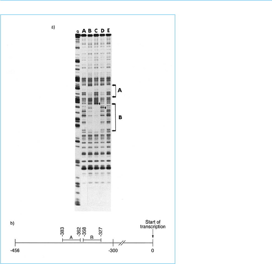
Figure 2.5
Panel (a): DNAseI footprinting assay
carried out on a region of the human
immunodeficiency virus (HIV) control
element. The two footprints (A and
B) are not observed when no cell
extract is added to the reaction
(track A) but are observed when
cellular extract is added in the
absence of competitor (track B).
Addition of unlabelled oligonucleotide
competitor containing the DNA
sequence of site A removes the site
A footprint without affecting site B
(track C) while an unlabelled
oligonucleotide containing the site B
DNA sequence has the opposite
effect (track D). Both footprints are
removed by a mixture of unlabelled
site A and B oligonucleotides (track
E). Arrows indicate the position of
sites at which cleavage with DNAseI
is enhanced in the presence of
protein bound to an adjacent site
indicating the existence of
conformational changes induced by
protein binding. The track labelled G
represents a marker track consisting
of the same DNA fragment
chemically cleaved at every guanine
residue. Panel (b): Position of sites
A and B within the HIV control
element. The arrow indicates the
start site of transcription.

piperidene, whereas no cleavage occurs at unmethylated G residues (Maxam
and Gilbert, 1980). A protein bound to the DNA will protect the guanine
residues which it contacts from methylation and hence they will not be cleaved
upon subsequent piperidene treatment. As in the other footprinting techni-
ques, therefore, specific bands produced by such treatment of naked DNA are
absent in the protein–DNA sample. Unlike the other methods, however,
because cleavage occurs at specific guanine residues, this method identifies
specific bases within the DNA that are contacted by the transcription factor
protein.
These footprinting techniques therefore offer an advance on the mobility
shift assay, allow ing a more precise visualiz ation of the DNA–protein interac-
tion. (For methodological details see Spiro and McMurray, 1999.)
2.2.3 METHYLATION INTERFERENCE ASSAY
The pattern of DNA–protein interaction can also be studied in more detail
using the methylation interference assay (Siebenlist and Gi lbert, 1980). Like
methylation protection, this method relies on the ability of DMS to methylate
G residues which can then be cleaved with piperidene. However, methylation
interference is based on assessing whether the prior methylation of specific G
residues in the target DNA affects subsequent protein binding. Thus, the
target DNA is first partially methylated using DMS so that on average only
one G residue per DNA molecule is methylated (Maxam and Gilbert, 1980).
Each individual DNA molecule will therefore contain some methylated G
residues with the particular residues which are methylated being different
in each molecul e. These partially methylated DNAs are then used in a DNA
mobility shift experiment with an appropri ate cell extract containing the DNA
binding protein. Following electrophoresis the band produced by the DNA
which has bound protein and that produced by the DNA which has not, are
excised from the gel and treated with piperidine to cleave the DNA at the
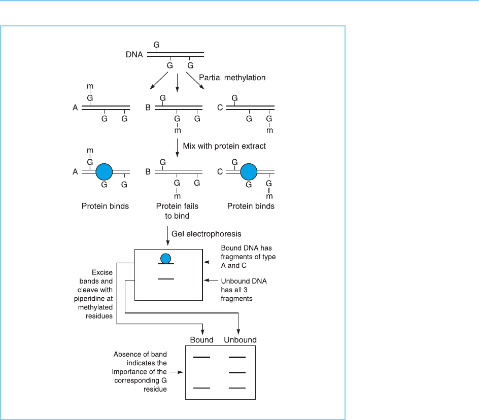
methylated G residues and not at unmethylated Gs. Clearly, if methylation of
a particular G prevents protein binding then cleavage at this particular methy-
lated G will be observed only in the DNA which failed to bind the protein.
Conversely, if a particular G residue plays no role in binding, then cleavage at
this G residue will be observed equally in both the DNA which bound the
protein and that which failed to do so (Fig. 2.6).
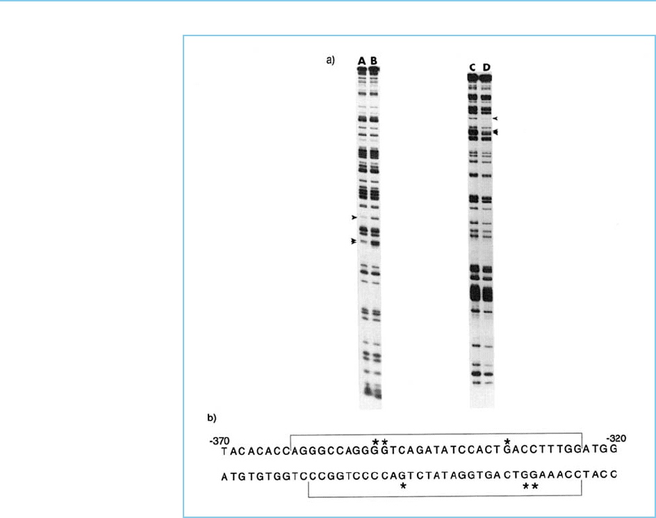
Figure 2.7 shows this type of analy sis applied to the protein binding to site
B within the negatively acting element in the human immunodeficiency virus
promoter (for the footprint produced by the binding of this protein see
Fig. 2.5). In this case the footprinted seque nce was palindromic (Fig. 2.7)
suggesting that the DNA–protein interaction may involve similar binding to
30 EUKARYOTIC TRANSCRIPTION FACTORS
the two halves of the palindrome. The methylation interference analysis of site
B confirms this by showing that methylation of equivalent G residues in each
half of the palind rome interferes with binding of the protein, indicating that
these residues are critical for binding.
Although the DMS method only studies contacts of the protein with G
residues, interference analysis can also be used to study the interaction of
DNA binding proteins with A residues in the binding site. This can be done
either by methylating all purines to allow study of interference at A and G
residues simultaneou sly (see for example Ares et al., 1987) or by using diethyl-
pyrocarbonate specifically to modify A residues (probably by carboxyethyla-
tion) rendering them susceptible to piperidine cleavage (see for example
METHODS FOR STUDYING TRANSCRIPTION FACTORS 31
Figure 2.6
Methylation interference
assay. Partially
methylated DNA is used
in a DNA mobility shift
assay and both the DNA
that has failed to bind
protein and that which
has bound protein and
formed a retarded band
are subsequently cleaved
at methylated G residues
with piperidine. If
methylation at a specific
G residue has no effect
on protein binding (types
A and C) the bound and
unbound DNA will
contain equal amounts of
methylated G at this
position. In contrast, if
methylation at a particular
G prevents binding of the
protein (type B), only the
unbound DNA will
contain methylated G at
this position.
Sturm et al., 1988). These techniques are of particular value when studying
sequences such as the octamer motif in which there are relatively few G
residues, hence limiting the information which can be obtained by studying
interference at G residues alone (Sturm et al., 1987; Baumruker et al., 1988).
Chemical interference techniques can therefore be used to supplement foot-
printing methodologies and identify the precise DNA–protein interactions
within the foo tprinted region. (For methodological details see Spiro and
McMurray, 1999.)
2.2.4 IN VIVO FOOTPRINTING ASSAY
Although the metho ds described so far can provide considerable information
about DNA–protein contacts they all suffer from the deficiency that the DNA–
protein interaction occurs in vitro when cell extract and the DNA are mixed.
32 EUKARYOTIC TRANSCRIPTION FACTORS
Figure 2.7
Panel (a): Methylation
interference assay applied
to the DNA of site B in
the HIV control element
as defined in the
footprinting experiment
shown in Figure 2.5.
Both the upper (tracks A
and B) and lower (tracks
C and D) strands of the
double-stranded DNA
sequence were analysed.
Tracks B and C show the
methylation pattern of the
unbound DNA that failed
to bind protein, whereas
tracks A and D show the
methylation pattern of
DNA that has bound
protein. The arrows show
G residues whose
methylation is
considerably lower in the
bound compared to the
unbound DNA and which
are therefore critical for
binding the specific
cellular protein that
interacts with this DNA
sequence. Panel (b):
DNA sequence of site B.
The extent of the
footprint region is
indicated by the square
brackets and the critical
G residues defined by the
methylation interference
assay in panel (a) are
asterisked. Note the
symmetrical pattern of
critical G residues within
the palindromic DNA
sequence.
Hence they indicate what factors can bind to the DNA rather than whether
such factors actually do bind to the DNA in the intact cell where a particular
factor may be sequestered in the cytoplasm or where its binding may be
impeded by the association of DNA with other proteins such as histones.
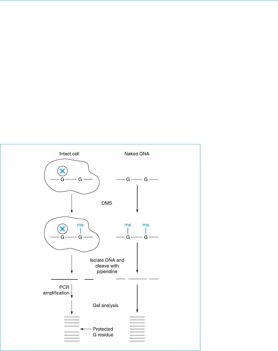
These problems are overcome by the technique of in vivo footprinting,
which is an extension of the in vitro DMS protection footprinting technique
described in section 2.2.2. Thus intact cells are freely permeable to DMS
which can therefore be used to methylate the DNA withi n its native chromatin
structure in such cells. Exactly as in the in vitro technique, G residues, to which
a protein has bound, will be protected from such methylation and will there-
fore not be cleaved when the DNA is subsequently isolated and treated with
piperidene. Hence the bands produced by cleavage at these residues will be
absent when the pattern produced by intact chromatin is compared to that
produced by naked DNA (Fig. 2.8).
METHODS FOR STUDYING TRANSCRIPTION FACTORS 33
Figure 2.8
In vivo footprinting using
the methylation
protection assay in which
specific G residues are
protected by bound
protein (X) from
methylation by DMS
treatment of intact cells.
Hence following DNA
isolation, cleavage of
methylated G residues
with piperidine and
subsequent amplification
by the polymerase chain
reaction (PCR), the band
corresponding to
cleavage at this protected
residue will be absent. In
contrast, cleavage at this
position will be observed
in naked DNA where no
protein protects this
residue from methylation.

Obviously the amounts of any specific DNA sequence obtained from total
chromatin in this procedure are vanishingly small compared to when a cloned
DNA fragment is used in the in vitro procedure. It is thus necessary to amplify
the DNA of interest from within total chromatin by the polymerase chain
reaction in order to obtain sufficient material for analysis by this method.
When this is done, however, in vivo footprinting provides an excellent
means for analysing DNA–protein contacts within intact cells in vivo as well
as determining the changes in such contacts which occur in response to
specific treatmen ts (see Herrera et al., 1989; Mueller and Wold, 1989 for
examples of this approach and Spiro and McMurray, 1999 for a full descrip-
tion of the methodologies involved).
Taken together, therefore, the three m ethods of DNA mobility shift, foot-
printing and methylation interference can provide considerable information
on the nature of the interaction between a particular DNA sequence and a
transcription factor. They serve as an essential prelude to a detailed study of
the transcription factor itself.
2.3 METHODS FOR PURIFYING AND/OR CLONING
TRANSCRIPTION FACTORS
2.3.1 PROTEIN PURIFICATION
As discussed above, once a particular DNA sequenc e has been shown to be
involved in transcriptional regulatio n, a number of techniques are available
for characterizing the binding of transcription factors to this sequence.
Although such studies can be carried out on crude cellular extracts containing
the protein, ultimately they need to be supplemented by studies on the pro-
tein itself. This can be achieved by purifying the transcription factor from
extracts of cells containing it. Unfortunately, however, conventional protein
purification techniques such as conventional chromatography and high pres-
sure liquid chromatography (HPLC) result in the isolation of transcription
factors at only 1–2% purity (Kadonaga and Tjian, 1986).
To overcome this problem and purify the transcription factor Sp1,
Kadonaga and Tjian (1986) devised a method involving DNA affinity chroma-
tography. In this method (Fig. 2.9), a DNA sequence containing a high affinity
binding site for the transcription factor is synthesized and the individual
molecules joined to form a multimeric molecule. This very high affinity bind-
ing site is then coupled to an activated sepharose support on a column and
total cellular protein passed down the column. The Sp1 protein binds speci-
fically to its corresponding DNA sequence while all other cellular proteins do
34 EUKARYOTIC TRANSCRIPTION FACTORS

not bind. The bound Sp1 can be eluted simply by raising the salt concentra-
tion. Two successive affinity chroma tography steps of this type successfully
resulted in the isolation of Sp1 at 90% purity, 30% of the Sp1 in the original
extract being recovered, representing a 500–1000-fold purification (Kadonaga
and Tjian, 1986).
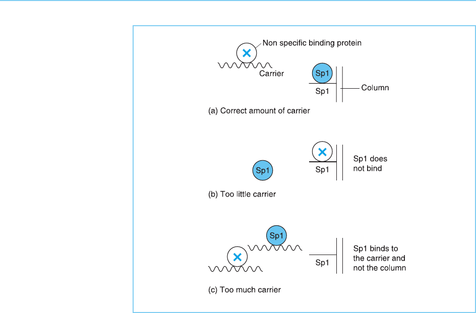
Although this simple one step method was successful in this case, it relies
critically on the addition of exactly the right amount of non-specific DNA
carrier to the cell extract. Thus this added carrier acts to remove proteins
which bind to DNA in a non-sequence specific manner and which would
hence bind non-specifically to the Sp1 affinity column and contaminate the
resulting Sp1 preparation. This contamination will occur if too little carrier is
added. If too much carrier is added, however, it will bind out the Sp1 since,
like all sequence specific proteins, Sp1 can bind with low affinity to any DNA
sequence. Hence in this case no Sp1 will bind to the column itself (Fig. 2.10).
To overcome this problem Rosenfeld and Kelley (1986) devised a method
in which proteins capable of binding to DNA with high affinity in a non-
sequence specific manner are removed prior to the affinity column. To do
this the bulk of cellular protein was removed on a Biorex 70 high capacity ion
METHODS FOR STUDYING TRANSCRIPTION FACTORS 35
Figure 2.9
Purification of
transcription factor Sp1
on an affinity column in
which multiple copies of
the DNA sequence
binding Sp1 have been
coupled to a sepharose
support (Kadonaga and
Tjian, 1986).
exchange column and proteins which can bind to any DNA with high affinity
were then removed on a cellulose column to which total bacterial DNA had
been bound. Subsequently the remaining protei ns which had bound to non-
sequence specific DNA only with low affinity were applied to a column con-
taining a high affinity binding site for transcription factor NF-1 (Fig. 2.11).
NF-1 bound to this site with high affinity and could be eluted in essentially
pure form by raising the salt concentration (Table 2.1). It should be noted
that in this and other purification procedures the fractions containing the
transcription factor can readily be identified by carrying out a DNA
mobility shift or footprinting assay with each fraction using the specific
DNA binding site of the transcription factor.
The purified protein obtained in this way can obviously be used to char-
acterize the protein, for example, by determin ing its molecular weight or by
raising an antibody to it to characterize its expression pattern in different cell
types. Similarly the activity of the protein can be assessed by adding it to
cellular extracts and assessing its effect on their ability to transcribe an exo-
genously added DNA in an in vitro transcription assay. Unfortunately, how-
ever, because of the very low abundance of transcription factors in the cell,
these purification procedures yield very small amounts of protein. For exam-
36 EUKARYOTIC TRANSCRIPTION FACTORS
Figure 2.10
Consequences of adding
different amounts of non-
specific carrier DNA to
the protein passing
through the Sp1 affinity
column. If the correct
amount of non-specific
carrier is added it will bind
proteins which interact
with DNA in a non-
sequence specific manner
allowing Sp1 to bind to
the column (A). However,
addition of too little carrier
will result in non-
sequence specific
proteins binding to the
column thereby
preventing the binding of
Sp1 (B), whereas in the
presence of too much
carrier both the non-
specific proteins and Sp1
will bind to the carrier (C).

METHODS FOR STUDYING TRANSCRIPTION FACTORS 37
Figure 2.11
Purification of
transcription factor NF-1
(Rosenfeld and Kelley,
1986). Following removal
of most cellular proteins
on a Biorex 70 ion
exchange column,
proteins that bind to all
DNA sequences with high
affinity were removed on
a bacterial DNA-cellulose
column. Subsequent
application of the
remaining proteins to a
column containing the
NF-1 binding site results
in the purification of NF-1
since it is the only protein
which binds with low
affinity to random DNA
but with high affinity to an
NF-1 site.
Table 2.1
Purification of transcription factor NF1 from HeLa cells
Total protein
(mg)
Specific binding of
32
P DNA (fmol/
mg protein) 10
3
Purification
(fold)
Yield
(%)
HeLa cell
extract*
4590 3.1 1.0 100
Biorex 70 column 550 27.1 8.7 104
E. coli DNA
cellulose
65.2 181 58.4 83
NF1 affinity
matrix
1st passage 2.1 4510 1455 67
2nd passage 1.1 7517 2425 57
* Prepared from 6 10
10
cells or 120 g cells

ple Treisman (1987) succeeded in purifying only 1.6 g of the serum response
factor starting with 2 10
10
cells or 40 g of cells. Such difficu lties clearly limit
the experiments that can be done with purified material. Indeed, the primary
use of purified factor in most cases has simply been to provide material to
isolate the gene encoding the protein. This gene can then be expressed either
in vitro or in bacteria to provide a far more abundant source of the corre-
sponding protein than could be obtained from cells that naturally express it.
2.3.2 GENE CLONING
Several methods are available for cloning the gene encoding a particular
transcription factor and these will be discussed in turn.
(a) Use of oligonucleotide probes predicted from the protein
sequence of the factor
If a particular transcription factor has been purified, it is possible to obtain
portions of its amino acid sequence. In turn, such sequences can be used to
predict oligonucleotides containing DNA sequences capable of encoding
these protein fragments. Due to the redundancy of the genetic code, whereby
several different DNA codons can encode a particular amino acid, there will
be multiple different oligonucleotides capable of encoding a particular amino
acid sequence. All these possible oligonucleotides are synthesized chemi cally,
made radioactive and used to screen a cDNA library prepared from mRNA
isolated from a cell type expressing the factor. The oligonucleotide in the
mixture which does correspo nd to the transcription factor amino acid
sequence will hybridize to the corresponding sequence in a cDNA clone
derived from mRNA encoding the factor. Hence such a clone can be readily
identified in the cDNA library (Fig. 2.12).
In cases where purified protein is available as in those discussed in the
previous section, this approach repres ents a relatively simple method for iso-
lating cDNA clones. It has therefore been widely used to isolate cDNA clones
corresponding to purified factors such as Sp1 (Kadonaga et al., 1987: Fig.
2.12), NF1 (Santoro et al., 1988) and the serum response factor (Norman
et al., 1988) (for methodological details see Nicolas et al., 1999).
(b) Use of oligonucleotide probes derived from the DNA binding site
of the f actor
Although relatively simple, the use of oligonucleotides derived from protein
sequences does require purified protein. As we have seen, purification of a
transcription factor requires a vast quantity of cells and is technically difficult.
38 EUKARYOTIC TRANSCRIPTION FACTORS
