Latchman. Eukariotic Transciption Factors
Подождите немного. Документ загружается.


Black
p
late
(
2,1
)
[13:25 16/10/03 N:/4056 LATCHMAN.751/0124371787 Eukaryotic/application/Colour-Plates.3d] Ref: 4056 Auth: Latchman Title: Eukaryotic Colour plate Page: 2 1-4
Plate 2 Binding of the
al (blue)/2 (red)
homeodomain heterodimer
to DNA. -helices are
shown as cylinders. Note
the three helical structure
of the homeodomains of
a1 and 2 and the C-
terminal region of 2
which forms an additional
-helix in the presence of
a1 and packs against the
2 homeodomain forming
the dimerization interface.
Plate 3 Structure of the
Cys
2
His
2
zinc finger
from Xfin. The Cys
residues are shown in
yellow and the His
residues in dark blue.

Black
p
late
(
3,1
)
[13:26 16/10/03 N:/4056 LATCHMAN.751/0124371787 Eukaryotic/application/Colour-Plates.3d] Ref: 4056 Auth: Latchman Title: Eukaryotic Colour plate Page: 3 1-4
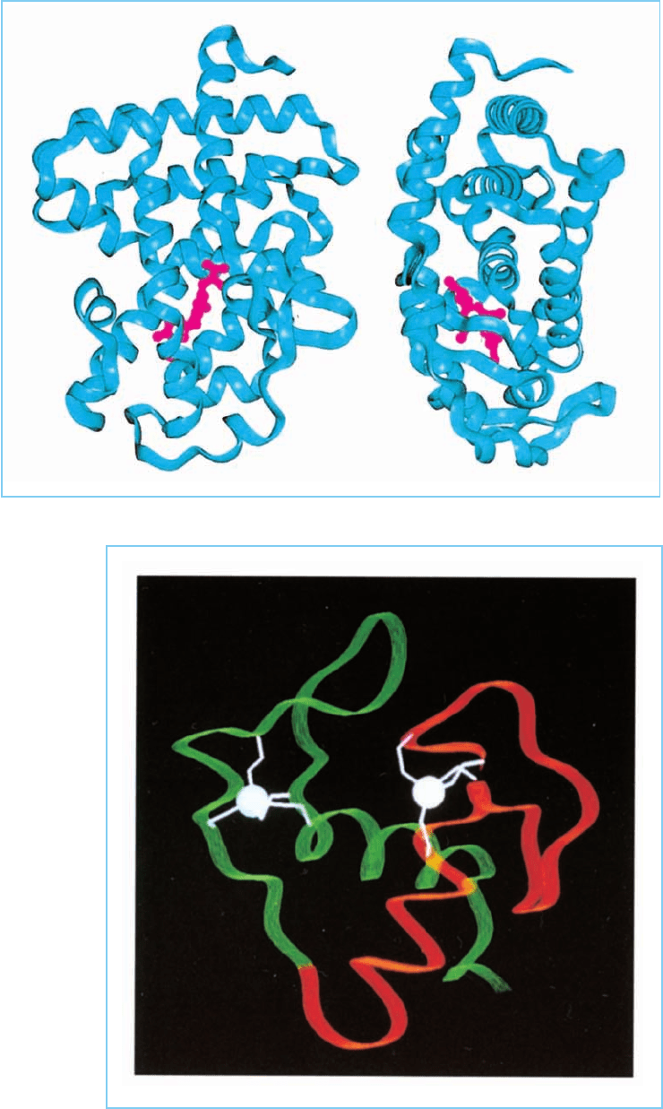
Plate 4 Two views
of the structure of the
thyroid hormone receptor
ligand binding domain
(blue) with bound thyroid
hormone ligand (red).
Note that the ligand is
completely buried in the
interior of the protein.
Plate 5 Structure of
the two Cys
4
zinc
fingers in a single
molecule of the
glucocorticoid receptor.
The first finger is shown
in red and the second
finger in green with the
zinc atoms shown
white.

Black
p
late
(
4,1
)
[13:26 16/10/03 N:/4056 LATCHMAN.751/0124371787 Eukaryotic/application/Colour-Plates.3d] Ref: 4056 Auth: Latchman Title: Eukaryotic Colour plate Page: 4 1-4
(7a) (7b)
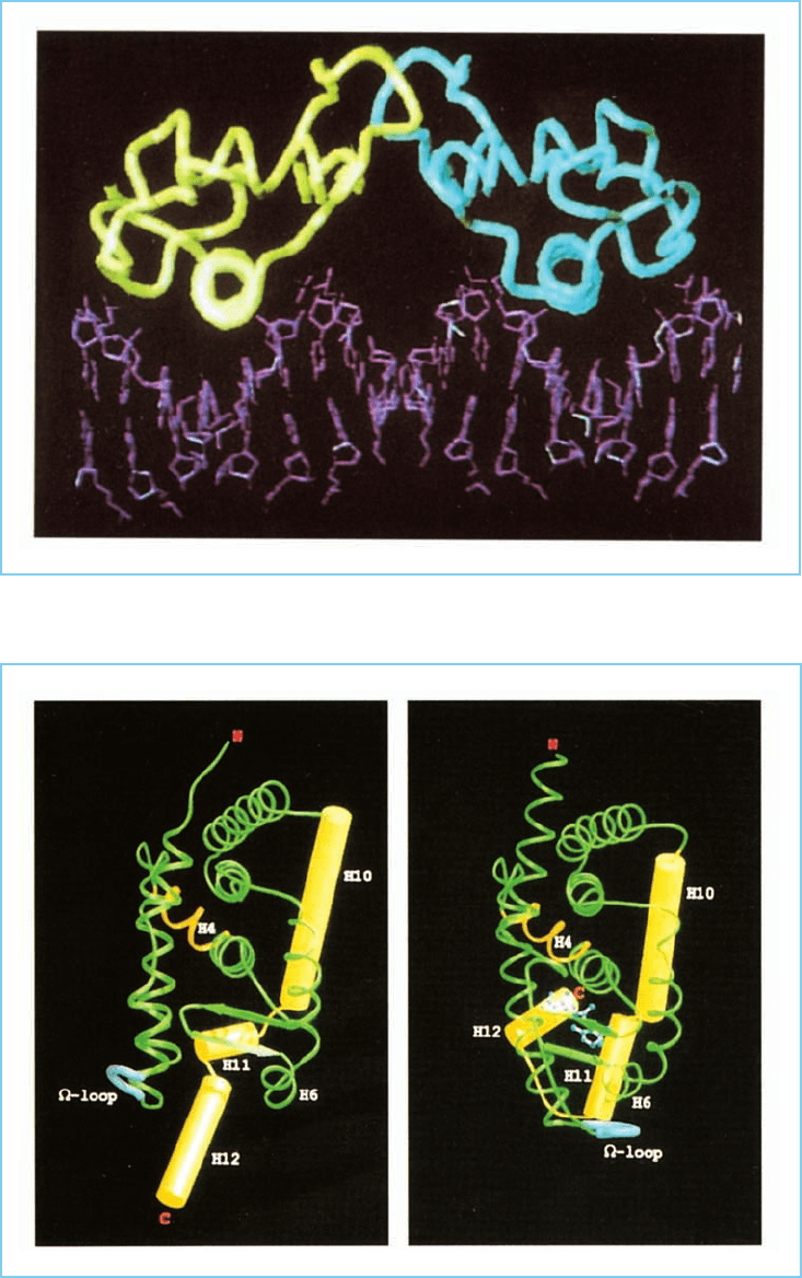
Plate 6 Structure of the
oestrogen receptor dimer
consisting of two receptor
molecules bound to DNA.
The two molecules of the
receptor are shown green
and blue respectively and
the DNA is shown in
purple.
Plate 7 Structure of (a)
the RXR receptor in the
absence of ligand and (b)
the closely related RAR
receptor following binding
of ligand (light blue atoms
joined by white bonds).
Note the structural
change induced by the
binding of ligand involving
the movement of the
H12 helix towards the
ligand binding core so
creating a sealed pocket
in which the ligand is
trapped.
