Latchman. Eukariotic Transciption Factors
Подождите немного. Документ загружается.

However, the multi-cysteine finger cannot be converted into a functional
cysteine-histidine finger by substituting two of its cysteine residues with histi-
dines indicating that the two types of finger are functionally distinct (Green
and Chambon, 1987). Moreover, unlike the cysteine-histidine zinc finger
which is present in multiple copies within the proteins which contain it, the
unit of two multi-cysteine fingers present in the steroid receptors is found
only once in each receptor. Interestingly, structural studies of the two multi-
cysteine fingers in the glucocorticoid and oestrogen receptors (for review see
Schwabe and Rhodes, 1991; Klug and Schwabe, 1995) have indicated that the
two fingers form one single structural motif consisting of two alpha helices
perpendicular to one another with the cysteine-zinc linkage holding the base
of a loop at the N terminus of each helix (Fig. 4.31; see Plate 5; Hard et al.,
1990). This is quite distinct from the modular structure of the two cysteine
two histidine finger where each finger constitutes an independent structural
element whose configuration is unaffected by the presence or absence of
adjacent fingers.
110 EUKARYOTIC TRANSCRIPTION FACTORS
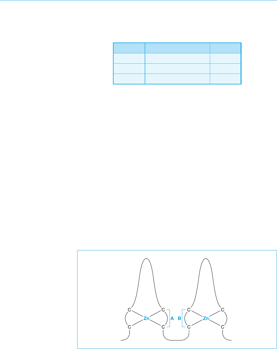
Figure 4.30
Schematic representation
of the four cysteine zinc
finger. Regions labelled A
and B are of critical
importance in determining
respectively the DNA
sequence which is bound
by the finger and the
optimal spacing between
the two halves of the
palindromic sequence
which is recognized.
Table 4.3
Transcriptional regulatory proteins with multiple cysteine
fingers
Finger type Factor Species
Cys
4
–Cys
5
Steroid, thyroid receptors Mammals
Cys
4
E1A Adenovirus
Cys
6
Gal4, PPRI, LAC9 Yeast
Thus, although these two DNA binding motifs are similar in their coordi-
nation of zinc, they differ in the lack of histidines and of the conserved
phenylalanine and leucine residues in the multi-cysteine finger, as well as
structurally. It is clear therefore that they represent distinct functional
elements and are unlikely to be evolutionarily related (for review see
Schwabe and Rhodes, 1991; Rhodes and Klug, 1993; Klug and Schwabe,
1995).
Whatever the precise relationship between these motifs, it is clear that the
multi-cysteine finger mediates the DNA binding of the nuclear receptors.
Thus mutations which eliminate or alter critical amino acids in this motif
interfere with DNA binding by the receptor (Fig. 4.32).
The role of the cysteine fingers in mediating DNA binding by the nuclear
receptors can also be demonstrated by taking advantage of the observation
that the different steroid receptors bind to distinct but related palindromic
sequences in the DNA of hormone responsive genes (see Khorasanizadeh and
Rastinejad, 2001 for review and Table 4.2 for a comparison of these binding
sites). Thus, if the cysteine-rich region of the oestrogen receptor is replaced by
that of the glucocorticoid receptor, the resulting chimaeric receptor has the
DNA binding specificity of the glucocorticoid receptor but continues to bind
oestrogen since all the other regions of the molecule are derived from the
oestrogen receptor (Green and Chambon, 1987; Fig. 4.33). Hence the DNA
binding specificity of the hybrid receptor is determined by its cysteine-rich
region, resulting in the hybrid receptor inducing the expression of gluco-
corticoid responsive genes (which carry its DNA binding site) in response
to oestrogen (to which it binds).
These so-called ‘finger swop’ experiments therefore provide further evi-
dence in favour of the critical role for the multi-cysteine fingers in DNA
binding, exchanging the fingers of two receptors exchanging the DNA bind-
FAMILIES OF DNA BINDING TRANSCRIPTION FACTORS 111
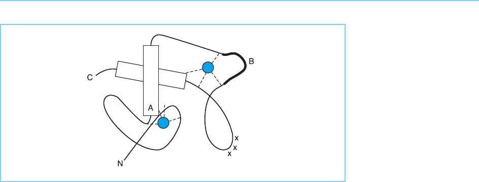
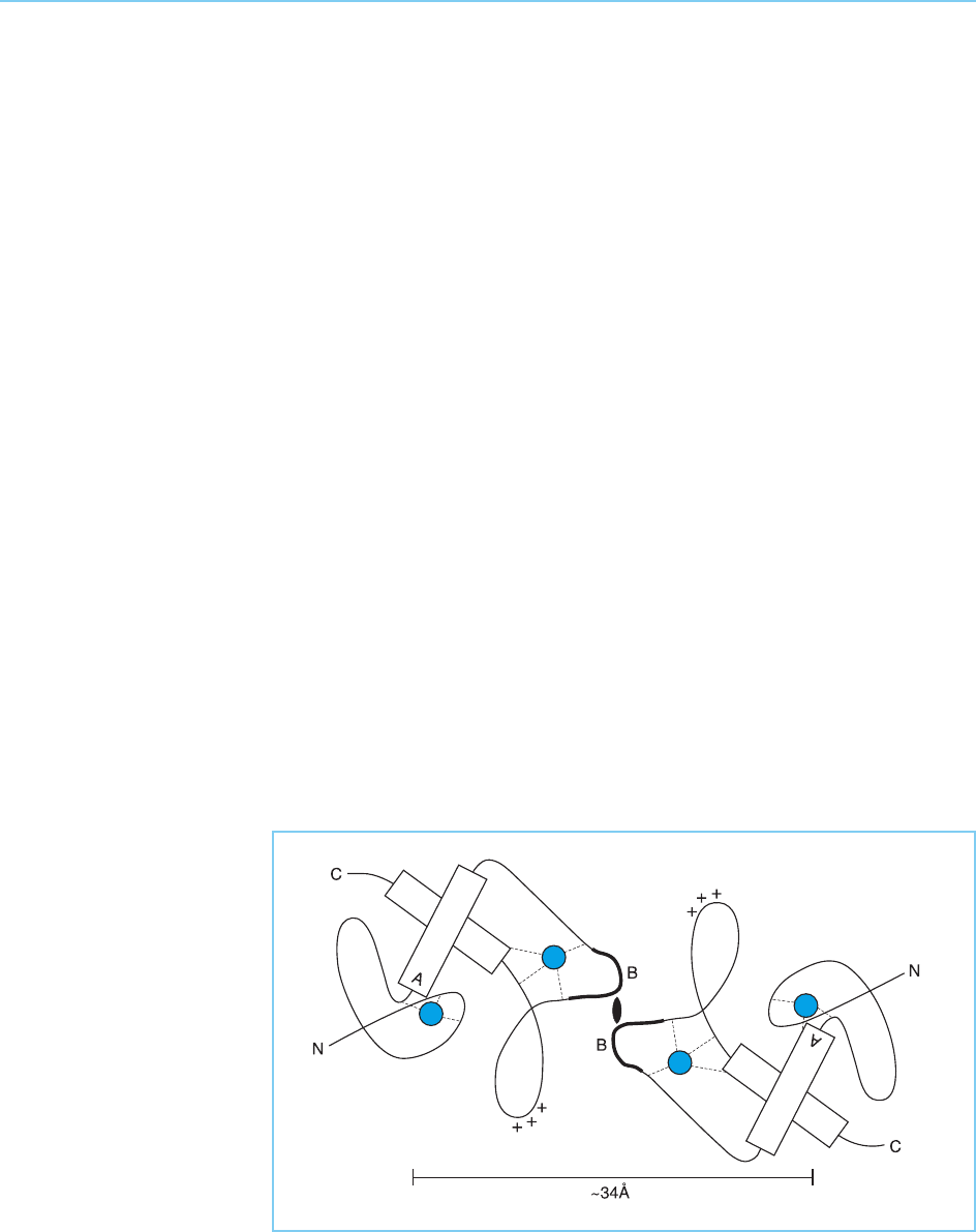
Figure 4.31
Schematic model of a pair
of zinc fingers in a single
molecule of the oestrogen
receptor. Note the helical
regions (indicated as
cylinders) with the critical
residues for determining
the DNA sequence which
is bound located at the
terminus of the
recognition helix (indicated
as A), the zinc atoms
(blue), conserved basic
residues (+++) and the
region that interacts with
another receptor molecule
and determines the
optimal spacing between
the two halves of the
palindromic sequence that
is recognized (indicated as
B). Note that A and B
indicate the same regions
as in Figure 4.30.
112 EUKARYOTIC TRANSCRIPTION FACTORS
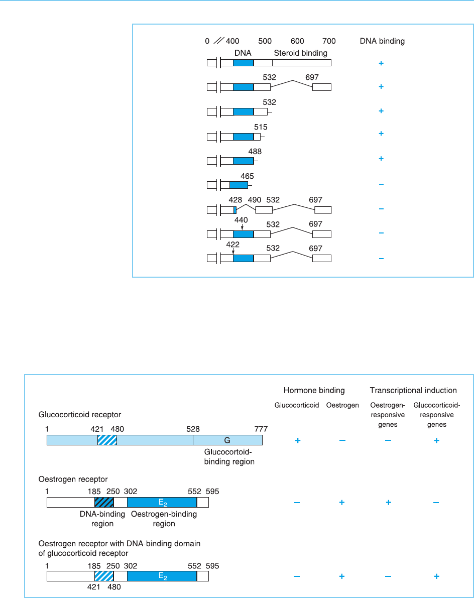
Figure 4.33
Effect of exchanging the
DNA binding domain
(shaded) of the oestrogen
receptor with that of the
glucocorticoid receptor on
the binding of hormone
and gene induction by the
hybrid receptor.
Figure 4.32
Effect of various deletions
or mutations on the DNA
binding of the
glucocorticoid receptor.
Note that DNA binding is
only prevented by
deletions that include part
of the DNA binding
domain (shaded) or by
mutations within it
(arrows), but not by
deletions in other regions
such as the steroid-
binding domain. Numbers
indicate amino acid
residues.
ing specificity. In addition, however, because of the existence of short distinct
DNA binding regions of this type in receptors which bind to distinct but
related DNA sequences, they provide a unique opportunity to dissect the
elements in a DNA binding structure which mediate binding to specific
sequences.
Thus by exchanging one or more amino acids between two different recep-
tors it is possible to investigate the effects of these changes on DNA binding
specificity and hence elucidate the role of individual amino acid differences in
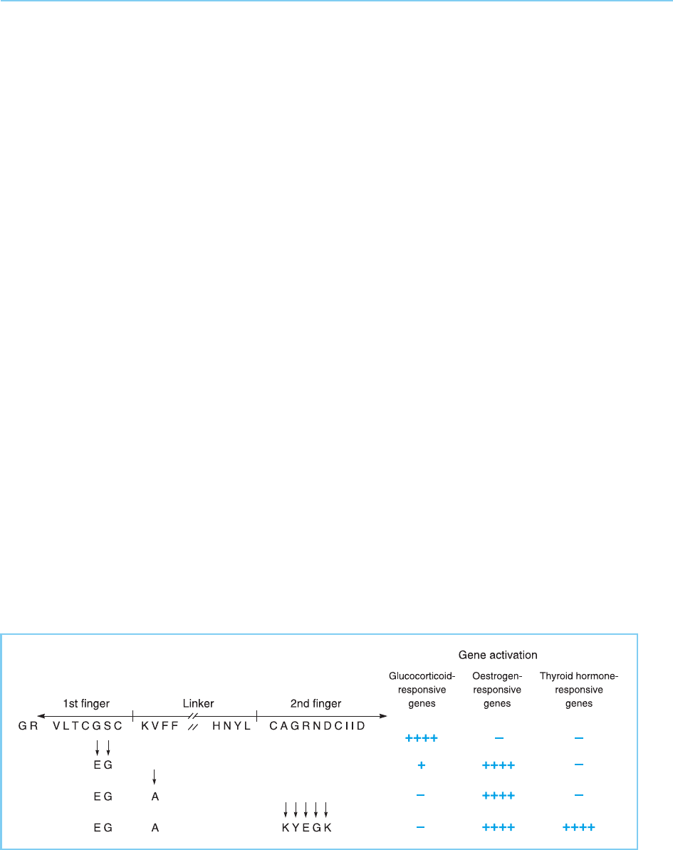
producing the different patterns of sequence specific binding. For example,
the alteration of the two amino acids between the third and fourth cysteines of
the N terminal finger in the glucocorticoid receptor for their equivalents in
the oestrogen receptor changes the DNA binding specificity of the chimaeric
receptor to that of the oestrogen receptor (Umesono and Evans, 1989;
Fig. 4.34). Hence the exchange of two amino acids in a critical region of a
protein of 777 amino acids (indicated as A in Fig. 4.30) can completely change
the DNA binding specificity of the glucocorticoid receptor resulting in it
binding to and activating genes that are normally oestrogen responsive. The
specificity of this hybrid receptor for such oestrogen responsive genes can be
further enhanced by exchanging another amino acid located between the two
fingers (Fig. 4.34) indicating that this region also plays a role in controlling the
specificity of DNA binding.
As noted above (section 4.4.1), the steroid receptors bind to palindromic
recognition sequences within DNA, with the receptor binding to DNA as a
homodimer in which each receptor molecule interacts with one half of the
palindrome. In addition to differences in the actual sequence recognized,
steroid/thyroid hormone receptors can also differ in the optimal spacing
between the two separate halves of the palindromic DNA sequence that is
recognized (see Table 4.2a). Thus the oestrogen receptor and the thyroid
FAMILIES OF DNA BINDING TRANSCRIPTION FACTORS 113
Figure 4.34
Effect of amino acid
substitutions in the zinc
finger region of the
glucocorticoid receptor
on the ability to bind to
and activate genes that
are normally responsive
to different steroid
hormones.
hormone receptor both recognize the identical palindromic sequence in the
DNA but differ in that in the thyroid receptor binding sites the two halves of
the palindrome are adjacent whereas in the oestrogen receptor binding sites
they are separated by three extra bases. The further alteration of the chimae-
ric receptor illustrated in Figure 4.34 by changing five amino acids in the
second finger to their thyroid hormone receptor equivalents is sufficient to
allow the receptor to recognize thyroid hormone receptor binding sites
(Umesono and Evans, 1989; Fig. 4.34). These amino acids in the second finger
(indicated as B in Fig. 4.30) appear to play a critical role therefore in deter-
mining the optimal spacing of the palindromic sequence that is recognized.
As discussed above, structural studies of the two zinc fingers in the oestro-
gen and glucocorticoid receptors suggest that they form a single structural
motif with two perpendicular alpha helices (see Fig. 4.31). In this structure,
the critical amino acids for determining the spacing in the palindromic
sequence recognized are located on the surface of the molecule allowing
them to interact with equivalent residues on another receptor monomer dur-
ing dimerization (indicated as B in Fig. 4.35; see Plate 6; Schwabe et al., 1993).
Hence differences in the interaction of these regions in the different recep-
tors determine the spacing of the two monomers within the receptor dimer
and thus the optimal spacing in the palindromic DNA sequence that is
recognized.
Interestingly, within this structure, the critical residues for determining the
precise DNA sequence that is recognized are located at the N terminus of the
first alpha helix (indicated as A in Fig. 4.31 and Fig. 4.35), further supporting
the critical role of such helices in DNA binding. Moreover, in the proposed
114 EUKARYOTIC TRANSCRIPTION FACTORS
Figure 4.35
Interaction of two
oestrogen receptor
molecules to form a DNA
binding dimer. Compare
with Figure 4.31 and note
the interaction of the B
regions on each molecule.
The resulting dimer has a
spacing of 34 Angstroms
between the two DNA-
binding regions allowing
binding in successive
major grooves of the DNA
molecule.
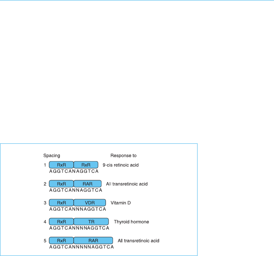
structure of the oestrogen receptor dimer, the DNA binding helices in each
monomer will be separated by 34 Angstroms allowing each of these recogni-
tion helices to make sequence specific contacts in adjacent major grooves of
the DNA molecule.
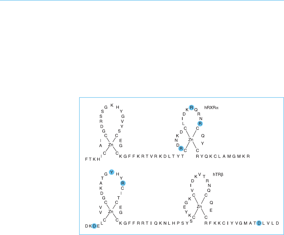
Differences in the DNA binding domain also regulate the binding of
members of the nuclear receptor family to directly repeated sequences
with different spacings between the two halves of the repeat (see Table
4.2b). Thus, when the direct repeats are separated by only one base, they
can bind a homodimer of the retinoid X-receptor (RXR) and hence confer
a response to 9-cis retinoic acid which binds to this receptor (Fig. 4.36). In
contrast the RXR homodimer cannot bind to the direct repeats when they are
separated by between two and five base pairs. Rather, on these elements RXR
forms a heterodimer with other members of the nuclear receptor family
(Fig. 4.36).
Moreover, the nature of the heterodimers that form on a particular
response element controls the response it mediates with the nature of the
non-RXR component determining the response. Thus a spacing of two or five
base pairs binds a heterodimer of RXR and the retinoic acid receptor (RAR)
and therefore mediates responses to all transretinoic acid that binds to RAR.
In contrast, a spacing of four base pairs binds a heterodimer of RXR and the
thyroid hormone receptor (TR) and therefore can mediate responses to
thyroid hormone.
FAMILIES OF DNA BINDING TRANSCRIPTION FACTORS 115
Figure 4.36
Binding of different
nuclear receptor
heterodimers to directly
repeated elements with
different spacings
between the repeats
determines the response
mediated by each
element.
As on the palindromic repeats, it is the DNA binding domain of the recep-
tors that controls which heterodimers can form on particular spacings of the
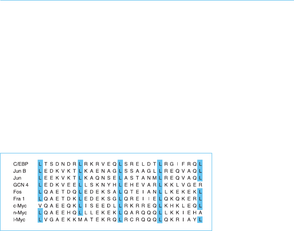
direct repeat. Interestingly, the crystal structure of the RXR-TR heterodimer
bound to a direct repeat with a four base spacing indicates that the dimeriza-
tion interface involves amino acids in the first finger of the thyroid hormone
receptor and the second finger of RXR rather than only residues in the
second finger as occurs for homodimerization of receptors on palindromic
repeats (Rastinejad et al., 1995) (Fig. 4.37).
The definition of the DNA binding domain of the nuclear receptors as a
short sequence containing two multi-cysteine fingers has therefore allowed
the elucidation of the features in this motif which mediate the different
sequence specificities of the different receptors and their relationship to the
structure of the motif. In particular, a helical region of the first finger plays a
critical role in determining the precise DNA sequence that is recognized by
binding in the major groove of the DNA. Similarly, other regions in either the
first or second fingers control the spacing of adjacent palindromic or directly
repeated sequences which is optimal for the binding of receptor homo- or
heterodimers by interacting with another receptor monomer and hence
affecting the structure of the receptor dimer that forms.
116 EUKARYOTIC TRANSCRIPTION FACTORS
Figure 4.37
Zinc fingers in the
retinoid X-receptor and
the thyroid hormone
receptor . The residues
in each receptor that are
involved in heterodimer
formation with the other
receptor are indicated.
4.5 THE BASIC DNA BINDING DOMAIN
4.5.1 THE LEUCINE ZIP PER AND THE BASIC DNA BINDING
DOMAIN
As discussed in the preceding sections of this chapter, the study of motifs
common to several different transcription factors has led to the identification
of the role of these motifs in DNA binding. A similar approach led to the
identification of the leucine zipper motif (for reviews see Lamb and
McKnight, 1991; Hurst, 1996; Kerppola and Curran, 1995). Thus this struc-
ture has been detected in several different transcription factors such as the
CAAT box binding protein C/EBP, the yeast factor GCN4 and the oncogene
products Myc, Fos and Jun (see Chapter 9, sections 9.3.1 and 9.3.3). It consists
of a leucine-rich region in which successive leucine residues occur every
seventh amino acid (Fig. 4.38).
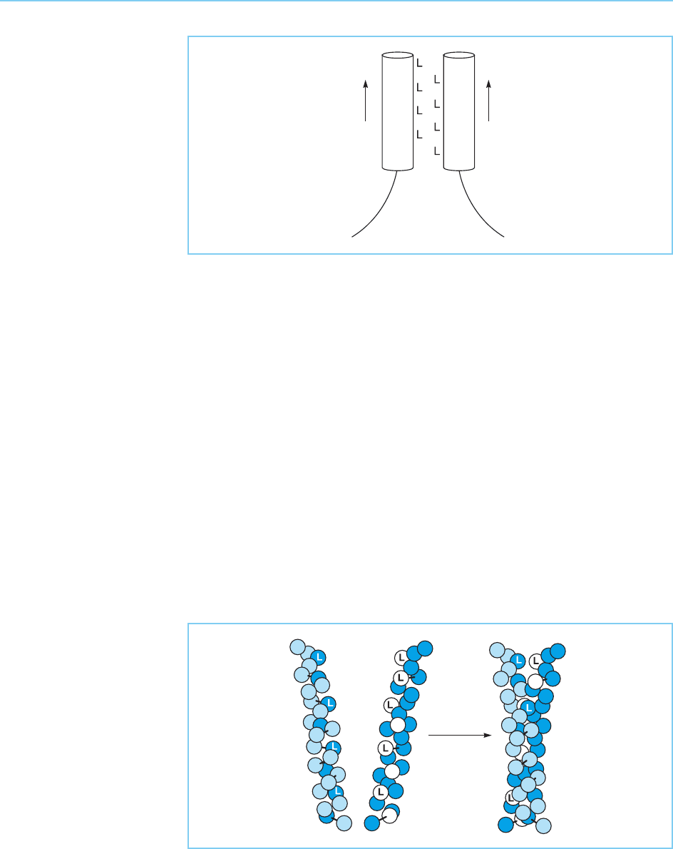
In all these cases, the leucine-rich region can be drawn as an alpha-helical
structure in which adjacent leucine residues occur every two turns on the
same side of the helix. Moreover, these leucine residues appear to play a
critical role in the functioning of the protein. Thus, with one exception (a
single methionine in the Myc protein), the central leucine residues of the
motif are conserved in all the factors that contain it (Fig. 4.38). It was there-
fore proposed (Landshultz et al., 1988) that the long side chains of the leucine
residues extending from one polypeptide would interdigitate with those of
the analogous helix of a second polypeptide, forming a motif known as the
leucine zipper which would result in the dimerization of the factor (Fig. 4.39).
This effect could also be achieved by a methionine residue which, like leucine,
has a long side chain with no lateral methyl groups but not by other hydro-
phobic amino acids such as valine or isoleucine which have methyl groups
extending laterally from the beta carbon atom.
In agreement with this idea, substitutions of individual leucine residues in
C/EBP or other leucine zipper-containing proteins such as Myc, Fos and Jun
FAMILIES OF DNA BINDING TRANSCRIPTION FACTORS 117
Figure 4.38
Alignment of the leucine-
rich region in several
cellular transcription
factors. Note the
conserved leucine
residues (L) which occur
every seven amino acids.
with isoleucine or valine, abolish the ability of the intact protein to form a
dimer, indicating the critical role of this region in dimerization. A comparison
of the effects of various mutations of this type on the ability of the mutant
protein to dimerize, suggested that the two leucine-rich regions associate in a
parallel manner with both helices oriented in the same direction (as illustrated
in Fig. 4.39) rather than in an anti-parallel configuration as originally sug-
gested (Landshultz et al., 1989). This idea was confirmed by structural studies
of the leucine zipper regions in GCN4 and in the Fos/Jun dimer bound to
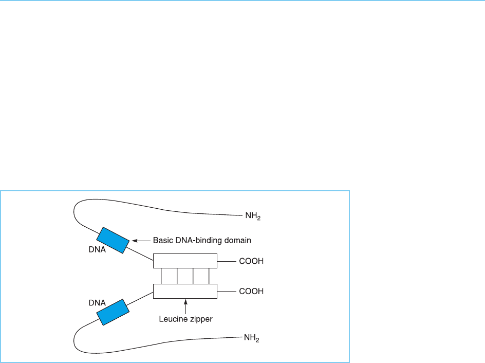
DNA (Glover and Harrison, 1995). These studies indicated that each zipper
motif forms a right-handed alpha-helix with dimerization occurring via the
association of two parallel helices that coil around each other to form a coiled
coil motif similar to that found in fibrous proteins such as the keratins and
myosins (Fig. 4.40).
In addition to its role in dimerization, the leucine zipper is also essential for
DNA binding by the intact molecule. Thus mutations in the zipper which
118 EUKARYOTIC TRANSCRIPTION FACTORS
Figure 4.39
Model of the leucine
zipper and its role in the
dimerization of two
molecules of a
transcription factor.
Figure 4.40
Coiled coil structure of
the leucine zipper formed
by two helical coils
wrapping around each
other. L indicates a
leucine residue.
prevent dimerization also prevent DNA binding from occurring (Landshultz
et al., 1989). Unlike the zinc finger or helix-turn-helix motifs, however, the
zipper is not itself the DNA binding domain of the molecule and does not
directly contact the DNA. Rather it facilitates DNA binding by an adjacent
region of the molecule which in C/EBP, Fos and Jun is rich in basic amino
acids and can therefore interact directly with the acidic DNA. The leucine
zipper is believed therefore to serve an indirect structural role in DNA bind-
ing, facilitating dimerization which in turn results in the correct positioning of
the two basic DNA binding domains in the dimeric molecule for DNA binding
to occur (Fig. 4.41).
In agreement with this idea mutations in the basic domain abolish the
ability to bind to DNA without affecting the ability of the protein to dimerize
as expected for mutations that directly affect the DNA binding domain
(Landshultz et al., 1989). Similarly, exchange of the basic region of GCN4
for that of C/EBP results in a hybrid protein with the DNA binding specificity
of C/EBP while exchange of the leucine zipper region has no effect on the
DNA binding specificity of the hybrid molecule (Fig. 4.42).
Hence the DNA binding specificity of leucine zipper-containing transcrip-
tion factors is determined by the sequence of their basic domain with the
leucine zipper allowing dimerization to occur and hence facilitating DNA
binding by the basic domain. As expected from this idea, the basic DNA
binding domain can interact with DNA in a sequence specific manner in
the absence of the leucine zipper if it is first dimerized via an intermolecular
disulphide bond (Fig. 4.43). Interestingly, the basic DNA binding domain can
bind to DNA as a monomer in the case of the Skn-1 factor which lacks a
leucine zipper (Blackwell et al., 1994). In this factor, however, the basic
FAMILIES OF DNA BINDING TRANSCRIPTION FACTORS 119
Figure 4.41
Model for the structure of
the leucine zipper and
the adjacent DNA
binding domain following
dimerization of the
transcription factor
C/EBP.
