Kaplan Cynthia G., MD Color Atlas of Gross Placental Pathology
Подождите немного. Документ загружается.

38 Chapter 3 Umbilical Cord
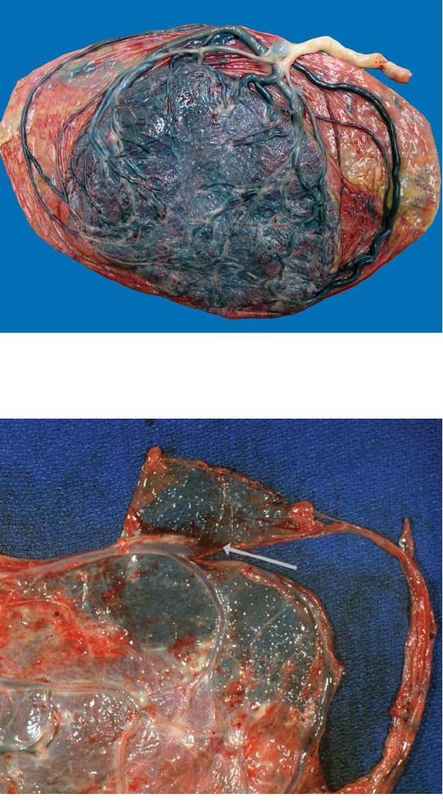
Figure 3.23. Cord
insertion into the
free membranes
(velamentous
insertion) occurs
in about 1% of
deliveries. The
vessels must be
carefully examined
for integrity
and extent of
membranous
passage. Old
hemorrhage behind
the membranes is
sometimes found
where the cord
attaches.
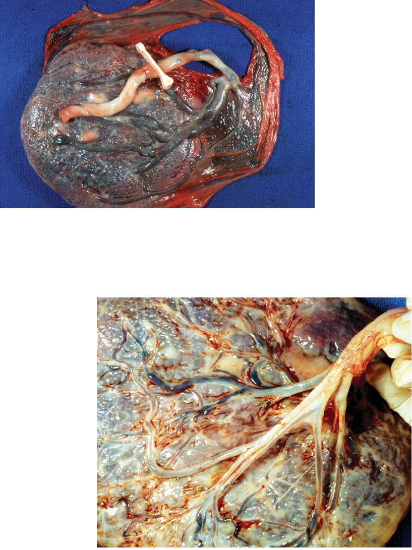
Figure 3.24.
Intrapartum rupture
of a velamentous
vessel occurred in
this case (arrow)
supported by
maternal history and
neonatal anemia.
This infant survived.
Hemorrhage from a
ruptured vessel is
usually found in the
sub-amniotic region.
Vessels may also be
torn after the infant
has been born,
during placental
delivery. While this
has no clinical
significance, it should
be noted since
complete historical
information may not
be available at the
time of placental
examination.
Insertion 39
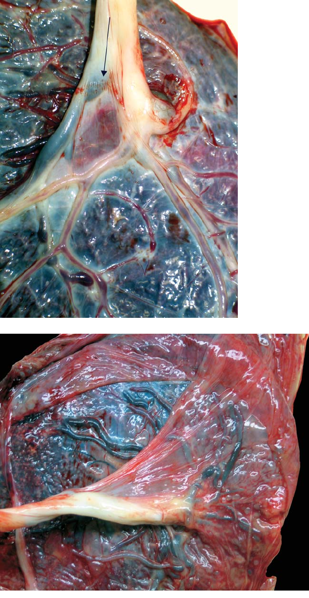
Figure 3.25. Despite the close proximity of the cord insertion and velamentous
vessels to the site of membrane rupture in this vaginal delivery, no vascular
disruption occurred. Vessels overlying the cervical os (vasa previa) are at the
greatest risk of tearing and causing catastrophic fetal blood loss. Velamentous
vessels may also be compressed during labor. There is a fleck of calcium in one
velamentous vessel which is likely old thrombosis (arrow).
The vessels in the cord may separate before it reaches the surface
(furcate). The unattached surface amnion may also attach to the cord
several centimeters before the cord reaches the placental surface (Figure
3.26 to Figure 3.29).
Figure 3.26. This
cord divides into
vessels which lose
their protective
covering of
Wharton’s jelly
above the surface of
the placenta. Such
furcate insertions
will have the risks
attendant to
velamentous vessels.
40 Chapter 3 Umbilical Cord
Figure 3.27. At times the cord is par-
tially encased by a fold of amnion at its
placental end, a “chorda” or amniotic
web. This cord is minimally furcate with
a web of amnion enclosing the vessels. A
clamp mark is present (arrow).
Figure 3.28.
Amniotic webs are
very common and
can extend for
several centimeters
up the cord. They
may be loose or
relatively tight and
bind the cord to the
fetal surface of the
placenta. If the
amnion is largely
detached from the
chorion, a web is
likely.
Infections 41
Figure 3.29. A long
tight amniotic web is
present. Webs may
limit the mobility of
the cord, potentially
compromising blood
flow. There is also
subamniotic
hemorrhage.
Although bleeding
from vessels between
the amnion and
chorion is usually
an artifact arising
during placental
delivery, it is
common when webs
are present,
suggesting abnormal
stress in this area.
Infections
The cord inflammation seen with most ascending membranous infections
(see page 56) is usually not recognizable grossly. Candida is an excep-
tion and shows characteristic micro abscesses on the cord surface (Figure
3.30, Figure 3.31). This infection may cause a rash on the infant at birth.
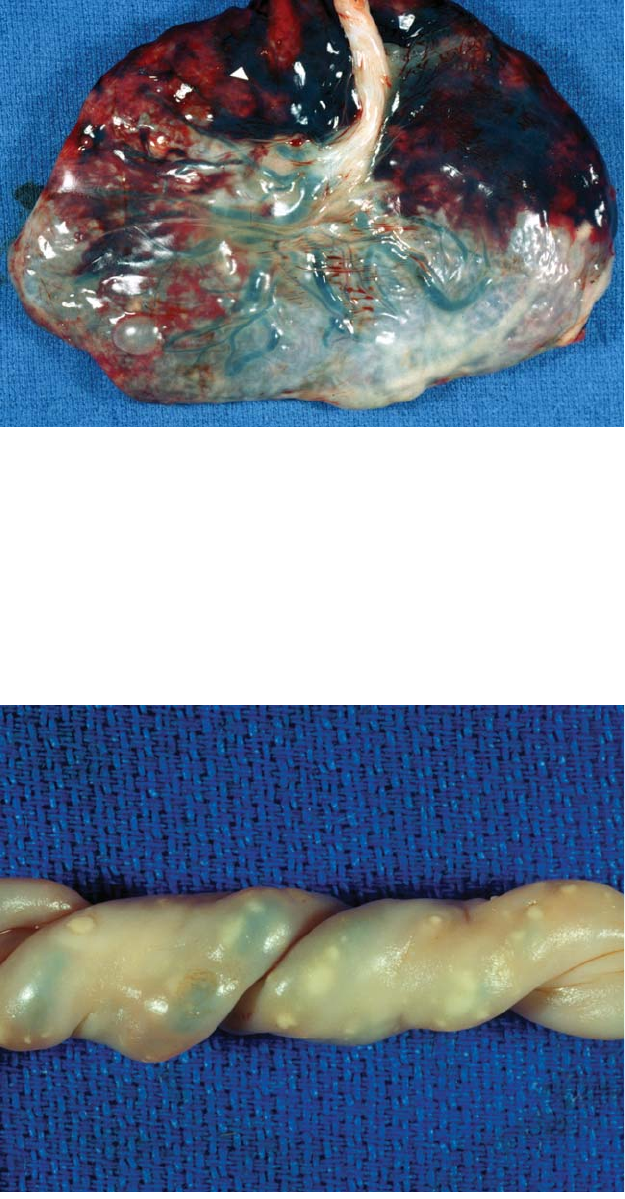
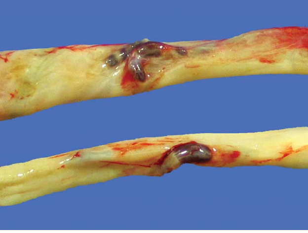
Figure 3.30.
Scattered yellow to
white 2-mm to 3-mm
plaques on the
cord are virtually
pathognomonic of
Candida funisitis.
They are not seen
elsewhere on
the placental
membranes. There is
usually an associated
chorioamnionitis.

42 Chapter 3 Umbilical Cord
A
B
Figure 3.31. (A) Histologically Candida funisitis shows micro abscesses just
under the cord surface. These are filled with necrotic debris, in which it is diffi-
cult to identify organisms, (B) numerous fungal pseudohyphae and yeasts are
present on methenamine silver stain.
Infections 43
Figure 3.32. Necrotizing funisitis represents a chronic inflammatory process in
the umbilical cord, apparently infectious in origin. There is calcification sur-
rounding the vessels leading to an extremely rigid cord. This is at times visible
along the length as a white stripe.
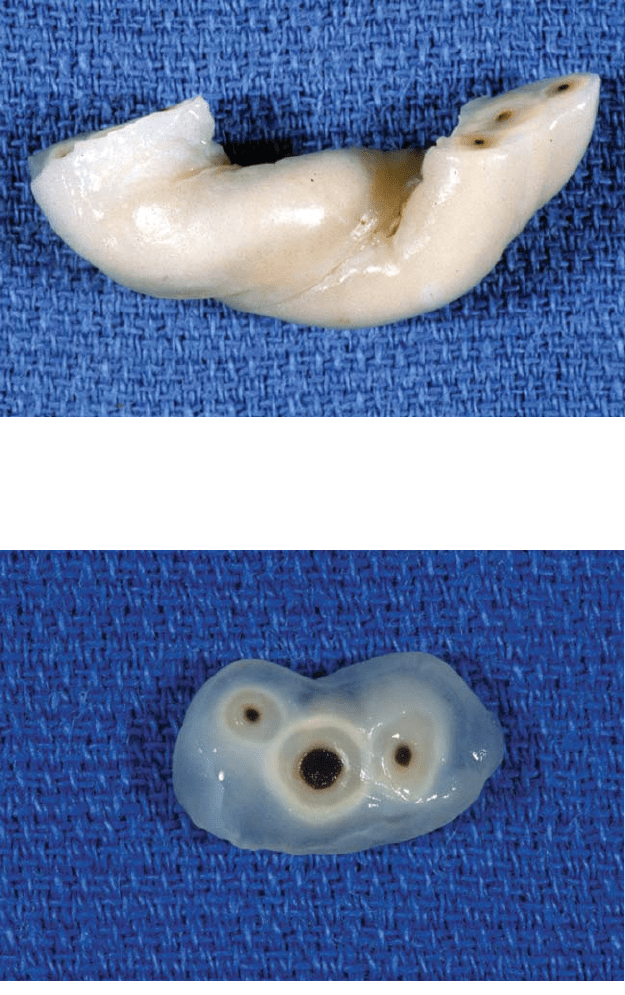
Figure 3.33. Cross-section of a cord with necrotizing funisitis shows white bands
suggesting diffusion rings surrounding each of the three vessels. These are com-
posed of necrotic inflammatory cells and calcification.
It is more likely to cause sepsis in premature infants. The “barber-pole”
configuration of chronic necrotizing funisitis is felt to represent a chronic
and sometimes healed intra-uterine infection with organisms of low vir-
ulence (Figure 3.32, Figure 3.33).
44 Chapter 3 Umbilical Cord
Ulceration
Loss of Wharton’s jelly with ulceration of the cord surface is uncommon,
but the associated infants may have significant problems. It is usually
related to bile or meconium exposure (Figure 3.34).
Figure 3.34. Ulceration of the cord occurs in some cases of upper gastrointesti-
nal tract atresia. This infant had proximal jejunal atresia. The exposed vessels
may hemorrhage leading to fetal compromise or death. The intestinal obstruc-
tions are located below the Ampulla of Vater as the bile, appears to be etiologic.
Ulceration also may occur with longstanding meconium exposure.
4
Fetal Membrances and Surface
45
Layers
The peripheral membranes and fetal placental surface are continuous,
and most processes are seen in both. The layer of membrane closest
to the fetus is amnion. External is the chorion, which is minimal on the
peripheral membranes and more extensive on the disk. The remnant of
the yolk sac lies between the amnion and chorion (Figure 4.1). The
chorion is continuous with all the villous tissue. There is close proximity
of the surface membranes to the maternal blood of the intervillous space,
while the peripheral membranes abut the decidua and its blood vessels.
This relationship permits maternal cells access to the membranes.
Subchorionic Fibrin and Hemorrhage
Deposits of fibrin from the maternal circulation and thrombosis are
common beneath the fetal surface.As pregnancy progresses, the amounts
of these materials generally increase (Figure 4.2, Figure 4.3). Subchori-
onic thrombi eventually become compacted fibrin. The quantity of sub-
chorionic fibrin has been associated with fetal activity. Large nodular
subchorionic hematomas, sometimes called “Breus moles,” are seen in
both liveborns and spontaneous abortions (Figure 4.4). Unusually thick
layers of subchorionic hemorrhage can be associated with chronic bleed-
ing and prematurity (Figures 4.5).
Extrachorial Placentation
The membranes normally insert at the peripheral margin of the villous
tissue which is usually the outer limit of the vascular plate. Extrachorial
placentation exists when villous tissue extends outward beyond the vas-
cular plate. This takes two forms, circummargination and circumvallation
(Figure 4.7). In circumvallation there is a redundant, doubled-back mem-
brane fold with enclosed debris and old hemorrhage at the point of mem-
brane insertion (Figure 4.8, Figure 4.9). In circummargination there is a

46 Chapter 4 Fetal Membrances and Surface
Figure 4.1. The yellow 4-mm nodule is the calcified remnant of the yolk sac. It
lies free between the amnion and chorion. These are quite commonly found in
normal term placentas and are usually located near the edge of the placenta or
in the membranes.
Figure 4.2. Subchorionic fibrin tends to increase with gestational age, although
it is quite variable. An immature placenta shows minimal subchorionic fibrin,
leading to the deep blue surface coloration commonly seen. The minimal fibrin
present appears as tiny nodules under the fetal surface (arrow).

Extrachorial Placentation 47
Figure 4.3. Abundant fibrin deposition is often a striking feature of the fetal
surface in term placentas. It is composed of larger nodular aggregates of fibrin
and old subchorionic hemorrhages which have lost their pigmentation. It may
become quite dense, as seen in this term placenta. When possible, histologic sec-
tions should not be taken from areas with thick fibrin. Inflammatory processes
are often masked in such areas. An increased number of early amniotic sac infec-
tions will be diagnosed if thin, more transparent surface areas are sampled.
Figure 4.4. Extensive thick clot and hemorrhage undermine the fetal surface in
this case, a change which may be seen as early as midtrimester. The membranes
are discolored from hemosiderin pigment, and the amniotic fluid may be thick
and brown. Placentas such as this are sometimes called “Breus moles.”