Kaplan Cynthia G., MD Color Atlas of Gross Placental Pathology
Подождите немного. Документ загружается.

28 Chapter 3 Umbilical Cord
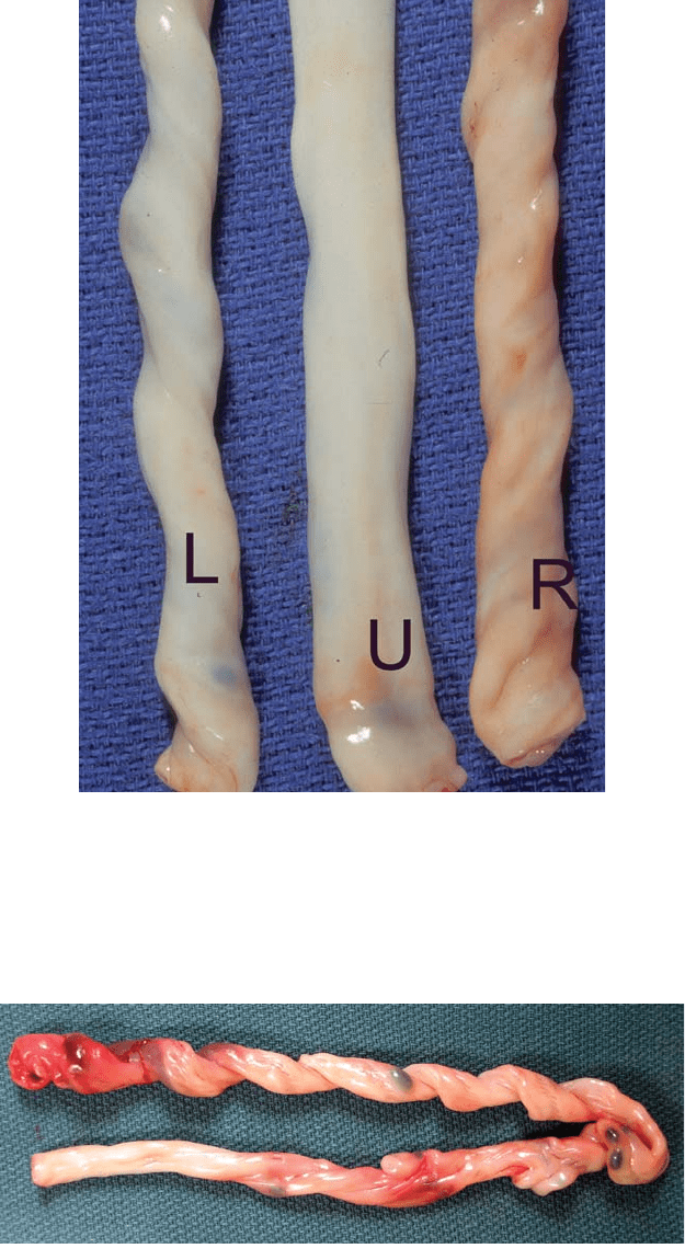
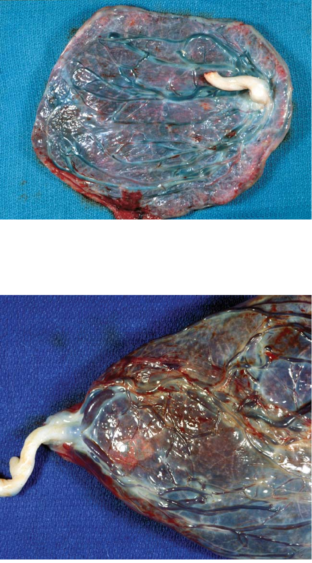
Figure 3.5. Umbilical cords showing left (L), absent (A), and right (R) twists.
Infants whose cords lack a twist exhibit more perinatal morbidity. Cords missing
one umbilical artery are also more frequently untwisted. No other correlations
with fetal outcome have been identified.
Figure 3.6. It is not unusual for a cord to have regions with differing directions
and density of twisting. Here a right twisted cord becomes an untwisted one.
Length 29
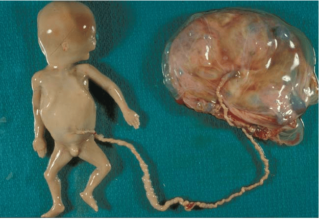
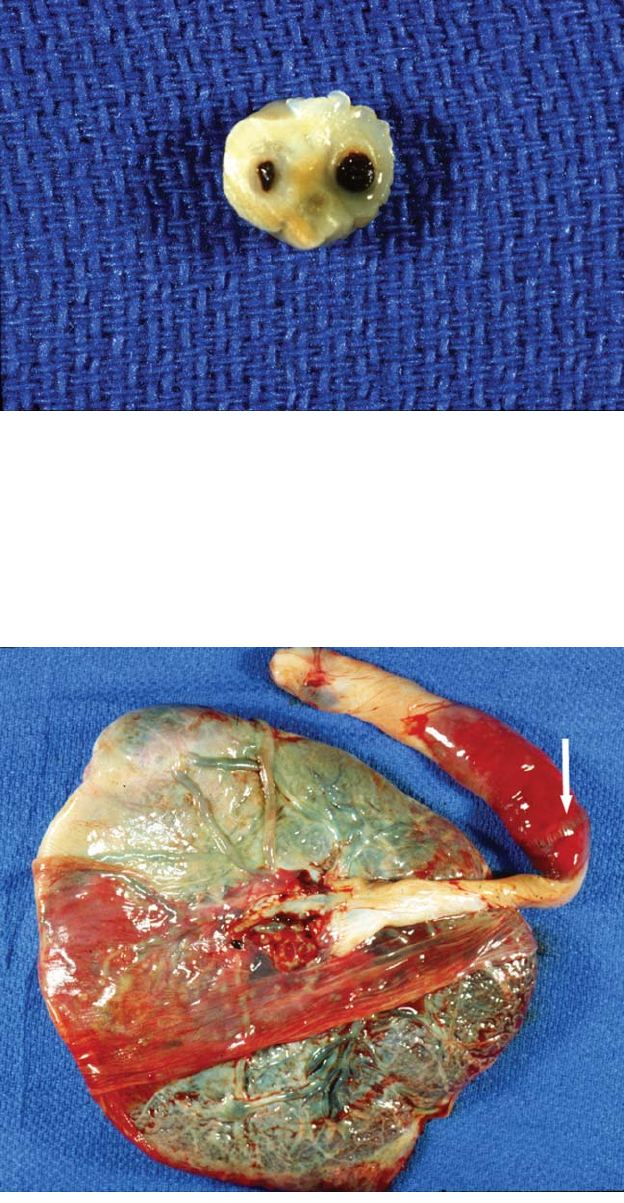
Figure 3.7. This mid-trimester fetal demise shows an excessively long and
twisted cord. Markedly twisted cords may be associated with fetal compromise
or death. Such twisting is not a postmortem artifact and is seen throughout
gestation. No other cause of fetal death was found on complete autopsy with
karyotype.
associated with fetal morbidity and mortality (Figure 3.7). In general, one
should be cautious in attributing fetal death to this or other cord prob-
lems particularly if congestion and/or thrombosis are absent. It may one
of several factors, or truly incidental.
Length
One of the most obvious features of the umbilical cord is the length. This
increases throughout gestation, although the growth rate slows in the
third trimester. Fetal activity and stretch on the cord are major factors
determining length. There is a genetic component. Normal tables have
been developed (Appendices B-4 and B-5), based on the entire length.
Both abnormally long and short cords have significant clinical correlates.
Long cords (>75 cm) are well associated with knots and fetal entangle-
ments. They may correlate with later hyperactivity. Congestion and
thrombosis in cord vessels are important signs of true obstruction (Figure
3.8 to Figure 3.14).
30 Chapter 3 Umbilical Cord
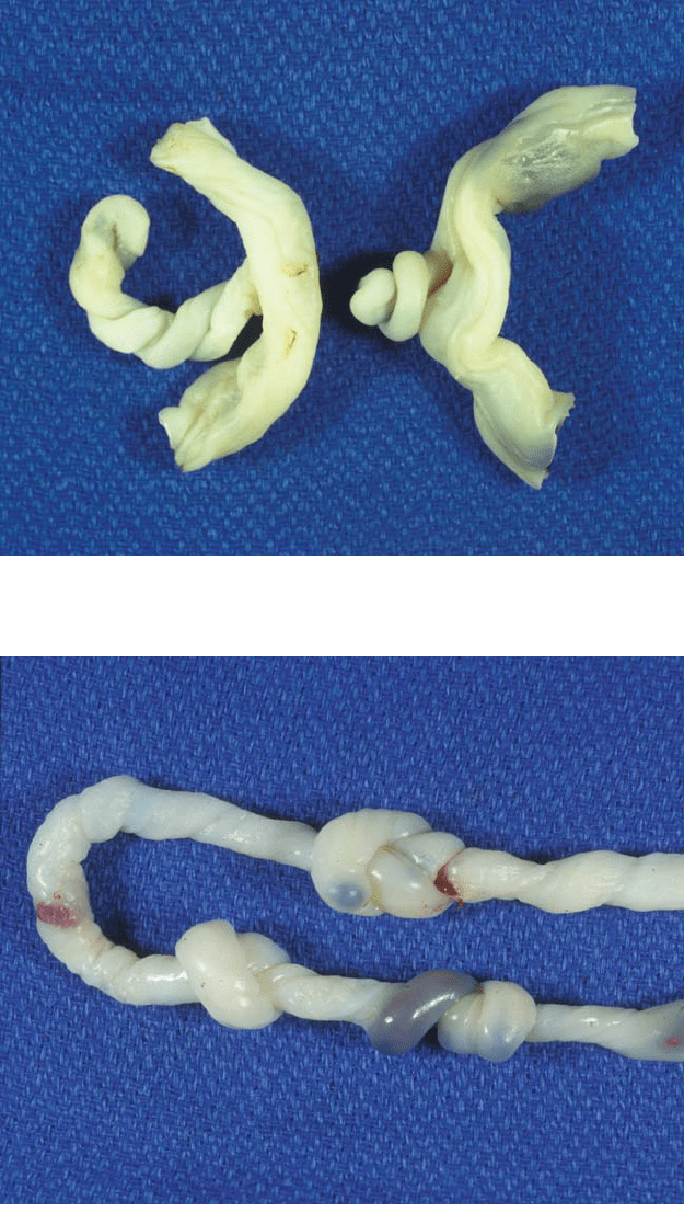
Figure 3.8. The vein redundancy in false knots can be quite impressive. These
are of no clinical significance and are not prone to thrombosis or hemorrhage.
Figure 3.9. These complicated knots occurred in a 31-week infant.There is slight
congestion, but no thrombosis was noted. There were no clinical signs of cord
compromise. True knots and entanglements are common. Most are not associ-
ated with problems. They do occasionally cause fetal distress and death. Knots
should be carefully examined for changes which suggest functionally significant
obstruction.

Length 31
Figure 3.10. In fatal cord compressions, flow in the vein has usually been com-
promised, leading to congestion on the placental side. Such was the case in this
intrauterine demise.
Figure 3.11. This midtrimester loss was thought to be due to true cord entan-
glement and occlusion. A complete autopsy including cytogenetics failed to
reveal other significant pathology.
32 Chapter 3 Umbilical Cord
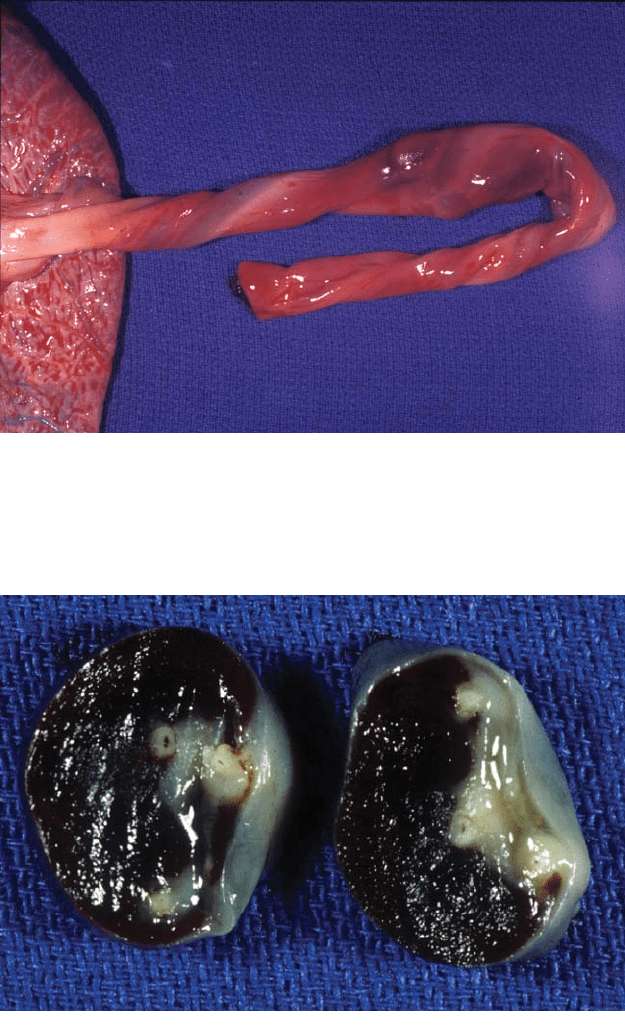
Figure 3.13. This three-vessel cord shows thrombosis of one artery. An occlusive
thrombus in one of two umbilical arteries can occur without fetal problems
because there are usually vascular anastomoses between the two arteries. This
enables perfusion of the entire placenta.
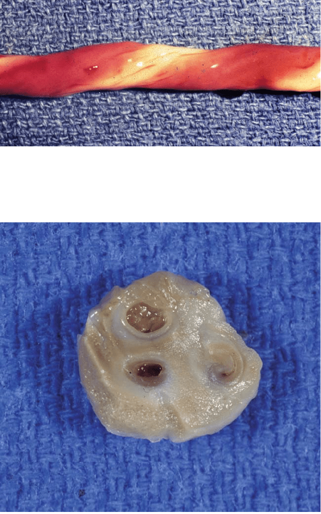
Figure 3.12. The only source of nutrients to the umbilical arteries is the blood
flow. If an artery is totally thrombosed the muscle will become necrotic allowing
leakage of blood pigments which discolor the cord stroma along its course.
Length 33
Figure 3.14. This
cord cross-section
shows a very small
artery with pigment
in the surrounding
tissue. Thrombosis
and eventual
disappearance of
that vessel is a
common etiology
of single umbilical
artery.
A minimum cord length of 32 cm is felt to be necessary for normal
vaginal delivery. Undue traction on the cord can cause fetal distress, cord
tearing with hemorrhage, and possibly placental separation. The major-
ity of hemorrhages in the cord will be associated with clamp marks and
are artifact (Figure 3.15 to Figure 3.17). Entanglement can lead to a func-
tionally short cord. Short cords are known to occur in disorders with
Figure 3.15. The
area of hemorrhage
shows a clamp mark
(arrow). It is
unlikely this is a true
rupture of the cord
and is probably not
the cause of fetal
distress. The majority
of cord hemorrhages
are an artifact,
associated with cord
clamping. Ideally,
microscopic sections
are not taken from
such areas. These
marks are often
quite numerous from
cord traction with
a clamp during
placental delivery.
34 Chapter 3 Umbilical Cord
Figure 3.16. This hemorrhage occurred in a stillborn infant with a short cord,
complete length of 32 cm. The occlusive hematoma appeared to be arterial in
origin and compressed the umbilical vein. Early thrombosis was present.
Figure 3.17. Both true cord hemorrhages and artifactual ones have a similar
appearance on cross-section. The blood often tracks for considerable distances
along the vessels. In problematic cases, multiple microscopic sections from the
area may show vital changes.
Insertion 35
decreased fetal movement (oligohydramnios, arthrogryposis). They are
also associated with more problems in neurological development, sug-
gesting the associated infants may have had longstanding in utero prob-
lems compromising mobility. Because of these associations, it is
extremely important to measure the entire cord length, including that
left on the baby or taken for cord gases. Ideally this is done in the deliv-
ery room.
Diameter
Premature infants tend to have thicker umbilical cords than more mature
babies, while cord substance is often lacking and cords are thin in utero-
placental insufficiency. Edema of the cord can be impressive. It is incon-
sistently seen in a variety of pathologic states (Figure 3.16). Isolated
areas of true cord stricture also occur, particularly near the fetal body
wall and at the placenta cord insertion (Figure 3.17).
Insertion
The insertion of the cord into the placental disk occurs in a variety of
sites. It may be into the placental substance or into membranes.The posi-
tion of insertion is due to the plane of implantation of the conception
and/or differential placental growth from uterine conditions. Placentas
with velamentous vessels are particularly important to evaluate and doc-
ument, since such vessels can be associated with compression or rupture
(Figure 3.18 to Figure 3.25). While these are usually seen with velamen-
tous insertions, small membranous vessels can be present along the edge
with normal insertions.
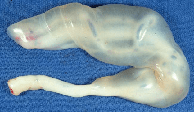
Figure 3.18. Marked edema is present in this umbilical cord. The vessels become
cordlike strands within the very loose Wharton’s jelly. While such edema may be
seen with a variety of perinatal diseases, it is most often an impressive inciden-
tal finding.
36 Chapter 3 Umbilical Cord
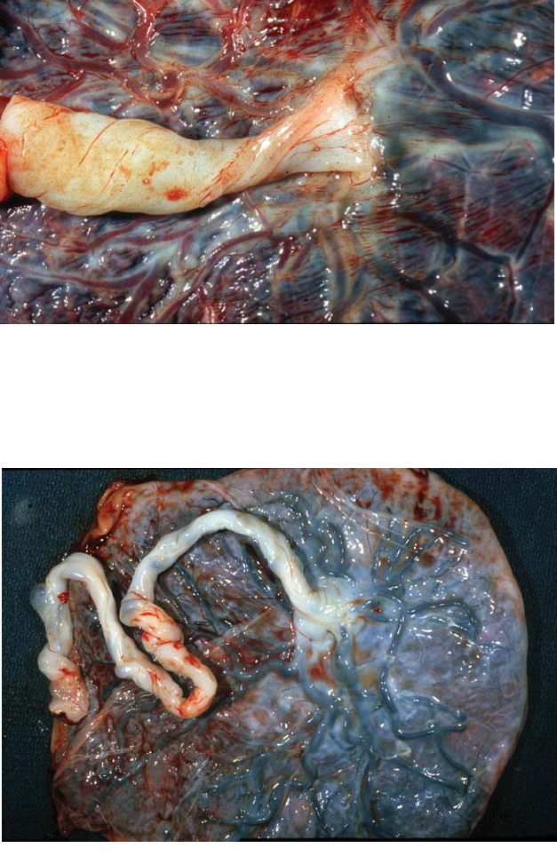
Figure 3.19. This cord becomes narrowed near its insertion into the placenta
with reduced Wharton’s jelly. Differences in diameter are common along the
length. It is usually wider close to the infant. Strictures which are physiologically
significant often show microscopic thrombosis.
Figure 3.20. Most placentas will have a cord insertion in the center or slightly
eccentric in the disk, the latter shown here. The surface vessels disperse from the
cord in a relatively even circumferential manner. Even when the cord has been
torn from the placenta, examination of the distribution of surface blood vessels
usually reveals the site of insertion.
Insertion 37
Figure 3.21. This cord inserts close, but not quite at the margin of the placenta.
The vessels course in one direction away from the cord. Such a vascular distrib-
ution is found in 38% of placentas and is likely somewhat less effective in per-
fusing the fetus.
Figure 3.22. A true marginal insertion is present (Battledore placenta). Mem-
branous vessels adjacent to marginal cord insertions are common. Marginal and
velamentous cords may be less mobile and more prone to compromise. The
infants are slightly smaller on average. In many of the very peripheral insertions,
there are a reduced number of fetal surface vessel branches.